This website uses cookies to ensure you get the best experience on our website.


- Table of Contents
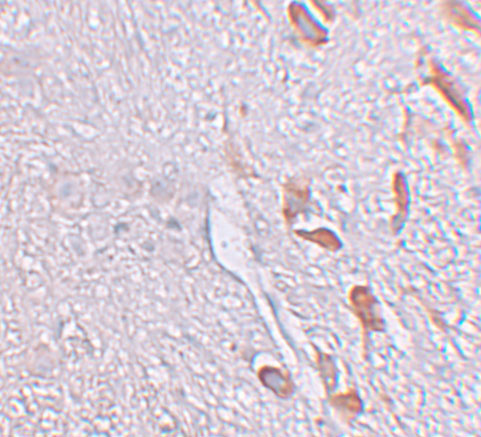
This LRRTM1 IHC guide covers cytoplasmic brain staining, including neuronal staining in caudate, cerebral cortex and hippocampus, and Purkinje cell staining in cerebellum (HPA tissue IHC). The catalog antibody is validated for human IHC-P at a starting concentration of 5 μg/mL (datasheet: A08006).

Expected localisation, validated protocols, controls and antibodies — the at-a-glance facts below, then the full design guide.
| Expected localisation | Brain cytoplasm (HPA tissue IHC); postsynaptic membrane (UniProt) | |
| Staining pattern | Cytoplasmic staining in brain neuronal cells (HPA tissue IHC) | |
| Antigen retrieval | Citrate pH 6.0 HIER, 95–98 °C, 20 min (rule: cytoplasmic / membrane antigen) | |
| Positive control | Testis+4 more · see all | |
| Negative control | Adipose tissue+4 more · see all |
| Fixation | Keep fixation consistent across sections. (standard IHC practice; not target-specific) | |
| Caveat | Presumed off-target staining has been observed (HPA tissue IHC) | |
| Regulation | Regulation not reported (UniProt) | |
| Isoform / epitope | No isoforms annotated; extracellular vs cytoplasmic epitope matters (UniProt) |
The catalog antibody protocol is paired with one published LRRTM1 IHC protocol using paraffin sections of human teeth (PMC3620385).
| Sample | Tissue sections; selected-image fixative not specified (standard IHC workflow) |
| Fixation | Image fixative and duration unreported (datasheet A08006); verify before use. |
| Sectioning | 4–5 µm sections on charged slides (standard) |
| Deparaffinisation | Xylene, graded ethanol series to water (standard) |
| Antigen retrieval | Heat-induced epitope retrieval in citrate buffer, pH 6.0, 20 min at 95–98 °C (standard rule: cytoplasmic / membrane antigen) |
| Peroxidase block | 3% H2O2, 10 min, room temperature (standard) |
| Blocking | 10% normal serum of the secondary host, 30 min, room temperature (standard) |
| Primary antibody | Rabbit anti-LRRTM1, 5 μg/mL (datasheet A08006) |
| Primary incubation | Overnight at 4 °C (standard) |
| Detection | HRP-polymer secondary, DAB chromogen 5–10 min (standard) |
| Counterstain | Hematoxylin, blue, dehydrate and mount (standard) |
| Expected result | LRRTM1-positive staining in elongated or late spermatids of testis (HPA tissue IHC: High). HPA tissue profile: Cytoplasmic expression in brain. No signal in the no-primary control. |
LRRTM1 is a cell membrane protein with a postsynaptic location and an extracellular region spanning residues 35–427 (UniProt Q86UE6 topology). In tissue IHC, expect cytoplasmic staining in brain, including neuronal cells in the cerebral cortex and caudate (HPA tissue IHC). HPA rates its tissue staining “Approved,” with medium agreement between staining and RNA data and presumed off-target binding disregarded (HPA tissue IHC).
| Medium staining in cerebral cortex or caudate neuronal cells, with a cytoplasmic pattern. | This matches the reported brain tissue pattern (HPA tissue IHC: Medium in neuronal cells; cytoplasmic expression in brain). Interpret the visible compartment as tissue IHC staining; UniProt’s postsynaptic membrane annotation describes protein location, not a requirement that chromogenic sections resolve synapses (UniProt Q86UE6 subcellular location; standard IHC practice). |
| Strong nuclear-only staining in brain cells, with little staining in the expected neuronal cytoplasm. | Nuclear-only staining is outside the reported brain pattern (HPA tissue IHC: cytoplasmic expression in brain). Review the no-primary control and tissue morphology for detection artefact (standard IHC practice). A compartment mismatch alone does not identify the offending reagent or establish that LRRTM1 is absent. |
| Prominent staining in cells reported as not detected, such as adipocytes in adipose tissue or glandular cells in adrenal gland. | These cells are negative comparators in the HPA tissue survey (HPA tissue IHC: Not detected in adipocytes and adrenal glandular cells). Consider antibody cross-reactivity or endogenous chromogenic activity, then compare controls and localisation (standard IHC practice). Staining outside the survey pattern warrants investigation, not automatic exclusion. |
| A uniform haze across tissue and blank areas, obscuring differences between cells. | A haze without anatomical structure is difficult to score as LRRTM1 staining (standard IHC practice). Check the no-primary slide for detection background and review blocking, washes and chromogen exposure (standard IHC practice). HPA’s reported neuronal and spermatid staining is cell-specific, so a uniform haze lacks that supporting distribution (HPA tissue IHC). |
| No visible signal in cerebral cortex neuronal cells on an otherwise intact section. | This conflicts with the reported medium cortical neuronal staining (HPA tissue IHC: Cerebral cortex, Medium). First verify that the positive control and detection system worked; then review antibody use and the section’s neuronal content (standard IHC practice). A single negative section cannot overturn the HPA pattern or establish target absence. |
| Compartment and topology | UniProt places LRRTM1 at the cell and postsynaptic membranes, with one transmembrane span at 428–448 and a cytoplasmic region at 449–522 (UniProt Q86UE6 topology). HPA nevertheless describes tissue IHC as cytoplasmic in brain (HPA tissue IHC). Score the observed IHC pattern against that tissue evidence; do not demand a resolved synaptic outline in routine chromogenic sections (standard IHC practice). |
| Choice of tissue and cell population | The strongest listed staining is in elongated or late spermatids in testis; listed brain neuronal populations are medium, including cortex, caudate and hippocampus (HPA tissue IHC). Select and score the specified cell population, because a tissue-wide intensity label would obscure these differences (standard IHC practice). |
| Strength of tissue evidence | The tissue profile is “Approved,” with medium staining–RNA consistency and presumed off-target binding disregarded (HPA tissue IHC). The listed patterns guide expectations, but an isolated positive cell population needs morphological and control review (standard IHC practice). HPA062660 has IHC “Approved”; HPA046607 has ICC “Approved,” with no IHC status listed (HPA antibody validation). |
| IF/ICC Q&A: Should its localisation match IHC? | HPA reports an approved Golgi apparatus location in ICC-IF images from SH-SY5Y and U2OS, while its tissue IHC summary reports cytoplasmic expression in brain (HPA subcellular; HPA tissue IHC). These are different assay contexts; use the tissue IHC profile to judge paraffin sections (standard IHC practice). |
| Situation | Likely cause | Next action |
|---|---|---|
| Cortical neurons are unstained. | The result differs from the reported medium cortical neuronal signal (HPA tissue IHC); a failed stain or poorly represented target cells is possible (standard IHC practice). | Check section morphology and neuronal content, then compare a known-positive section and detection controls processed in the same run (standard IHC practice). Review the antibody’s IHC validation before interpreting a negative sample (HPA antibody validation). |
| Brain staining is predominantly nuclear. | The compartment differs from HPA’s cytoplasmic brain profile and UniProt’s membrane annotation (HPA tissue IHC; UniProt Q86UE6 subcellular location). The cause remains unresolved from localisation alone. | Inspect morphology, compare the no-primary control and repeat the stain with appropriate controls if needed (standard IHC practice). Do not score nuclear-only signal as the expected LRRTM1 pattern (HPA tissue IHC). |
| Adipose adipocytes stain strongly. | HPA reports these cells as not detected (HPA tissue IHC). Cross-reactivity or chromogenic background is possible (standard IHC practice). | Compare matched no-primary and negative-tissue controls; check whether colour follows cells or appears broadly across the section (standard IHC practice). Record the mismatch with the HPA tissue profile before assigning target specificity (HPA tissue IHC). |
| Colour appears throughout tissue and on the no-primary control. | Detection background, including endogenous activity, is plausible when signal persists without primary antibody (standard IHC practice). | Review blocking and detection controls, washing and chromogen development using the assay’s instructions (standard IHC practice). Reassess cell-specific staining only after the background is controlled (standard IHC practice). |
| Testis stains strongly but brain appears weaker. | That ranking matches the listed High signal in elongated or late spermatids and Medium signal in several neuronal populations (HPA tissue IHC). | Compare the specified cell types rather than whole-section darkness (standard IHC practice). Accept a weaker neuronal signal when its distribution and controls fit the reported brain pattern (HPA tissue IHC; standard IHC practice). |
| ICC-IF shows Golgi signal, but the paraffin IHC slide looks cytoplasmic. | HPA reports approved Golgi localisation for ICC-IF and cytoplasmic expression for brain tissue IHC (HPA subcellular; HPA tissue IHC). | Judge the paraffin slide against tissue IHC evidence and its controls (standard IHC practice). Treat the ICC-IF image as assay-specific localisation evidence, not a requirement for Golgi-shaped chromogenic staining (HPA subcellular; standard IHC practice). |


Comprehensive Human Protein Atlas IHC scoring per tissue (reliability: Approved — Medium consistency between antibody staining and RNA expression data. Presumed off target binding observed and disregarded.). Rows are taken directly from the HPA tissue chart — click any row's HPA link to view the source.
| Tissue | Cell type | Level | Evidence | Source |
|---|---|---|---|---|
| Testis | Elongated or late spermatids | High | Protein (IHC) | HPA → |
| Caudate | Neuronal cells | Medium | Protein (IHC) | HPA → |
| Cerebellum | Purkinje cells | Medium | Protein (IHC) | HPA → |
| Cerebral cortex | Neuronal cells | Medium | Protein (IHC) | HPA → |
| Hippocampus | Neuronal cells | Medium | Protein (IHC) | HPA → |
| Tissue | Cell type | Level | Evidence | Source |
|---|---|---|---|---|
| Adipose tissue | Adipocytes | Not detected | Protein (IHC) | HPA → |
| Adrenal gland | Glandular cells | Not detected | Protein (IHC) | HPA → |
| Appendix | Glandular cells | Not detected | Protein (IHC) | HPA → |
| Bone marrow | Hematopoietic cells | Not detected | Protein (IHC) | HPA → |
| Breast | Adipocytes | Not detected | Protein (IHC) | HPA → |
Troubleshoot LRRTM1 chromogenic IHC in paraffin sections using the product image, protein topology, and tissue staining evidence as reference points.
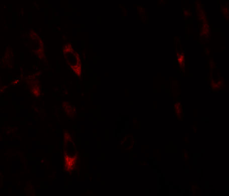
The catalog antibody has IHC and IF images in human brain tissue (IHC/IF image captions) and lists Human, Mouse and Rat reactivity (catalog reactivity).
A08006 has an IHC image in human brain tissue at 5 μg/mL (IHC image caption). A08006 also has an IF image in human brain tissue at 20 μg/mL (IF image caption).
Which to pick: For paraffin-section IHC, choose A08006: IHC-P is listed (catalog applications), and its own IHC image shows human brain tissue at 5 μg/mL; the fixative is unreported (IHC image caption). For IF, A08006 has a human brain tissue image at 20 μg/mL (IF image caption); ICC validation is unreported (catalog applications). For cross-species work, A08006 lists Human, Mouse and Rat reactivity (catalog reactivity), but the supplied IHC and IF images show human brain tissue only (IHC/IF image captions); it is rabbit-hosted, with clonality unreported (catalog host/clone).