LZTS2 / Leucine zipper putative tumor suppressor 2 · IHC design guide

Design Immunohistochemistry for LZTS2

Plan chromogenic LZTS2 IHC-P around cytoplasmic tissue staining (HPA tissue IHC). Use prostate glandular cells as a high-staining reference (HPA tissue IHC), and start antibody A07806 at 5 μg/mL (datasheet: A07806).

Evidence assembled Oct 2026 · For research use; verify linked source records and product datasheet before use
Immunohistochemistry protocol sheet for LZTS2 (IHC for LZTS2): expected localisation Cytoplasmic tissue staining (HPA tissue IHC); centrosomes (UniProt), antibody A07806, validated IHC image, and IHC protocol steps
Printable LZTS2 IHC protocol sheet — expected localisation Cytoplasmic tissue staining (HPA tissue IHC); centrosomes (UniProt), antibody A07806, controls and protocol steps. Open the full LZTS2 IHC guide →

LZTS2 Immunohistochemistry Experimental Design Guide

Expected localisation, validated protocols, controls and antibodies — the at-a-glance facts below, then the full design guide.

Must know before staining
Expected localisation Cytoplasmic tissue staining (HPA tissue IHC); centrosomes (UniProt)
Staining pattern Cytoplasmic staining in several tissue cell types (HPA tissue IHC)
Antigen retrieval Citrate pH 6.0 HIER, 95–98 °C, 20 min (rule: cytoplasmic / membrane antigen)
Positive control ⓘ Bronchus+4 more · see all
Negative control ⓘ Adipose tissue+4 more · see all
Important caveats
Reasons your staining may differ from the expected pattern.
Fixation Keep fixation consistent across sections. (standard IHC practice; not target-specific)
Caveat Antibody staining and RNA show low consistency (HPA tissue IHC)
Regulation Prostate-enriched expression (UniProt)
Isoform / epitope No isoforms annotated; one 1–669 chain (UniProt)
Section 1

Recommended LZTS2 IHC & IF Protocols

Compare the catalog antibody’s IHC-P protocol with three published LZTS2 IHC workflows (PMC5360334; PMC7892348; PMC9013323).

Recommended immunohistochemistry (IHC-P) protocol parameters
SampleTissue sections; selected-image fixative not specified (standard IHC workflow)
FixationImage fixative and duration unreported (datasheet A07806); verify before use.
Sectioning4–5 µm sections on charged slides (standard)
DeparaffinisationXylene, graded ethanol series to water (standard)
Antigen retrievalHeat-induced epitope retrieval in citrate buffer, pH 6.0, 20 min at 95–98 °C (standard rule: cytoplasmic / membrane antigen)
Peroxidase block3% H2O2, 10 min, room temperature (standard)
Blocking10% normal serum of the secondary host, 30 min, room temperature (standard)
Primary antibodyRabbit anti-LZTS2, 5 μg/mL (datasheet A07806)
Primary incubationOvernight at 4 °C (standard)
DetectionHRP-polymer secondary, DAB chromogen 5–10 min (standard)
CounterstainHematoxylin, blue, dehydrate and mount (standard)
Expected resultLZTS2-positive staining in ciliated cells (cell body) of bronchus (HPA tissue IHC: High). HPA tissue profile: Cytoplasmic expression in several tissues. No signal in the no-primary control.
💡Decision noteStart with citrate pH 6.0 HIER at 95–98 °C for 20 min (page antigen retrieval).
Section 2

What Is the Expected LZTS2 Staining Pattern?

LZTS2 is reported in the cytoplasm, centrosome and, during cytokinesis, midbody (UniProt Q9BRK4). In tissue IHC, expect cytoplasmic staining in selected cells, including prostate glandular cells and bronchial ciliated cell bodies (HPA tissue IHC). LZTS2 has no transmembrane segment (UniProt Q9BRK4 topology). The tissue profile is Approved, with low consistency between antibody staining and RNA expression, so interpret intensity cautiously (HPA tissue IHC).

What am I looking at on my slide?
Cytoplasmic stain in prostate glands or bronchial ciliated cell bodies.This matches High staining reported for those cell types (HPA tissue IHC). Score the named cells and their cytoplasm; staining elsewhere in the section does not establish the same pattern. The cytoplasmic location also agrees with UniProt Q9BRK4.
Predominantly nuclear stain with little cytoplasmic signal.A nuclear dominant pattern does not match the reported tissue cytoplasmic profile (HPA tissue IHC) or the annotated cytoplasmic and centrosomal locations (UniProt Q9BRK4). Treat it as suspect, then compare a positive control and a no-primary control before assigning it to LZTS2 (general IHC practice).
Strong stain in adipocytes or lung alveolar cells.Those specific cells were Not detected in the HPA tissue survey (HPA tissue IHC). Unexpected staining can indicate cross-reactivity or endogenous chromogenic activity; check the no-primary control and the cellular boundaries before calling it positive (general IHC practice).
Diffuse brown haze obscures cell boundaries.The reported result is cellular cytoplasmic staining, so haze that cannot be assigned to cells is not interpretable as the expected pattern (HPA tissue IHC). Compare background across the section and a no-primary control; nonspecific staining or detection background can mimic a weak positive (general IHC practice).
No stain in a prostate glandular-cell positive control.Prostate glandular cells showed High staining in the HPA survey (HPA tissue IHC). A blank control therefore raises a staining-run or specimen question, but does not alone prove antibody failure. Verify that the expected cells are present and review the IHC workflow (general IHC practice).
💡Expected LZTS2 appearanceCall a section positive when defined cell bodies show cytoplasmic stain, such as High staining in prostate glandular cells (HPA tissue IHC); nuclear dominant stain or uniform brown haze is suspect against that pattern (HPA tissue IHC; general IHC practice).
How each factor affects the staining
Cell-specific tissue patternHPA reports High staining in prostate glands, liver cholangiocytes and pancreatic exocrine cells, but Not detected in adipocytes (HPA tissue IHC). Identify the cell type before scoring a tissue.
Tissue-profile confidenceThe tissue profile is Approved, yet antibody staining has low consistency with RNA expression data (HPA tissue IHC). Use the observed IHC pattern as a reference with that limitation.
Intracellular locationUniProt annotates centrosomes throughout the cell cycle and midbodies during cytokinesis (UniProt Q9BRK4). These locations may inform interpretation of focal cellular staining; the HPA tissue summary describes cytoplasmic expression (HPA tissue IHC).
IF/ICC location evidenceCytosol is supported, while plasma membrane is uncertain in the HPA subcellular record (HPA subcellular ICC-IF). Membrane signal alone is therefore a weak basis for an IHC call.
Topology and processingLZTS2 has no transmembrane segment, signal peptide or propeptide; the annotated chain spans residues 1–669 (UniProt Q9BRK4). The supplied record gives no target-specific fixation effect.
Why is my staining missing, weak or wrong?
SituationLikely causeNext action
Positive-control prostate glands are blank.This conflicts with their reported High staining (HPA tissue IHC); a failed detection step or missing target cells are possible (general IHC practice).Confirm glandular cells are present, then check the antibody, detection reagents and retrieval steps used for the IHC run (general IHC practice).
Nuclei dominate the staining.That compartment disagrees with the reported cytoplasmic tissue pattern (HPA tissue IHC) and UniProt localization (UniProt Q9BRK4).Compare the same run with a known-positive section and a no-primary control; withhold a positive call if cytoplasmic cellular staining cannot be resolved (general IHC practice).
Adipocytes stain as strongly as expected-positive cells.Adipocytes were Not detected in the HPA survey (HPA tissue IHC); cross-reactivity or chromogenic background is possible (general IHC practice).Inspect whether signal follows cell boundaries and check a no-primary control for endogenous detection activity (general IHC practice).
Brown signal spreads across cells and empty spaces.Noncellular haze cannot be scored as the reported cytoplasmic cellular pattern (HPA tissue IHC); excess background is possible (general IHC practice).Review blocking, washes, antibody concentration and detection exposure using the same control section (general IHC practice).
Testis is faint despite its reported high expression.UniProt reports high testis expression, while HPA records Low staining in Leydig cells (UniProt Q9BRK4; HPA tissue IHC). The measures do not specify the same cellular readout.Score the identified Leydig cells against the HPA IHC observation; use a reported high-staining cell type to assess run performance (HPA tissue IHC; general IHC practice).
IF/ICC question: does membrane signal confirm LZTS2?HPA lists plasma membrane as uncertain and cytosol as supported in ICC-IF (HPA subcellular ICC-IF).Give greater weight to cytosolic cellular signal and assess membrane-only signal cautiously; use the separate IF/ICC guide for assay-specific decisions (HPA subcellular ICC-IF).

Sample controls for LZTS2 IHC & IF

🧪Run bronchus first: ciliated cells should stain (HPA: High in bronchial ciliated cells). Use adipose tissue as the negative comparator (HPA: adipocytes Not detected). On the bronchus slide, look for little or no specific staining in cells outside the ciliated-cell population, but do not assume every neighboring cell is a validated negative (HPA: ciliated cells High).
Positive control tissue: Bronchus (Ciliated cells (cell body), HPA High)
Negative control tissue: Adipose tissue (HPA Not detected)
ICC-IF cell lines (HPA subcellular resource): HPA ICC-IF images show LZTS2 in A-431, U-251MG, U2OS, with annotated localisation: Plasma membrane (uncertain), Cytosol (supported) (HPA subcellular).
Technical controls: Include a no-primary, secondary-only control; a host-species- and antibody-class-matched isotype control appropriate to the primary antibody’s clonality; and LZTS2-knockout material as a biological negative (standard IHC practice). For chromogenic bronchus IHC, block endogenous peroxidase and check mucus or pigment for background signal (standard IHC practice).
⚠️Feasibility: A LZTS2-specific fixation window and retrieval dependence are unreported in the supplied evidence; the fixative for the A07806 placenta IHC image is also unreported (caption: 5 μg/mL; fixative not stated). The supplied evidence does not establish that frozen sections or IF are easier; ICC-IF images exist for A-431, U-251MG and U2OS, but they do not establish tissue IF conditions (HPA subcellular). In bronchus, distinguish ciliated-cell staining from signal associated with mucus or pigment (HPA: bronchial ciliated cells High; standard IHC practice).

HPA tissue IHC evidence for LZTS2

Comprehensive Human Protein Atlas IHC scoring per tissue (reliability: Approved — Low consistency between antibody staining and RNA expression data.). Rows are taken directly from the HPA tissue chart — click any row's HPA link to view the source.

Positive expression · recommended positive controls

TissueCell typeLevelEvidenceSource
Bronchus Ciliated cells (cell body) High Protein (IHC) HPA →
Endometrium Glandular cells Medium Protein (IHC) HPA →
Fallopian tube Ciliated cells (cell body) High Protein (IHC) HPA →
Gallbladder Glandular cells High Protein (IHC) HPA →
Liver Cholangiocytes High Protein (IHC) HPA →

Undetected expression · recommended negative controls

TissueCell typeLevelEvidenceSource
Adipose tissue Adipocytes Not detected Protein (IHC) HPA →
Bone marrow Hematopoietic cells Not detected Protein (IHC) HPA →
Caudate Glial cells Not detected Protein (IHC) HPA →
Cerebellum Cells in granular layer Not detected Protein (IHC) HPA →
Cerebral cortex Glial cells Not detected Protein (IHC) HPA →
Section 3

Advanced LZTS2 IHC Tips

Troubleshoot LZTS2 staining in chromogenic IHC of paraffin sections, with IF guidance for checking cellular distribution.

How should I retrieve LZTS2 in paraffin sections when staining is weak?
Start with citrate pH 6.0 HIER at 95–98 °C for 20 min (page retrieval setting). Run a known positive section, such as placenta with trophoblastic cells, alongside the study sections (HPA tissue IHC: High in placental trophoblastic cells). The selected antibody image documents placental IHC at 5 μg/mL, but its caption does not document retrieval conditions (A07806 caption). If staining remains weak, compare a modestly longer retrieval time on adjacent sections while keeping detection conditions fixed (standard IHC practice). Judge improvement by cellular cytoplasmic staining and preserved morphology, rather than by stronger stain at section edges (HPA tissue IHC: cytoplasmic profile; standard IHC practice).
Could fixation explain weak or uneven LZTS2 staining?
Target-specific sensitivity of LZTS2 to fixation is unknown from the supplied evidence; the selected placental IHC caption does not state a fixative (A07806 caption). Record the fixative, fixation duration, processing history and section age for each specimen before comparing staining intensity (standard IHC practice). Compare sections with matched processing and the same citrate pH 6.0 retrieval run to assess whether the variation tracks specimen handling (page retrieval setting; standard IHC practice). Include a positive control section in each run, then inspect tissue preservation and edge staining before adjusting antibody concentration (HPA tissue IHC: High in placental trophoblastic cells; standard IHC practice).
Which LZTS2 staining patterns are credible in tissue sections?
Expect predominantly cytoplasmic staining in tissue IHC, while allowing that centrosomal or midbody signal may occupy only a small part of a cell (HPA tissue IHC: cytoplasmic profile; UniProt Q9BRK4: centrosome and midbody). The HPA subcellular summary also supports cytosol and labels plasma membrane localisation uncertain (HPA subcellular: Cytosol supported; Plasma membrane uncertain). Assess staining within morphologically intact cells and compare the compartment with the negative control on the same run (standard IHC practice). A strong, exclusively nuclear DAB pattern needs independent validation before assignment to LZTS2, whose annotated localisation is cytoplasmic (UniProt Q9BRK4: subcellular localisation; standard IHC practice).
How should epitope uncertainty affect antibody selection and staining interpretation?
The supplied record lists 0 annotated isoforms and a 669 amino acid chain, but gives no antibody epitope (UniProt Q9BRK4: isoforms and processing; A07806 caption). It also lists phosphoserines at residues 249, 296 and 570, without evidence that they affect this antibody (UniProt Q9BRK4: modified residues). Check the catalog antibody’s immunogen or epitope documentation before drawing conclusions about variant or phosphorylation dependent staining (standard IHC practice). If an epitope remains undisclosed, compare staining with an independently validated antibody against a distinct region and retain the same tissue controls (standard IHC practice).
How can IF help check an LZTS2 IHC pattern?
Use IF as a separate localisation check, multiplexing LZTS2 with a validated marker for the cell population being assessed, such as the placental trophoblastic population seen in tissue IHC (HPA tissue IHC: High in placental trophoblastic cells; standard IF practice). Choose a fluorophore channel after checking tissue autofluorescence and include single stain controls for bleed through (standard IF practice). Because LZTS2 has no annotated transmembrane segment and cytosol is a supported location, test permeabilisation when seeking intracellular signal (UniProt Q9BRK4: topology; HPA subcellular: Cytosol supported; standard IF practice). Compare the IF compartment with chromogenic IHC, while treating plasma membrane localisation cautiously because its HPA assignment is uncertain (HPA subcellular: Plasma membrane uncertain).
What should I change when diffuse DAB obscures LZTS2 staining?
First inspect a no primary control to distinguish detection background from antibody associated staining (standard IHC practice). For chromogenic detection, check the peroxidase block and shorten DAB development if the control also darkens; these are general workflow adjustments (standard IHC practice). If background appears only with primary antibody, titrate around the documented 5 μg/mL placental image concentration and compare blocking and wash conditions on adjacent sections (A07806 caption; standard IHC practice). Preserve interpretable cytoplasmic signal in expected cells while reducing diffuse extracellular stain and section edge deposits (HPA tissue IHC: cytoplasmic profile; standard IHC practice).
How should I score LZTS2 IHC across specimens? ⚠ ANSWER MARKED FOR VERIFICATION
Define the cell population and cytoplasmic compartment before scoring, because tissue IHC reports cytoplasmic expression in several tissues (HPA tissue IHC: cytoplasmic profile). Record the percentage of positive cells and an intensity based H-score on intact tissue, or positive cell density per mm² when cell numbers vary (standard IHC practice). Normalise each result to the relevant viable cell population or viable tissue area, and apply one threshold and imaging setup across the comparison (standard IHC practice). Report controls and separate cell types rather than pooling them, since HPA reports different cell level staining within tissues (HPA tissue IHC: cell level profile; standard IHC practice).
How can I distinguish genuine LZTS2 staining from artefact?
Look for reproducible cytoplasmic staining in intact expected cells, such as placental trophoblastic cells or prostatic glandular cells (HPA tissue IHC: cytoplasmic profile; High in those cells). Check whether the signal persists away from section edges and necrotic areas and disappears in a no primary control (standard IHC practice). Investigate exclusively nuclear staining, staining in an unexpected cell population or DAB signal present after omitting primary antibody before calling a specimen positive (UniProt Q9BRK4: cytoplasmic localisation; standard IHC practice). Interpret discordance cautiously: HPA marks tissue staining Approved but reports low consistency between antibody staining and RNA expression (HPA tissue IHC: reliability description).
Boster reagents

Best LZTS2 / Leucine zipper putative tumor suppressor 2 IHC Antibodies

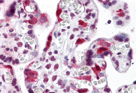
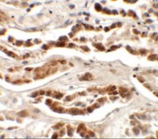
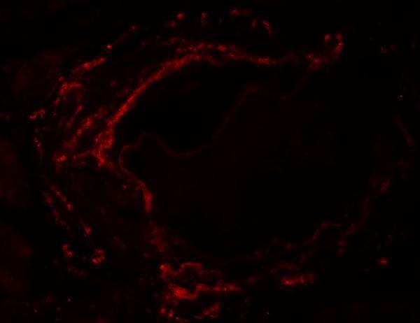
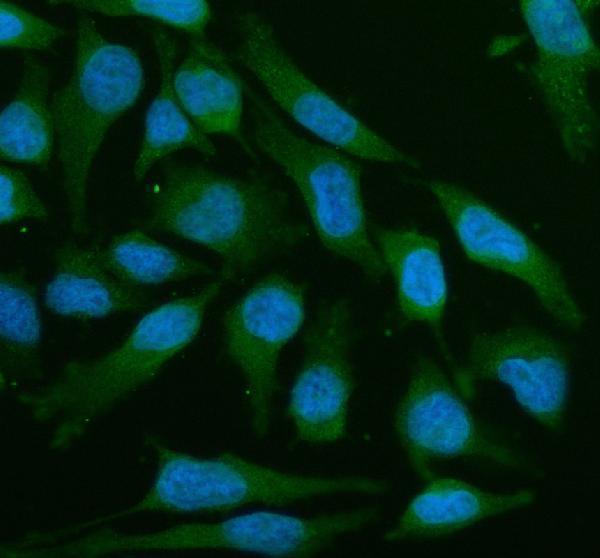
The catalog shows LZTS2 IHC data in human placenta and kidney tissue and IF data in human kidney tissue and HeLa cells (image captions). Listed reactivity also includes mouse and rat (catalog reactivity).

Real IHC data Immunohistochemistry of LZTS2 in human placenta tissue with LZTS2 antibody at 5 μg/mL.
Anti-LZTS2 Antibody
Cat # A07806
Real IHC data Immunohistochemistry of LZTS2 in human kidney tissue with LZTS2 antibody at 2.5 μg/mL.
Anti-LZTS2 Antibody
Cat # A07806-1
Real IF data IF analysis of LZTS2 using anti-LZTS2 antibody (A07806-3). LZTS2 was detected in an immunocytochemical section of HELA cells. Enzyme antigen retrieval was performed using IHC enzyme antigen retrieval reagent (AR0022) for 15 mins. The cells were blocked with 10% goat serum. And then incubated with 5 μg/mL rabbit anti-LZTS2 Antibody (A07806-3) overnight at 4°C. DyLight®488 Conjugated Goat Anti-Rabbit IgG (BA1127) was used as secondary antibody at 1:500 dilution and incubated for 30 minutes at 37°C. The section was counterstained with DAPI. Visualize using a fluorescence microscope and filter sets appropriate for the label used.
Anti-LZTS2 Antibody ®
Cat # A07806-3

A07806 has human placenta IHC data (A07806 image caption); A07806-1 has human kidney IHC and IF data (A07806-1 image captions). A07806-3 has IF/ICC data in HeLa cells (A07806-3 image caption).

Which to pick: For paraffin-section IHC, choose polyclonal A07806 for human placenta or A07806-1 for human kidney (catalog: IHC-P; respective image captions); the captions do not report a fixative (image captions). For IF/ICC, A07806-3 has a HeLa cell IF example, while A07806-1 has a human kidney IF example (catalog applications; respective image captions). For planned mouse work, A07806-1 lists IHC-P and IF reactivity; A07806-3 lists IF/ICC reactivity in human, mouse, and rat, though its pictured example is human cells (catalog applications and reactivity; A07806-3 image caption).

Each figure is that product's own IHC / IF validation image from its datasheet.

References

  1. UniProt Consortium. UniProt entry Q9BRK4 (LZTS2_HUMAN, Leucine zipper putative tumor suppressor 2).
  2. Human Protein Atlas. LZTS2 tissue IHC expression (reliability: Approved).
  3. Human Protein Atlas. LZTS2 subcellular location (ICC-IF): Localized to the plasma membrane and cytosol..
  4. Human Protein Atlas. LZTS2 antibody validation summary (1 antibodies).
  5. LZTS2 and PTEN collaboratively regulate ß-catenin in prostatic tumorigenesis. PloS one 2017 — PMC5360334.
  6. β-Trcp and CK1δ-mediated degradation of LZTS2 activates PI3K/AKT signaling to drive tumorigenesis and metastasis in hepatocellular carcinoma. Oncogene 2021 — PMC7892348.
  7. Long non-coding RNA linc00921 suppresses tumorigenesis and epithelial-to-mesenchymal transition of triple-negative breast cancer via targeting miR-9-5p/LZTS2 axis. Human cell 2022 — PMC9013323.
  8. PubMed PMID:11347906 — UniProt-cited evidence.
  9. PubMed PMID:15164054 — UniProt-cited evidence.
  10. PubMed PMID:15489334 — UniProt-cited evidence.