This website uses cookies to ensure you get the best experience on our website.
- Table of Contents
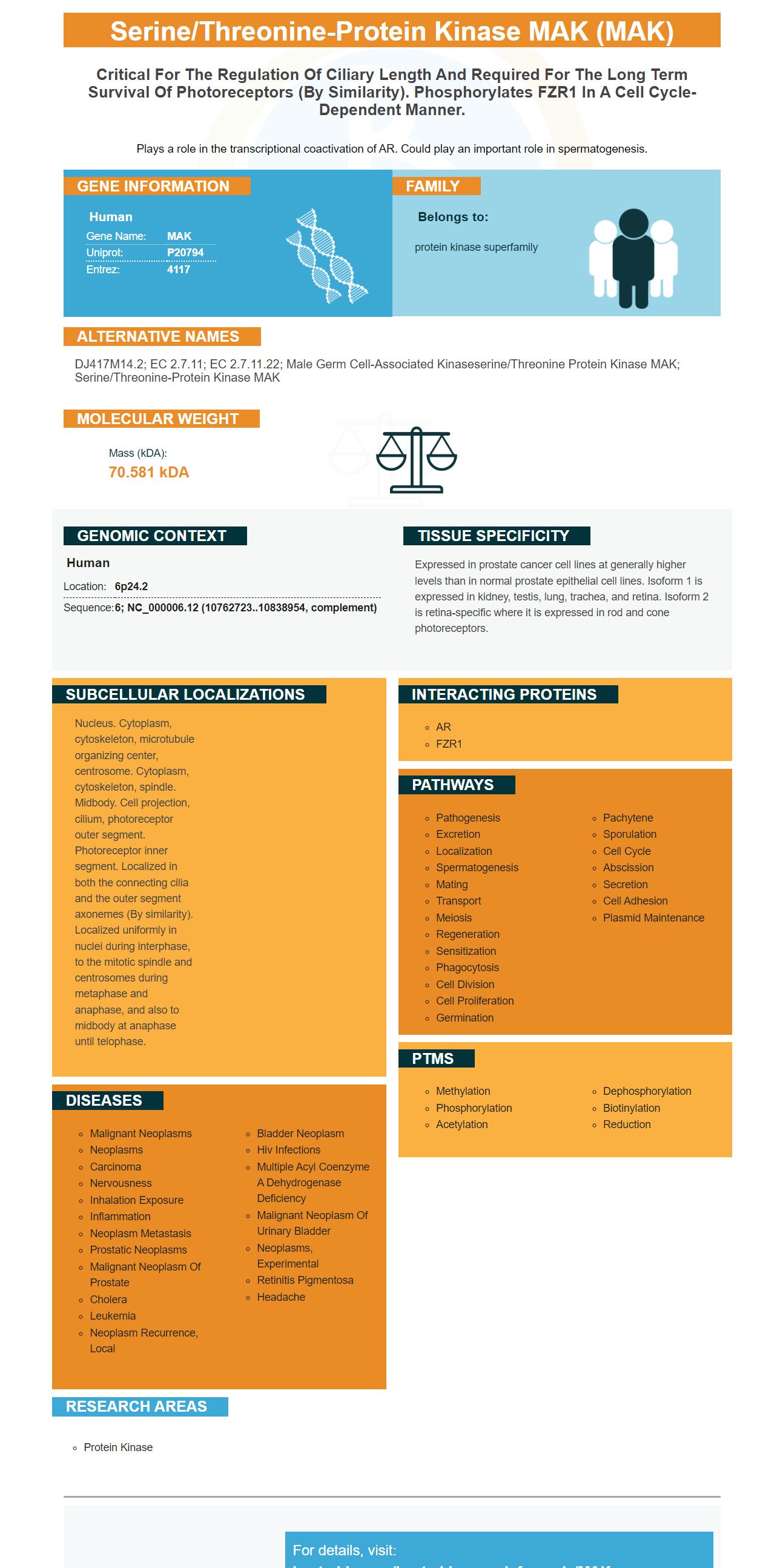
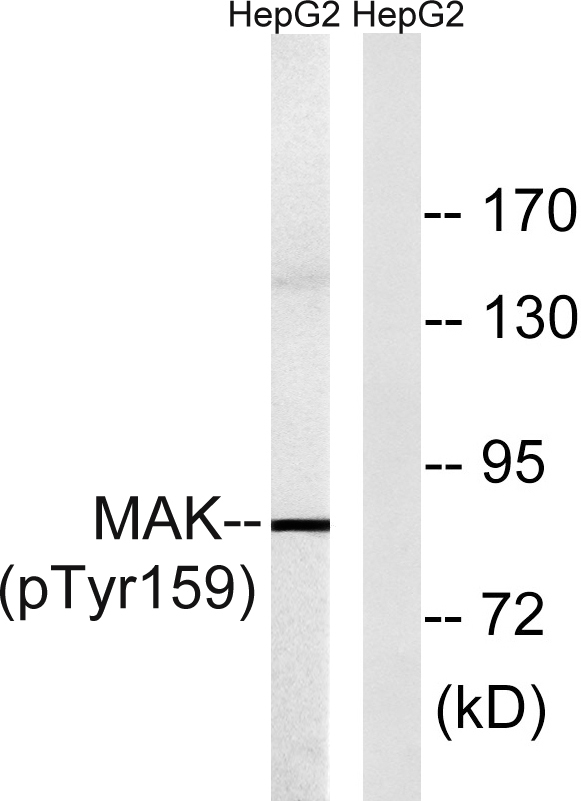
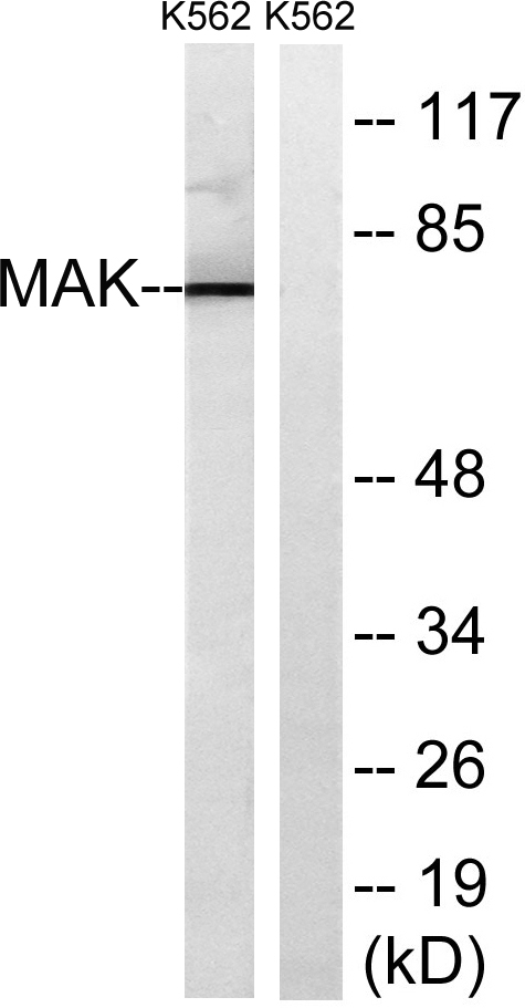
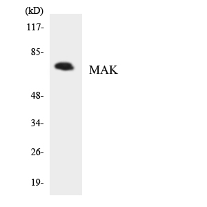
Facts about Serine/threonine-protein kinase MAK.
Plays a role in the transcriptional coactivation of AR. Could play an important role in spermatogenesis.
Human | |
---|---|
Gene Name: | MAK |
Uniprot: | P20794 |
Entrez: | 4117 |
Belongs to: |
---|
protein kinase superfamily |
dJ417M14.2; EC 2.7.11; EC 2.7.11.22; male germ cell-associated kinaseserine/threonine protein kinase MAK; serine/threonine-protein kinase MAK
Mass (kDA):
70.581 kDA
Human | |
---|---|
Location: | 6p24.2 |
Sequence: | 6; NC_000006.12 (10762723..10838954, complement) |
Expressed in prostate cancer cell lines at generally higher levels than in normal prostate epithelial cell lines. Isoform 1 is expressed in kidney, testis, lung, trachea, and retina. Isoform 2 is retina-specific where it is expressed in rod and cone photoreceptors.
Nucleus. Cytoplasm, cytoskeleton, microtubule organizing center, centrosome. Cytoplasm, cytoskeleton, spindle. Midbody. Cell projection, cilium, photoreceptor outer segment. Photoreceptor inner segment. Localized in both the connecting cilia and the outer segment axonemes (By similarity). Localized uniformly in nuclei during interphase, to the mitotic spindle and centrosomes during metaphase and anaphase, and also to midbody at anaphase until telophase.
If you've been using Boster Bio's CD2 and IL6 markers for cancer research, you've probably wondered how to use the MAK Marker as well. Both markers are very similar and serve a similar purpose in the body. But which is the most important use for this marker? Learn how to make the best of this marker when conducting research.
Boster Bio's CD2 marker binds to the extracellular tail of CD2. The intracellular tail of CD2 is linked to the extracellular domain by a transmembrane loop. It contains five SH3 binding regions, two of them acting as GYF binding motifs. These domains play an important role in CD2 function.
The CD2 cytoplasmic domain interacts with the extracellular domain of CD5. The CD2 cytoplasmic domain is responsible for interaction with the extracellular domain of CD5.
Antigen CD2 can be found on the cell surfaces of most human T-cells and NK cells. It is found on approximately 95%, as well as some natural killer cell cells. However it is absent from Blymphocytes. CD2 is a co-stimulatory molecule involved in signaling between cells. It is also a specific marker of T cells and can be used to distinguish neoplastic T cells from B-cell lymphomas.
Researchers are using IL-6 to diagnose cancer. This cytokine modulates the response to radiation and oxidative stress in cancer cells. It acts as a biomarker in the detection of SARS, an illness caused by COVID-19 virus. The antibodies from Boster Bio target the covid-19 protein. The covid-19 protein is associated with the TMPRSS2 enzyme, which activates ACE2-receptors.
IL-6 is a key cytokine for the acute phase and also plays a significant role in the development of chronic inflammatory disorders. Scientists have recognized IL-6 family members in multiple fluids as cancer biomarkers, including serum, saliva, bronchoalveolar and lavage fluid. Scientists have also discovered how monoclonal anti-bodies and chemopreventive medicines can modulate cytokine levels.
Researchers can use this marker to determine if a patient will progress to stage III or I of the disease. This group includes a significant proportion of patients who will develop respiratory problems and then be treated with antibiotics. These patients may go on to reach stage IIa. In either case, the IL-6 marker is relevant to the diagnosis of cancer. Therefore, physicians should consider using IL-6 in their practice.
Boster Bio has developed a novel method of measuring IL-6 using the IL6 marker. This marker offers a unique view of the role of IL-6 in the immune system. IL-6 promotes Th27 cells development and neutrophil recruitment during cancer. However, it also has detrimental effects on the development of certain types of cancer. It is therefore important to assess whether patients are receiving the right treatment.
Boster Bio's Complement C3 is a revolutionary biomarker for the detection of cancer. C3 enhances the presence complement proteins in cancer cell cells. It is highest in liver cancer patients. The C3 marker serves both a diagnostic and therapeutic purpose. Although clinical applications for C3 are still under investigation there are many promising indications that the C3's effectiveness. Here are just some of them.
The C3 marker levels vary according to age, sex, and overall health. The normal range of this blood marker (mg/dL) is between 80 and 160 milligrams. After an injury, injection or disease, the supplement levels rise, but can fall after a time. People with inherited component C3 deficiency are at a higher risk for developing certain autoimmune diseases.
Numerous studies have shown the C3 marker may have different functions in individuals with allotype F. Those with allotype F have a higher chance of developing MI. C3*F-derived products are more likely than to be surrounded monocytes. Further, the C3 marker is highly specific, with high affinity for the human cell. However, more research is needed to understand how C3 works in the body.
In vitro studies have shown that C3 can regulate neuronal function as well as behavior. However, C3/EBPb plays a role in angiogenesis. It is an essential component to the immune system. Complement C3 is a central component of the complement system and is important in the proinflammatory effects of C/EBPb in the central nervous system. So, Boster Bio's Complement C3 marker can help scientists understand how this protein affects a human's body.
Monoclonal antibody against the epithelial cells adhesion molecule Epcam reacts with human Ho-3, which has been shown that EpCAM can be bound to hypoglycosylated EpCAM. EpCAM is an epithelial glycoprotein that is transmembrane and is expressed on the surfaces of most epithelial cellular cells. It is a well-known antigen in pancarcinoma. It has been used for immunotherapy of human carcinoma. In addition to EpCAM, it also binds to LAIR-1 (CD305) and LAIR-2 (CD306), which are both expressed on epithelial cells.
Boster Bio's EpCAM Ab, named adecatumumab by the company, is a humanized form of anti-EpCAM antibodies. It shares a human Fcg1 with the mouse. The superior CDC, ADCC and human IgG1 areotypes were the reasons for choosing the human IgG1 version. Similarly, human/murine chimeric edrecolomab has been used in limited studies, but the chimeric version is now available. This antibody can be used in combination with GM-CSF.
Clinical studies on mice and human cells were done to determine the monoclonal anti-EpCAM/CD326 monoclonal antibody levels. The three concentrations were determined after adding 10% of human serum to cultured KATO III biliary carcinoma cells. Fluorescence was used to detect the TDA that was released from the lysed cells. Controls were human serum, heat-inactivated human serum, and murine IgG2a isotype control antibodies.
The EpCAM / CD326 anti-MAK monoclonal antibody from Boster Bio reacts with normal tissue. This monoclonal antibodies is highly specific for the MAK marker. EpCAM is a glycosylated protein found in the brains of mice and humans. The monoclonal antibody anti-EpCAM antibody recognizes their fusion protein and reacts with these cells.
Boster Bio's anti Macrossialin CD68 antibody detects CD68, which is a highly glycosylated transmembrane protein that is found on macrophages, dendritic and myeloid-derived osteoclasts. It is also useful in immunohistochemistry studies. It can distinguish Myelomonocytic Tumors versus Malignant Fibrous Histiocytoma.
The 110-kD transmembrane GPOprotein encoded in the human CD68 gene. It belongs to LAMP and is highly expressed by tissue macrophages, monocytes, and tissue macrophages. It is found on the cell surface of most cells and has one LAMP domain as well as a Mucin domain. Its murine ortholog contains a similar amino acid sequence, with 80.6% sequence identity.
The anti-Macrosialin anti-MacrosialinCD68 protein is a member in the Scavenger receptive family. It is upregulated with inflammatory stimuli. It binds modified LDL (phosphatidylserine) and other LDL. It can also shuttle between plasma membranes and endosomes and has been linked with atherogenesis. It is intended for research purposes and comes with a 100% guarantee from LSBio.
PMID: 12084720 by Xia L., et al. Identification of human male germ cell-associated kinase, a kinase transcriptionally activated by androgen in prostate cancer cells.
PMID: 21835304 by Ozgul R.K., et al. Exome sequencing and cis-regulatory mapping identify mutations in MAK, a gene encoding a regulator of ciliary length, as a cause of retinitis pigmentosa.