This website uses cookies to ensure you get the best experience on our website.


- Table of Contents
Plan MAP3K6 chromogenic IHC around the ubiquitous cytoplasmic pattern reported in tissue sections (HPA tissue IHC). The catalog antibody lists an IHC dilution of 1:100–1:300 (datasheet), and high staining is reported in respiratory epithelial and endometrial glandular cells (HPA tissue IHC).

Expected localisation, validated protocols, controls and antibodies — the at-a-glance facts below, then the full design guide.
| Expected localisation | Cytoplasmic tissue staining (HPA tissue IHC) | |
| Staining pattern | Widespread cytoplasmic staining across cell types (HPA tissue IHC) | |
| Antigen retrieval | Citrate pH 6.0 HIER, 95–98 °C, 20 min (rule: cytoplasmic / membrane antigen) | |
| Positive control | Bronchus+4 more · see all | |
| Negative control | None in HPA (detected in all 45 tissues); use no-primary + isotype controls |
| Fixation | Keep fixation consistent across sections (standard IHC practice; not target-specific) | |
| Caveat | Staining–RNA agreement is medium; verify the pattern (HPA tissue IHC) | |
| Regulation | Expression regulation is not annotated (UniProt) | |
| Isoform / epitope | 3 isoforms; epitope effects are unestablished (UniProt) |
The catalog antibody’s IHC-P protocol is followed by published chromogenic IHC methods for gastric cancer cells (PMC10913707) and rat lung sections (PMC13616702).
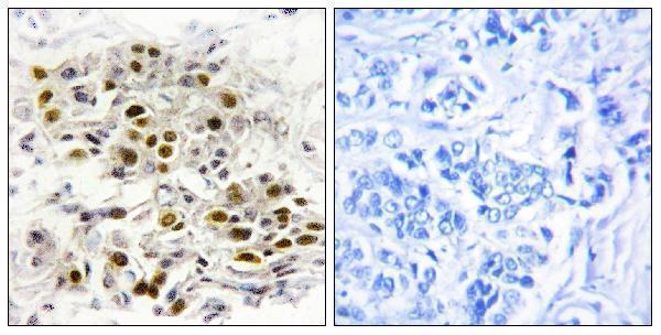
| Sample | Paraffin-embedded human breast carcinoma tissue; fixative not specified (datasheet A09063) |
| Fixation | Image fixative and duration unreported (datasheet A09063); verify before use. |
| Sectioning | 4–5 µm sections on charged slides (standard) |
| Deparaffinisation | Xylene, graded ethanol series to water (standard) |
| Antigen retrieval | Heat-induced epitope retrieval in citrate buffer, pH 6.0, 20 min at 95–98 °C (standard rule: cytoplasmic / membrane antigen) |
| Peroxidase block | 3% H2O2, 10 min, room temperature (standard) |
| Blocking | 10% normal serum of the secondary host, 30 min, room temperature (standard) |
| Primary antibody | Rabbit anti-MAP3K6, 1:100-1:300 (datasheet A09063) |
| Primary incubation | Overnight at 4 °C (standard) |
| Detection | HRP-polymer secondary, DAB chromogen 5–10 min (standard) |
| Counterstain | Hematoxylin, blue, dehydrate and mount (standard) |
| Expected result | MAP3K6-positive staining in respiratory epithelial cells of bronchus (HPA tissue IHC: High). HPA tissue profile: Ubiquitous cytoplasmic expression. No signal in the no-primary control. |
In paraffin IHC, expect mainly cytoplasmic MAP3K6 staining across tissues, with high staining reported in respiratory epithelium, several glandular cell populations, smooth muscle cells and other listed cells (HPA: tissue IHC). The tissue profile is Approved, with medium consistency between staining and RNA data and external verification pending (HPA: reliability). MAP3K6 has no annotated transmembrane segment or signal peptide (UniProt O95382: topology and processing).
| Cytoplasmic staining in respiratory epithelial cells, pancreatic exocrine cells or smooth muscle cells. | This matches the reported high tissue staining and ubiquitous cytoplasmic profile (HPA: tissue IHC). Compare cell identity and compartment before scoring intensity. |
| Only extracellular or luminal deposits appear, with no cytoplasmic staining in the expected cells. | This is a compartment mismatch and suggests an IHC artefact; the tissue profile is cytoplasmic (HPA: tissue IHC). Check deposits and detection controls before calling it MAP3K6. |
| Strong staining appears only in a cell population outside the reported tissue pattern. | Consider antibody cross-reactivity or endogenous detection activity (general IHC practice). HPA reports low tissue specificity, so an unlisted cell type alone does not establish a false positive (HPA: tissue IHC). |
| Brown haze covers cells and surrounding tissue without clear cell boundaries. | Diffuse background prevents a reliable compartment call (general IHC practice). It cannot be scored as the cytoplasmic pattern reported for tissue IHC (HPA: tissue IHC). |
| No staining appears in a section containing a reported high-staining cell population. | An absent signal in that candidate positive control calls for a run check; it does not establish that the specimen lacks MAP3K6 (HPA: tissue IHC; general IHC practice). |
| Tissue and cell choice | HPA reports high staining in bronchial respiratory epithelium, endometrial and stomach glandular cells, pancreatic exocrine cells, smooth muscle cells, peripheral nerve and cells in seminiferous ducts; adipocytes are medium (HPA: tissue IHC). |
| Strength of evidence | The tissue profile is Approved but has medium staining–RNA consistency and awaits external verification; HPA051192 is IHC Approved, with no Enhanced IHC status supplied (HPA: tissue reliability and antibody status). |
| Compartment and topology | Tissue IHC is described as ubiquitously cytoplasmic (HPA: tissue IHC). UniProt gives no subcellular annotation and reports no transmembrane segment or signal peptide; topology alone cannot settle every stain location (UniProt O95382). |
| Isoforms and epitope scope | Three isoforms are listed, but the supplied record does not map the IHC antibody epitope or establish which isoforms it detects (UniProt O95382: isoforms; HPA: antibody status). |
| IF/ICC Q&A: should it match IHC? | HPA ICC-IF places MAP3K6 mainly in the nucleoplasm, with additional plasma membrane, cytosol and actin filament localization (HPA: subcellular). Interpret that assay separately from cytoplasmic tissue IHC (HPA: tissue IHC). |
| Situation | Likely cause | Next action |
|---|---|---|
| No signal in a reported high-staining control cell population. | The run may have failed, or the positive cells may be absent from the examined area (general IHC practice). | Confirm cell identity and section integrity; inspect the run controls, retrieval record, primary-antibody step and detection reagents before interpreting a negative (general IHC practice; HPA: tissue IHC). |
| Extracellular or luminal staining dominates while expected cell cytoplasm is clear. | A deposit or other staining artefact may mimic positivity (general IHC practice); this distribution conflicts with the tissue profile (HPA: tissue IHC). | Compare with a no-primary control and a reported high-staining cell population; score only localized cellular staining (general IHC practice; HPA: tissue IHC). |
| Unexpected cells stain more strongly than the reported cell population. | Cross-reactivity or endogenous detection activity is possible (general IHC practice); HPA does not provide a formal negative-cell list (HPA: tissue IHC). | Check morphology, no-primary and detection controls, and a reported positive cell type before assigning specificity (general IHC practice; HPA: tissue IHC). |
| The entire section has diffuse brown background. | Nonspecific reagent binding or incomplete endogenous-enzyme blocking can obscure cellular staining (general IHC practice). | Review blocking, washes and detection controls; reassess whether cytoplasmic signal remains distinguishable (general IHC practice; HPA: tissue IHC). |
| Only nuclei stain in paraffin IHC. | This differs from the cytoplasmic tissue profile, although nucleoplasm is the main approved ICC-IF location (HPA: tissue IHC and subcellular). | Check tissue morphology and IHC controls before calling an artefact or a new IHC pattern; do not transfer the ICC-IF location directly to IHC scoring (HPA: tissue IHC and subcellular; general IHC practice). |
| Weak staining in Purkinje cells, caudate neurons or non-germinal-center lymph-node cells. | These are reported low-staining populations, so weak signal alone is inconclusive (HPA: tissue IHC). | Use a reported high-staining population to judge run performance, then score the low-staining cells by compartment and background (HPA: tissue IHC; general IHC practice). |


Comprehensive Human Protein Atlas IHC scoring per tissue (reliability: Approved — Medium consistency between antibody staining and RNA expression data. Pending external verification.). Rows are taken directly from the HPA tissue chart — click any row's HPA link to view the source.
| Tissue | Cell type | Level | Evidence | Source |
|---|---|---|---|---|
| Bronchus | Respiratory epithelial cells | High | Protein (IHC) | HPA → |
| Endometrium | Glandular cells | High | Protein (IHC) | HPA → |
| Pancreas | Exocrine glandular cells | High | Protein (IHC) | HPA → |
| Smooth muscle | Smooth muscle cells | High | Protein (IHC) | HPA → |
| Soft tissue | Peripheral nerve | High | Protein (IHC) | HPA → |
| Tissue | Cell type | Level | Evidence | Source |
|---|---|---|---|---|
| None in HPA: MAP3K6 is detected in all 45 scored tissues. Use a no-primary (secondary-only) and an isotype control instead. | ||||
Troubleshoot MAP3K6 staining in paraffin sections by checking retrieval, cell type, compartment and controls before comparing chromogenic signal across samples.
A09063 lists IHC and IF applications for human and mouse samples (catalog: applications/reactivity); its IHC image shows a paraffin-embedded human breast carcinoma section (IHC image caption).
A09063 will render with an IHC image of paraffin-embedded human breast carcinoma tissue and a peptide-blocked comparison (IHC image caption). The catalog lists IHC and IF applications and human and mouse reactivity for A09063 (catalog: applications/reactivity); no IF image is supplied (catalog: IF images).
Which to pick: For tissue IHC, choose A09063: its own image documents staining in a paraffin-embedded human breast carcinoma section, with fixative unreported (IHC image caption). For IF/ICC, A09063 lists IF, while ICC-specific validation and clonality are unreported (catalog: applications; clone field). For work involving human and mouse samples, A09063 lists reactivity with both species; its IHC image documents human tissue only (catalog: reactivity; IHC image caption).