MAPK7 / Mitogen-activated protein kinase 7 · IHC design guide

Design Immunohistochemistry for MAPK7

Plan chromogenic MAPK7 IHC on paraffin sections using the catalog antibody’s 2–5 μg/ml range (datasheet A02812-3). Compare cytoplasmic tissue staining (HPA tissue IHC) with the possibility of activation-linked nuclear localization (UniProt), while accounting for uncertain tissue staining reliability (HPA tissue IHC).

Evidence assembled Oct 2026 · For research use; verify linked source records and product datasheet before use
Immunohistochemistry protocol sheet for MAPK7 (IHC for MAPK7): expected localisation Cytoplasmic tissue staining (HPA tissue IHC); nuclear translocation upon activation (UniProt), antibody A02812-3, validated IHC image, and IHC protocol steps
Printable MAPK7 IHC protocol sheet — expected localisation Cytoplasmic tissue staining (HPA tissue IHC); nuclear translocation upon activation (UniProt), antibody A02812-3, controls and protocol steps. Open the full MAPK7 IHC guide →

MAPK7 Immunohistochemistry Experimental Design Guide

Expected localisation, validated protocols, controls and antibodies — the at-a-glance facts below, then the full design guide.

Must know before staining
Expected localisation Cytoplasmic tissue staining (HPA tissue IHC); nuclear translocation upon activation (UniProt)
Staining pattern Cytoplasmic staining; high in glandular cells and myocytes (HPA tissue IHC)
Antigen retrieval EDTA pH 8.0 HIER, heat-mediated (datasheet A02812-3)
Positive control ⓘ Adrenal gland+4 more · see all
Negative control ⓘ Adipose tissue+1 more · see all
Important caveats
Reasons your staining may differ from the expected pattern.
Fixation Keep fixation consistent (standard IHC practice; not target-specific); Matched tissue-IHC evidence does not establish the fixation claim. Validate the specimen-specific method before use. (selected-SKU IHC image A02812-3)
Caveat Tissue staining has low agreement with RNA expression (HPA tissue IHC)
Regulation EGF activates MAPK7 via MAP2K5 (UniProt)
Isoform / epitope 4 isoforms; no transmembrane segment; antibody epitope coverage unknown (UniProt; datasheet A02812-3)
Section 1

Recommended MAPK7 IHC & IF Protocols

The catalog antibody’s IHC-P protocol is paired with one published MAPK7 IHC protocol from human tumor samples and mouse tissues (PMC13411615).

Recommended immunohistochemistry (IHC-P) protocol parameters
SampleParaffin-embedded human breast cancer tissue; fixative not specified (datasheet A02812-3)
FixationImage fixative and duration unreported (datasheet A02812-3); verify before use.
Sectioning4–5 µm sections on charged slides (standard)
DeparaffinisationXylene, graded ethanol series to water (standard)
Antigen retrievalHeat retrieval: EDTA pH 8.0 (datasheet A02812-3); 20 min, 95–100 °C (standard)
Peroxidase block3% H2O2, 10 min, room temperature (standard)
Blocking10% goat serum (datasheet A02812-3)
Primary antibodyRabbit anti-MAPK7, 2-5μg/ml (datasheet A02812-3)
Primary incubationOvernight at 4 °C (datasheet A02812-3)
DetectionStreptavidin-biotin complex (SABC), DAB chromogen (datasheet A02812-3)
CounterstainHematoxylin, blue, dehydrate and mount (standard)
Expected resultMAPK7-positive staining in glandular cells of adrenal gland (HPA tissue IHC: High). HPA tissue profile: General cytoplasmic expression. No signal in the no-primary control.
💡Decision noteStart with heat-mediated EDTA retrieval at pH 8.0 for the catalog antibody (datasheet A02812-3); the published protocol does not specify retrieval (PMC13411615).
Section 2

What Is the Expected MAPK7 Staining Pattern?

MAPK7 is cytoplasmic and can enter the nucleus upon activation; HPA reports a general cytoplasmic IHC pattern (UniProt Q13164 localisation; HPA tissue IHC). Look for staining in glandular cells, neurons and skeletal myocytes reported as High by HPA (HPA tissue IHC). MAPK7 has no transmembrane segment, so a membrane rim is unexpected (UniProt Q13164 topology). Treat tissue staining cautiously: HPA rates the IHC profile Uncertain because antibody staining and RNA expression have low consistency (HPA tissue IHC).

What am I looking at on my slide?
Cytoplasmic chromogen in colon glandular cells, pancreatic exocrine cells or skeletal myocytes (HPA tissue IHC).This matches HPA's High cell-level staining and general cytoplasmic profile (HPA tissue IHC). Compare signal with adjacent cells and the negative control; High is an atlas category, not a required chromogen intensity on every section (HPA tissue IHC; general IHC practice).
Nuclear signal accompanies cytoplasmic signal, or signal appears exclusively as a sharp membrane rim.Nuclear MAPK7 can be plausible because activation promotes nuclear translocation (UniProt Q13164 localisation). A membrane-only rim conflicts with its reported cytoplasmic and nuclear locations and lack of a transmembrane segment; check morphology and controls before calling it an artefact (UniProt Q13164 localisation and topology; general IHC practice).
Strong chromogen appears in adipocytes or soft-tissue fibroblasts while expected positive cells are weak (HPA tissue IHC).HPA lists those cells as Not detected, making the pattern suspicious for cross-reactivity or endogenous detection activity (HPA tissue IHC; general IHC practice). These are comparative controls, not proof of absence: HPA rates its tissue IHC profile Uncertain (HPA tissue IHC).
Diffuse stain covers cells, extracellular spaces and the section edge without clear cellular boundaries.The distribution is less interpretable than HPA's general cytoplasmic pattern (HPA tissue IHC). Check the detection-only control and washing, then review blocking and chromogen development as general IHC troubleshooting steps (general IHC practice).
No signal appears in a well-preserved skeletal-muscle positive section (HPA: High in myocytes).A failed run, ineffective retrieval or detection, or antibody conditions may explain the absence (general IHC practice). Repeat with controls before judging the specimen; HPA's Uncertain reliability means one negative section cannot settle MAPK7 expression (HPA tissue IHC).
💡Expected MAPK7 appearanceCall a result plausible when cell-associated cytoplasmic staining is present in an HPA High cell population, such as skeletal myocytes or colon glandular cells; nuclear signal may also occur, whereas membrane-only rims or widespread extracellular chromogen warrant control review (HPA tissue IHC; UniProt Q13164 localisation and topology; general IHC practice).
How each factor affects the staining
Tissue and cell selectionHPA reports High staining in skeletal myocytes, colon glandular cells and several other listed populations, but Not detected in adipocytes and soft-tissue fibroblasts (HPA tissue IHC). Assess the named cell population, not an undifferentiated tissue-wide score (general IHC practice).
Atlas confidenceThe tissue IHC profile is Uncertain because staining has low consistency with RNA expression; one HPA antibody is IHC Uncertain and the other has no IHC rating (HPA tissue IHC; HPA antibodies). Use the pattern as a provisional expectation (HPA tissue IHC).
Activation and compartmentMAPK7 translocates to the nucleus upon activation, while UniProt also lists cytoplasm, nucleus and PML bodies (UniProt Q13164 localisation). Nuclear staining can be biologically plausible, but staining alone does not establish activation (UniProt Q13164 localisation; general IHC interpretation).
Isoforms and antibody epitopeUniProt lists 4 isoforms and a kinase domain at residues 55–347 (UniProt Q13164 isoforms and domains). No epitope is supplied, so this record cannot predict which isoforms the catalog antibody detects or whether retrieval changes recognition.
IF/ICC Q&A: Should nuclear fluorescence be rejected?No. HPA supports nucleoplasm and cytosol localisation in ICC-IF, and UniProt reports activation-related nuclear translocation (HPA subcellular ICC-IF; UniProt Q13164 localisation). Interpret IF/ICC with its own guide and controls; this section supplies no IF protocol.
Why is my staining missing, weak or wrong?
SituationLikely causeNext action
Positive-control cells show no chromogen.The staining run or selected antibody conditions may have failed; HPA's High designation is an observed pattern, not a guarantee for every run (HPA tissue IHC; general IHC practice).Confirm tissue preservation, retrieval, primary-antibody conditions and detection reagents against the established IHC workflow, then repeat with controls (general IHC practice).
Signal is confined to cell membranes.This conflicts with the reported cytoplasmic and nuclear locations and absence of a transmembrane segment (UniProt Q13164 localisation and topology).Review cell boundaries at higher magnification and compare the primary-omission control; reassess antibody specificity if the rim persists (general IHC practice).
Adipocytes or soft-tissue fibroblasts stain strongly.These cells are Not detected in HPA tissue IHC; nonspecific antibody binding or endogenous detection activity is possible (HPA tissue IHC; general IHC practice).Compare a detection-only control and a known-positive cell population, then review blocking and detection chemistry (general IHC practice). Treat the HPA negative category cautiously because its tissue profile is Uncertain (HPA tissue IHC).
The entire section has diffuse brown background.Poorly confined chromogen can obscure HPA's general cytoplasmic pattern; excess background may arise during blocking, washing or detection (HPA tissue IHC; general IHC practice).Check the detection-only control, wash steps and chromogen development time; judge MAPK7 only where cell boundaries remain interpretable (general IHC practice).
Nuclear staining appears alongside cytoplasmic staining.Nuclear localisation is compatible with MAPK7, which can translocate after activation (UniProt Q13164 localisation). IHC localisation alone does not demonstrate pathway activation (general IHC interpretation).Record nuclear and cytoplasmic compartments separately and compare controls; use an independent activation readout if activation is the research question (general IHC practice).
Two tissue sections give conflicting apparent expression.HPA's tissue IHC reliability is Uncertain, and its listed High or Not detected calls apply to specified cell populations (HPA tissue IHC).Recheck cell identity, section quality and controls before comparing scores; document compartment and cell population for each section (general IHC practice).

Sample controls for MAPK7 IHC & IF

🧪Run cerebral cortex first: neuronal cells should stain (HPA: High in cerebral cortex neuronal cells). Use adipose tissue as the negative tissue (HPA: adipocytes Not detected); on the cortex slide, morphologically non-neuronal cells with no signal can serve as an internal background reference, though their MAPK7-negative status is not established by the supplied HPA row.
Positive control tissue: Adrenal gland (Glandular cells, HPA High)
Negative control tissue: Adipose tissue (HPA Not detected)
ICC-IF cell lines (HPA subcellular resource): HPA ICC-IF images show MAPK7 in A-431, U-251MG, U2OS, HeLa, ASC52telo, hTERT-RPE1 (serum starved), NIH 3T3, with annotated localisation: Nucleoplasm (supported), Cytosol (supported) (HPA subcellular).
Technical controls: Include a no-primary (secondary-only) control, a concentration-matched rabbit IgG isotype control appropriate to the primary antibody’s clonality, and a validated MAPK7-knockout specimen or immunizing-peptide block as a biological specificity control (selected-SKU caption: rabbit primary antibody). Block endogenous peroxidase and check for endogenous biotin when using the caption’s biotin-based DAB detection; distinguish neuronal pigment from chromogen in cerebral cortex (selected-SKU caption: biotin-based DAB detection; HPA: High in cerebral cortex neuronal cells).
⚠️Feasibility: A target-specific fixation window or fixation effect is unreported, and the selected-SKU paraffin-section caption does not state its fixative (selected-SKU caption: fixative not stated). Heat retrieval in EDTA at pH 8.0 was used for the catalog antibody’s paraffin-section image, but the supplied evidence does not establish that retrieval is essential (selected-SKU caption: EDTA pH 8.0). Whether frozen sections or IF are easier is unreported; cerebral cortex neuronal lipofuscin can complicate interpretation of brown DAB signal (HPA: High in cerebral cortex neuronal cells).

HPA tissue IHC evidence for MAPK7

Comprehensive Human Protein Atlas IHC scoring per tissue (reliability: Uncertain — Low consistency between antibody staining and RNA expression data.). Rows are taken directly from the HPA tissue chart — click any row's HPA link to view the source.

Positive expression · recommended positive controls

TissueCell typeLevelEvidenceSource
Adrenal gland Glandular cells High Protein (IHC) HPA →
Cerebellum Cells in molecular layer High Protein (IHC) HPA →
Cerebral cortex Neuronal cells High Protein (IHC) HPA →
Colon Glandular cells High Protein (IHC) HPA →
Duodenum Glandular cells High Protein (IHC) HPA →

Undetected expression · recommended negative controls

TissueCell typeLevelEvidenceSource
Adipose tissue Adipocytes Not detected Protein (IHC) HPA →
Soft tissue Fibroblasts Not detected Protein (IHC) HPA →
Section 3

Advanced MAPK7 IHC Tips

Troubleshoot MAPK7 staining in paraffin sections by checking retrieval, compartment, controls, and scoring before interpreting chromogenic signal.

Which retrieval condition should I try first for MAPK7 in paraffin sections?
Use heat-mediated antigen retrieval in EDTA at pH 8.0 for paraffin sections (datasheet A02812-3). The selected tissue image used this condition before overnight incubation at 4°C with 2 µg/ml primary antibody (datasheet A02812-3). If staining is weak, check that sections remained covered by retrieval solution during heating and that runs used the same heating and cooling conditions (standard IHC practice). Compare any retrieval adjustment on adjacent sections alongside a known staining control, because stronger DAB signal alone does not establish improved specificity (standard IHC practice).
Could fixation explain weak or uneven MAPK7 staining?
Target-specific fixation sensitivity is unknown: the selected paraffin-section caption does not state a fixative (datasheet A02812-3). Record the actual fixative, fixation duration, section thickness, and storage history before comparing staining across specimens (standard IHC practice). For uneven signal, inspect whether weak areas track tissue thickness, folds, or poorly preserved regions, then compare adjacent sections processed together with EDTA retrieval at pH 8.0 (datasheet A02812-3; standard IHC practice). Do not infer a MAPK7-specific fixation effect from its intracellular location or phosphorylation sites; those features do not measure fixation sensitivity (UniProt Q13164).
How should I assess cytoplasmic and nuclear MAPK7 staining?
Score cytoplasmic and nuclear DAB separately, because MAPK7 occurs in both compartments and can translocate to the nucleus upon activation (UniProt Q13164). General cytoplasmic tissue staining has been reported, while nucleoplasm and cytosol localisation are supported in cellular imaging (HPA tissue IHC; HPA subcellular). Use a counterstain and inspect intact cells at higher magnification so overlapping cytoplasm is not mistaken for nuclear signal (standard IHC practice). Nuclear staining may warrant follow-up, but total MAPK7 IHC alone cannot establish activation or phosphorylation at Ser720 or Thr733 (UniProt Q13164; standard IHC interpretation).
Can this staining distinguish MAPK7 isoforms or phosphorylation states?
MAPK7 has 4 annotated isoforms, and its protein kinase domain spans residues 55–347 (UniProt Q13164). The supplied antibody caption gives no epitope map or isoform-reactivity data, so shared staining cannot be assigned to a particular isoform (datasheet A02812-3). Likewise, phosphorylation at Ser720 or Thr733 does not make a total-MAPK7 stain a phospho-specific assay (UniProt Q13164; standard IHC interpretation). If isoform or activation status is the research endpoint, obtain epitope and specificity evidence for the chosen reagent and score only the claim that evidence supports (standard IHC practice).
How can I investigate MAPK7 localisation by multiplex IF?
For IF/ICC, examine cytosol and nucleoplasm, both supported MAPK7 locations in cellular imaging (HPA subcellular). In skeletal muscle, pair MAPK7 with an independently validated myocyte marker to identify the expected positive cell population (HPA tissue IHC: High in myocytes; standard IF practice). Choose spectrally separated fluorophores and inspect unstained tissue channels for autofluorescence before assigning faint signal to MAPK7 (standard IF practice). MAPK7 has no transmembrane segment, so its relevant epitopes are intracellular; use permeabilisation appropriate to the antibody’s mapped epitope and verify that cell boundaries and nuclear morphology remain interpretable (UniProt Q13164; standard IF practice).
What should I check when DAB signal appears throughout the section?
The selected paraffin-section workflow used 10% goat serum, biotinylated secondary antibody, a streptavidin–biotin detection complex, and DAB (datasheet A02812-3). Include a no-primary control to reveal signal from the detection system, and review peroxidase blocking and endogenous biotin as possible background sources in this workflow (standard IHC practice). Compare a matched section with the same EDTA retrieval at pH 8.0, then assess whether background follows damaged edges, debris, or the whole slide rather than intact cells (datasheet A02812-3; standard IHC practice). Evaluate blocking and washing consistently before changing primary-antibody concentration (standard IHC practice).
How should I quantify MAPK7 DAB staining across specimens? ⚠ ANSWER MARKED FOR VERIFICATION
Define the tissue region and eligible cell population before scoring, and exclude folds, necrosis, and section edges consistently (standard IHC practice). For cellular staining, report the percentage of positive cells and an H-score: sum the percentages at intensities 0–3 multiplied by their intensity grades, yielding 0–300 (standard IHC practice). Record nuclear and cytoplasmic scores separately because MAPK7 occupies both compartments (UniProt Q13164). Normalise positive-cell counts to the number of eligible cells, or stained area to measured tissue area, and keep illumination, DAB development, and threshold rules consistent across specimens (standard IHC practice).
How can I distinguish plausible MAPK7 staining from artefact?
Plausible staining should resolve within intact cytoplasm or nuclei, consistent with known MAPK7 localisation, rather than follow section edges or necrotic debris (UniProt Q13164; standard IHC practice). Skeletal-muscle myocytes are reported as strongly stained, whereas adipocytes were not detected in the cited tissue survey; use those observations cautiously because its staining-to-RNA consistency is rated uncertain (HPA tissue IHC). Compare the same region with a no-primary control to expose endogenous enzyme or detection-system signal (standard IHC practice). Nuclear enrichment can fit MAPK7 translocation, but a chromogenic total-protein image alone does not prove pathway activation (UniProt Q13164; standard IHC interpretation).
Boster reagents

Best MAPK7 / Mitogen-activated protein kinase 7 IHC Antibodies

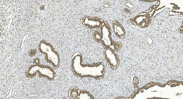
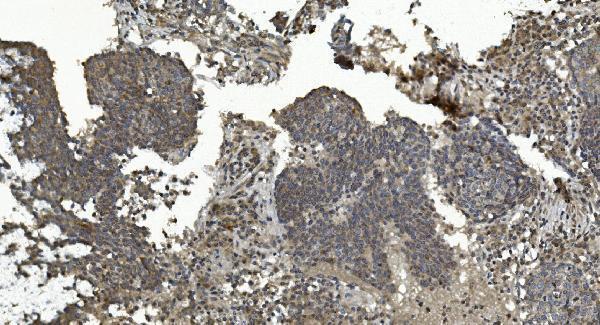
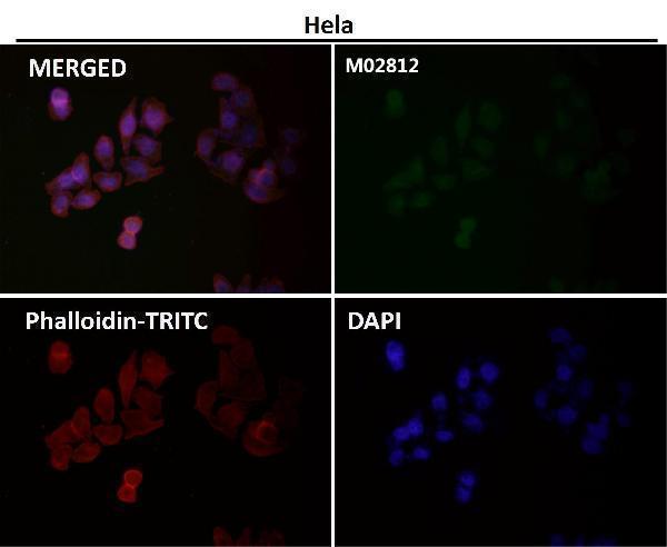
Anti-MAPK7 antibodies have human paraffin-section IHC images for breast and lung cancer tissue (A02812-3 image captions) and an IF image with no sample type reported (M02812 image caption).

Real IHC data IHC analysis of ERK5/MAPK7 using anti-ERK5/MAPK7 antibody (A02812-3). ERK5/MAPK7 was detected in paraffin-embedded section of human breast cancer tissue. Heat mediated antigen retrieval was performed in EDTA buffer (pH8.0, epitope retrieval solution). The tissue section was blocked with 10% goat serum. The tissue section was then incubated with 2μg/ml rabbit anti-ERK5/MAPK7 Antibody (A02812-3) overnight at 4°C. Biotinylated goat anti-rabbit IgG was used as secondary antibody and incubated for 30 minutes at 37°C. The tissue section was developed using Strepavidin-Biotin-Complex (SABC) (Catalog # SA1022) with DAB as the chromogen.
Anti-ERK5/MAPK7 Antibody ®
Cat # A02812-3
Real IF data Immunofluorescent analysis using the Antibody at 1:50 dilution.
Anti-ERK5 Rabbit Monoclonal Antibody
Cat # M02812

A02812-3 is listed for IHC and shows staining in human breast and lung cancer paraffin sections (catalog applications; A02812-3 image captions). M02812 is listed for ICC/IF and has an IF image at 1:50; its caption does not identify the sample (catalog applications; M02812 image caption).

Which to pick: Choose A02812-3 for tissue IHC: its images document human paraffin sections with EDTA retrieval at pH 8.0 and antibody at 2 μg/ml; the fixative is unreported (A02812-3 image captions). Choose M02812 for IF/ICC: it is a rabbit monoclonal listed for both applications, with an IF image at 1:50 (M02812 catalog; M02812 image caption). For broader listed species reactivity, A02812-3 includes human, monkey, mouse and rat, while M02812 includes human, mouse and rat; the A02812-3 IHC images show human tissue only (catalog reactivity; A02812-3 image captions).

Each figure is that product's own IHC / IF validation image from its datasheet.

References

  1. UniProt Consortium. UniProt entry Q13164 (MK07_HUMAN, Mitogen-activated protein kinase 7).
  2. Human Protein Atlas. MAPK7 tissue IHC expression (reliability: Uncertain).
  3. Human Protein Atlas. MAPK7 subcellular location (ICC-IF): Localized to the nucleoplasm and cytosol..
  4. Human Protein Atlas. MAPK7 antibody validation summary (2 antibodies).
  5. Targeting the Extracellular Signal-Regulated Kinase 5-Cellular Jun-Vimentin Axis to Inhibit Epithelial-Mesenchymal Transition and Metastasis in Patients with Triple-Negative Breast Cancer. International journal of medical sciences 2026 — PMC13411615.
  6. Identification and validation of dysregulated MAPK7 (ERK5) as a novel oncogenic target in squamous cell lung and esophageal carcinoma. BMC cancer 2015 — PMC4453990.
  7. miR-506-5p targets MAPK7 to impede glioma growth and invasion through suppressing epithelial-mesenchymal transition and matrix metalloproteinases. Translational cancer research 2026 — PMC13462558.
  8. Mapk7 enhances osteogenesis and suppresses adipogenesis by activating Lrp6/β-catenin signaling axis in mesenchymal stem cells. Communications biology 2025 — PMC11861680.
  9. PubMed PMID:7646528 — UniProt-cited evidence.
  10. PubMed PMID:7759517 — UniProt-cited evidence.
  11. PubMed PMID:15716121 — UniProt-cited evidence.