MARK3 / MAP/microtubule affinity-regulating kinase 3 · IHC design guide

Design Immunohistochemistry for MARK3

Plan MARK3 staining in paraffin sections with the IHC-validated antibody at 2–5 μg/ml (datasheet A05355-2). Use glandular cells with high reported staining as a reference for the expected cytoplasmic pattern (HPA tissue IHC).

Evidence assembled Oct 2026 · For research use; verify linked source records and product datasheet before use
Immunohistochemistry protocol sheet for MARK3 (IHC for MARK3): expected localisation Cytoplasmic tissue staining (HPA tissue IHC), antibody A05355-2, validated IHC image, and IHC protocol steps
Printable MARK3 IHC protocol sheet — expected localisation Cytoplasmic tissue staining (HPA tissue IHC), antibody A05355-2, controls and protocol steps. Open the full MARK3 IHC guide →

MARK3 Immunohistochemistry Experimental Design Guide

Expected localisation, validated protocols, controls and antibodies — the at-a-glance facts below, then the full design guide.

Must know before staining
Expected localisation Cytoplasmic tissue staining (HPA tissue IHC)
Staining pattern Glandular cells show cytoplasmic staining (HPA tissue IHC)
Antigen retrieval EDTA pH 8.0 HIER, heat-mediated (datasheet A05355-2)
Positive control ⓘ Appendix+4 more · see all
Negative control ⓘ Soft tissue
Important caveats
Reasons your staining may differ from the expected pattern.
Fixation Keep fixation consistent across sections (standard IHC practice; not target-specific)
Caveat Chondrocytes were not detected despite broad expression (HPA tissue IHC)
Regulation No stimulus-specific regulation reported (UniProt)
Isoform / epitope 7 isoforms; verify antibody epitope coverage (UniProt)
Section 1

Recommended MARK3 IHC & IF Protocols

The catalog antibody uses heat-mediated EDTA retrieval at pH 8.0 (datasheet A05355-2). Two published MARK3 IHC protocols provide comparison conditions (PMC8752757; PMC5269473).

Recommended immunohistochemistry (IHC-P) protocol parameters
SampleParaffin-embedded human lung cancer tissue; fixative not specified (datasheet A05355-2)
FixationImage fixative and duration unreported (datasheet A05355-2); verify before use.
Sectioning4–5 µm sections on charged slides (standard)
DeparaffinisationXylene, graded ethanol series to water (standard)
Antigen retrievalHeat retrieval: EDTA pH 8.0 (datasheet A05355-2); 20 min, 95–100 °C (standard)
Peroxidase block3% H2O2, 10 min, room temperature (standard)
Blocking10% goat serum (datasheet A05355-2)
Primary antibodyRabbit anti-MARK3, 2-5 μg/ml (datasheet A05355-2)
Primary incubationOvernight at 4 °C (datasheet A05355-2)
DetectionHRP-conjugated secondary, DAB chromogen (datasheet A05355-2)
CounterstainHematoxylin, blue, dehydrate and mount (standard)
Expected resultMARK3-positive staining in glandular cells of appendix (HPA tissue IHC: High). HPA tissue profile: General cytoplasmic expression. No signal in the no-primary control.
💡Decision noteStart with EDTA pH 8.0 for the catalog antibody (datasheet A05355-2); citrate pH 6 is a published alternative for femur sections (PMC5269473).
Section 2

What Is the Expected MARK3 Staining Pattern?

In paraffin-section IHC, expect predominantly cytoplasmic MARK3 staining in glandular cells, especially those of the appendix, duodenum, epididymis, gallbladder, rectum and small intestine (HPA: general cytoplasmic expression; High in listed glandular cells). Cell-membrane localisation is also reported, without a transmembrane segment (UniProt P27448: subcellular location and topology). HPA rates its tissue IHC profile Approved, with medium consistency between staining and RNA data (HPA: tissue IHC reliability).

What am I looking at on my slide?
Clear cytoplasmic staining in glandular cells of the appendix, duodenum or rectum.This matches the reported compartment and High staining in those cells (HPA: tissue IHC). Compare cells within the same section; staining throughout every cell is not required to recognise the reported pattern (general IHC practice).
Predominantly nuclear staining, with little or no cytoplasmic signal.A nuclear-dominant pattern is unsupported by the supplied localisation record (UniProt P27448: cytoplasm, cell membrane and dendrite; HPA: general cytoplasmic expression). Check controls and scoring before treating it as MARK3 localisation (general IHC practice).
Strong staining in chondrocytes while nearby expected-positive cells lack signal.HPA reports MARK3 as Not detected in soft-tissue chondrocytes, but High in several glandular-cell populations (HPA: tissue IHC). This reversal raises concern for cross-reactivity or detection activity; it cannot identify the cause by itself (general IHC practice).
Diffuse colour across cells, extracellular spaces and the negative control.Signal without a cellular pattern is difficult to assign to MARK3 (HPA: general cytoplasmic expression). Background shared with the negative control suggests nonspecific detection, endogenous activity or incomplete blocking (general IHC practice).
No staining in a section containing expected-positive glandular cells.Check the run before calling the sample negative: HPA reports High staining in glandular cells of several tissues, although its IHC reliability is Approved with medium RNA–staining consistency (HPA: tissue IHC). A failed positive control leaves the result uninterpretable (general IHC practice).
💡Expected MARK3 appearanceA convincing positive result is predominantly cytoplasmic staining in the reported glandular cells, potentially High in the listed tissues (HPA: tissue IHC); diffuse control-matched colour or nuclear-dominant staining warrants investigation (general IHC practice; UniProt P27448: subcellular location).
How each factor affects the staining
Tissue and cell choiceHPA reports High glandular-cell staining in six listed tissues, Medium in breast glandular cells and bone-marrow hematopoietic cells, and Not detected in chondrocytes (HPA: tissue IHC). Score the named cell population, not the whole section (general IHC practice).
CompartmentHPA describes general cytoplasmic tissue staining; UniProt also lists cell membrane and dendrite (HPA: tissue IHC; UniProt P27448: subcellular location). Membrane signal alone does not establish the expected paraffin-section pattern (HPA: tissue IHC).
Antibody validationThe listed antibody HPA024652 is IHC Approved, while the tissue profile has medium consistency with RNA data (HPA: antibody validation and tissue IHC). Neither rating establishes that every stained cell is MARK3-positive (general IHC interpretation).
Isoforms and epitope coverageUniProt lists seven MARK3 isoforms and kinase, UBA and KA1 domains (UniProt P27448: isoforms and domains). The supplied record gives no antibody epitope, so isoform coverage cannot be inferred (UniProt P27448: isoforms; HPA: supplied antibody entry).
Processing and phosphorylationUniProt lists a 1–753 chain, no signal peptide or propeptide, and multiple phosphorylation sites (UniProt P27448: processing and modified residues). These entries do not establish staining differences or a retrieval requirement for this antibody.
IF/ICC interpretationFor the separate IF/ICC guide: HPA summarises MARK3 as membrane-associated but supplies no main location or ICC-IF cell-line images (HPA: subcellular entry). That summary cannot validate an IF pattern or replace the tissue IHC observation.
Why is my staining missing, weak or wrong?
SituationLikely causeNext action
Expected-positive glandular cells are blank.The run may have failed, or the sampled section may lack the relevant cell population (HPA: High in listed glandular cells; general IHC practice).Confirm glandular cells on the counterstain, then review the positive control, antibody application, retrieval and detection steps (general IHC practice). No MARK3-specific retrieval condition is supplied.
Colour covers most of the section and the negative control.Shared background can reflect nonspecific detection, endogenous enzyme activity or incomplete blocking (general chromogenic IHC practice).Compare controls, review blocking and use an enzyme-control or quench appropriate to the chromogen system (general chromogenic IHC practice); do not score diffuse colour as cytoplasmic MARK3 (HPA: tissue IHC profile).
The strongest signal is nuclear.That compartment conflicts with the reported predominantly cytoplasmic tissue pattern (HPA: tissue IHC) and lacks support in the listed UniProt locations (UniProt P27448: subcellular location).Recheck counterstain and control sections, then assess whether the signal follows nuclei consistently (general IHC practice); report the discordance instead of assigning nuclear MARK3.
Chondrocytes stain strongly.HPA reports MARK3 as Not detected in soft-tissue chondrocytes (HPA: tissue IHC); unexpected colour may reflect cross-reactivity or detection background (general IHC practice).Compare a negative control and a section with expected-positive glandular cells (HPA: tissue IHC; general IHC practice). Treat isolated chondrocyte staining as uncertain until controls support specificity.
Only a narrow membrane rim stains.Membrane localisation is listed for MARK3, but HPA describes the paraffin-section tissue pattern as generally cytoplasmic (UniProt P27448: subcellular location; HPA: tissue IHC).Record the observed compartment and check whether cytoplasmic staining appears in expected-positive cells (HPA: tissue IHC); avoid calling a membrane-only pattern the established tissue result.
An IF/ICC image seems to contradict the IHC slide.HPA's subcellular entry says membrane but provides no main location or ICC-IF cell-line images; its tissue IHC entry reports general cytoplasmic expression (HPA: subcellular and tissue IHC entries).Evaluate IF/ICC in its separate guide and compare each result with its own controls (general IF/IHC practice). Do not use the limited IF summary to override the observed paraffin-section pattern.

Sample controls for MARK3 IHC & IF

🧪Run duodenum first; its glandular cells should stain for MARK3 (HPA: High in duodenal glandular cells). Use soft tissue chondrocytes as the biological negative (HPA: Not detected in chondrocytes); on the duodenal slide, treat non-glandular cells without detectable chromogen as background references only, since no validated MARK3-negative internal cell type is supplied (UniProt P27448: ubiquitous tissue specificity).
Positive control tissue: Appendix (Glandular cells, HPA High)
Negative control tissue: Soft tissue (HPA Not detected)
ICC-IF cell lines (HPA subcellular resource): HPA carries no ICC-IF cell line for MARK3; derive a cell-line control from the positive tissue's cell type (Glandular cells) and confirm it by RNA or western blot first.
Technical controls: Include a no-primary, secondary-only control and a concentration-matched rabbit IgG isotype control for the rabbit primary (selected-SKU IHC caption: rabbit anti-MARK3). A MARK3 knockout specimen, if available, provides a stronger biological specificity control; quench endogenous peroxidase for DAB detection and assess endogenous biotin if using avidin-biotin detection (selected-SKU IHC caption: HRP/DAB).
⚠️Feasibility: The selected-SKU paraffin-section example uses heat retrieval in EDTA at pH 8.0, followed by 2 μg/ml primary antibody overnight at 4°C; its fixative is unreported (selected-SKU IHC caption). A target-specific fixation window or fixation effect is unreported, and the supplied evidence does not establish whether frozen sections or IF are easier (selected-SKU IHC caption; HPA: no ICC-IF cell-line images). In duodenum, assess glandular background carefully, including endogenous biotin if an avidin-biotin method is chosen (HPA: High in duodenal glandular cells; standard IHC practice).

HPA tissue IHC evidence for MARK3

Comprehensive Human Protein Atlas IHC scoring per tissue (reliability: Approved — Medium consistency between antibody staining and RNA expression data.). Rows are taken directly from the HPA tissue chart — click any row's HPA link to view the source.

Positive expression · recommended positive controls

TissueCell typeLevelEvidenceSource
Appendix Glandular cells High Protein (IHC) HPA →
Duodenum Glandular cells High Protein (IHC) HPA →
Epididymis Glandular cells High Protein (IHC) HPA →
Gallbladder Glandular cells High Protein (IHC) HPA →
Rectum Glandular cells High Protein (IHC) HPA →

Undetected expression · recommended negative controls

TissueCell typeLevelEvidenceSource
Soft tissue Chondrocytes Not detected Protein (IHC) HPA →
Section 3

Advanced MARK3 IHC Tips

Troubleshoot MARK3 staining in paraffin sections by checking retrieval, compartment, cell type and controls before comparing chromogenic signal across specimens.

What retrieval should I start with for weak MARK3 staining?
Start with heat-mediated retrieval in EDTA pH 8.0 for paraffin sections (catalog caption A05355-2). The selected image documents this condition in human lung cancer tissue, followed by 2 μg/ml primary antibody overnight at 4°C; it does not establish an optimal heating duration (catalog caption A05355-2). If signal remains weak, compare a small range of heating times on adjacent sections while holding antibody concentration and detection constant (standard IHC practice). Include a known staining control and inspect tissue morphology, since excessive retrieval can damage sections and make apparent gains in signal hard to interpret (standard IHC practice).
Could fixation explain inconsistent MARK3 staining across paraffin blocks?
Target-specific MARK3 sensitivity to fixation is unknown from the supplied evidence; the selected paraffin-section caption does not state a fixative (catalog caption A05355-2). Record each block's fixative and fixation interval, then compare sections processed with the same EDTA pH 8.0 retrieval and detection conditions (catalog caption A05355-2; standard IHC practice). Use matched internal tissue structures or a consistently processed control section to distinguish processing variation from a change in cellular staining (standard IHC practice). Do not infer a MARK3-specific fixation effect from its reported cytoplasmic or membrane localisation or phosphorylation sites (UniProt P27448; HPA tissue IHC).
Which compartment should count as plausible MARK3 staining?
Evaluate cytoplasmic staining first: the tissue IHC profile describes general cytoplasmic expression (HPA tissue IHC). Membrane and dendritic projection localisation are also reported for MARK3, despite its lack of a transmembrane segment, so a discrete peripheral pattern needs cellular context and validation rather than automatic rejection (UniProt P27448). Score compartments separately on matched sections, using a counterstain to locate nuclei and cell boundaries (standard IHC practice). If staining is predominantly nuclear, check the secondary-only control and repeat the comparison with a positive control, because nuclear localisation is absent from the supplied MARK3 localisation record (UniProt P27448; standard IHC practice).
How can isoforms or epitope accessibility change the IHC result?
MARK3 has 7 listed isoforms, but the supplied caption does not identify this antibody's epitope or establish which isoforms it detects (UniProt P27448; catalog caption A05355-2). Map the immunogen sequence against the isoforms before attributing differences between cells to isoform expression (standard IHC practice). The protein contains a kinase domain at residues 56–307, a UBA domain at 326–365 and a KA1 domain at 704–753; its record also lists phosphorylation sites (UniProt P27448). Those features warrant an epitope review, but staining changes alone cannot identify an isoform or prove that phosphorylation masked the epitope (standard IHC practice).
How should I adapt this MARK3 IHC result for multiplex IF?
Treat IF as a separate assay: the supplied antibody example demonstrates chromogenic staining in a paraffin section, while no ICC/IF image is listed here (catalog caption A05355-2; HPA subcellular). Pair MARK3 with a marker that identifies the cell population being evaluated, and confirm each channel individually before interpreting overlap (standard IF practice). Select fluorophores after inspecting unstained tissue autofluorescence, reserving a spectrally distinct, low-background channel for the weaker signal (standard IF practice). If the mapped epitope faces the cytoplasm, titrate permeabilisation to admit antibody; MARK3 has no transmembrane segment, and the supplied antibody epitope is unspecified (UniProt P27448; standard IF practice).
How do I reduce diffuse brown signal without losing MARK3?
The selected paraffin-section example used 10% goat serum block, 2 μg/ml primary antibody overnight at 4°C, a peroxidase-linked secondary and DAB detection (catalog caption A05355-2). Start by checking a section without primary antibody, then compare background after a routine endogenous peroxidase block and thorough washing (standard IHC practice). If nonspecific staining persists, titrate the primary antibody below the documented concentration while keeping retrieval and DAB development comparable across sections (catalog caption A05355-2; standard IHC practice). Check whether brown deposits follow tissue edges, damaged areas or the entire section rather than plausible cellular compartments (standard IHC practice; UniProt P27448).
How should I quantify MARK3 across sections with different cell composition? ⚠ ANSWER MARKED FOR VERIFICATION
Define the cell population and compartment before scoring, because MARK3 has a general cytoplasmic tissue IHC profile and is reported in multiple cellular locations (HPA tissue IHC; UniProt P27448). For chromogenic sections, report the percentage of positive cells and an intensity-based H-score within that population, using the same thresholds and imaging conditions for all specimens (standard IHC practice). Normalise cell counts to the number of evaluable cells, or report positive-cell density per mm² of viable tissue when area is the comparison unit (standard IHC practice). Exclude necrotic or folded regions and document how ambiguous membrane-associated staining was scored (standard IHC practice).
What distinguishes true MARK3 signal from a staining artefact?
A plausible result shows cellular staining compatible with MARK3's reported cytoplasmic distribution, with membrane or dendritic projection signal interpreted in anatomical context (HPA tissue IHC; UniProt P27448). High staining in appendix or duodenal glandular cells offers a tissue comparison, while MARK3 is reported as low in lung macrophages; these patterns are reference observations, not controls for antibody specificity (HPA tissue IHC). Compare adjacent viable areas and a no-primary control to detect edge effects, necrotic deposits or endogenous peroxidase signal (standard IHC practice). Confirm an unexpected cell type or predominantly nuclear pattern with an independent specificity control before assigning it to MARK3 (standard IHC practice; UniProt P27448).
Boster reagents

Best MARK3 / MAP/microtubule affinity-regulating kinase 3 IHC Antibodies

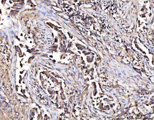
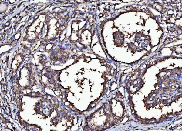
The IHC-validated antibody has a paraffin-section image from human lung cancer tissue (A05355-2 image caption); an IF/ICC antibody lists human and mouse reactivity, with no IF image supplied (M05355-1 catalog).

Real IHC data IHC analysis of MARK3 using anti-MARK3 antibody (A05355-2). MARK3 was detected in a paraffin-embedded section of human lung cancer tissue. Heat mediated antigen retrieval was performed in EDTA buffer (pH 8.0, epitope retrieval solution). The tissue section was blocked with 10% goat serum. The tissue section was then incubated with 2 μg/ml rabbit anti-MARK3 Antibody (A05355-2) overnight at 4°C. Peroxidase Conjugated Goat Anti-rabbit IgG was used as secondary antibody and incubated for 30 minutes at 37°C. The tissue section was developed using HRP Conjugated Rabbit IgG Super Vision Assay Kit (Catalog # SV0002) with DAB as the chromogen.
Anti-MARK3 Antibody ®
Cat # A05355-2

A05355-2 will render with its own IHC image from a paraffin-embedded human lung cancer section; its catalog lists IHC reactivity in human, mouse and rat (A05355-2 image caption; catalog). M05355-1 lists IF/ICC use in human and mouse, but has no supplied IF image (M05355-1 catalog).

Which to pick: Choose A05355-2 for chromogenic IHC on paraffin sections: its own caption documents EDTA retrieval at pH 8.0, 2 μg/ml primary antibody and DAB detection; the fixative is unreported (A05355-2 image caption). Choose the rabbit monoclonal M05355-1 for IF/ICC because IF/ICC is listed for that SKU; it has no supplied IF image or tissue IHC validation (M05355-1 catalog). For IHC across human, mouse and rat, A05355-2 lists all three species and has paraffin-section captions for each; M05355-1 lists human and mouse only (A05355-2 catalog and image captions; M05355-1 catalog).

Each figure is that product's own IHC / IF validation image from its datasheet.

References

  1. UniProt Consortium. UniProt entry P27448 (MARK3_HUMAN, MAP/microtubule affinity-regulating kinase 3).
  2. Human Protein Atlas. MARK3 tissue IHC expression (reliability: Approved).
  3. Human Protein Atlas. MARK3 subcellular location (ICC-IF): Membrane.
  4. Human Protein Atlas. MARK3 antibody validation summary (1 antibodies).
  5. The metabolic stress-activated checkpoint LKB1-MARK3 axis acts as a tumor suppressor in high-grade serous ovarian carcinoma. Communications biology 2022 — PMC8752757.
  6. Integrating GWAS and Co-expression Network Data Identifies Bone Mineral Density Genes SPTBN1 and MARK3 and an Osteoblast Functional Module. Cell systems 2017 — PMC5269473.
  7. MARK4 and MARK3 associate with early tau phosphorylation in Alzheimer's disease granulovacuolar degeneration bodies. Acta neuropathologica communications 2014 — PMC4046661.
  8. PubMed PMID:9543386 — UniProt-cited evidence.
  9. PubMed PMID:11433294 — UniProt-cited evidence.
  10. PubMed PMID:12508121 — UniProt-cited evidence.