This website uses cookies to ensure you get the best experience on our website.


- Table of Contents
Plan chromogenic MED8 IHC in paraffin sections with the catalog antibody’s documented workflow (datasheet A08094-1). Assess nuclear and cytoplasmic staining, using medium-staining glandular cells as a reference and controls to check the result (HPA tissue IHC).

Expected localisation, validated protocols, controls and antibodies — the at-a-glance facts below, then the full design guide.
| Expected localisation | Nuclear and cytoplasmic tissue staining (HPA tissue IHC) | |
| Staining pattern | Medium glandular-cell signal; nuclear/cytoplasmic pattern (HPA tissue IHC) | |
| Antigen retrieval | Citrate pH 6 HIER, heat-mediated (datasheet A08094-1) | |
| Positive control | Adipose tissue+4 more · see all | |
| Negative control | None in HPA (detected in all 45 tissues); use no-primary + isotype controls |
| Fixation | Keep paraffin-section fixation consistent (standard IHC practice; not target-specific); Matched tissue-IHC evidence does not establish the fixation claim. Validate the specimen-specific method before use. (selected-SKU IHC image A08094-1) | |
| Caveat | Staining–RNA agreement has medium consistency; use controls (HPA tissue IHC) | |
| Regulation | No expression regulator annotated (UniProt) | |
| Isoform / epitope | Three isoforms; isoform-specific epitope coverage unknown (UniProt; datasheet A08094-1) |
The catalog antibody protocol uses heat-mediated citrate retrieval at pH 6 (datasheet A08094-1). Two published MED8 IHC protocols provide additional tissue-specific examples (PMC12882893; PMC9086905).
| Sample | Paraffin-embedded mouse testis tissue; fixative not specified (datasheet A08094-1) |
| Fixation | Image fixative and duration unreported (datasheet A08094-1); verify before use. |
| Sectioning | 4–5 µm sections on charged slides (standard) |
| Deparaffinisation | Xylene, graded ethanol series to water (standard) |
| Antigen retrieval | Heat retrieval: Citrate pH 6, 20 min (datasheet A08094-1) |
| Peroxidase block | 3% H2O2, 10 min, room temperature (standard) |
| Blocking | 10% goat serum (datasheet A08094-1) |
| Primary antibody | Rabbit anti-MED8, 0.5-1μg/ml (datasheet A08094-1) |
| Primary incubation | Overnight at 4 °C (standard) |
| Detection | Streptavidin-biotin complex (SABC), DAB chromogen (datasheet A08094-1) |
| Counterstain | Hematoxylin, blue, dehydrate and mount (standard) |
| Expected result | MED8-positive staining in adipocytes of adipose tissue (HPA tissue IHC: Medium). HPA tissue profile: Nuclear and cytoplasmic expression in most tissues. No signal in the no-primary control. |
MED8 is a nuclear component of the Mediator transcription complex and has no transmembrane segment (UniProt Q96G25). In tissue IHC, expect predominantly nuclear staining across many cell types; HPA also reports cytoplasmic staining in most tissues (HPA tissue IHC). Interpret this as a broad pattern with variable intensity: HPA rates the tissue staining “Approved,” with medium consistency against RNA data and external verification pending (HPA tissue IHC).
| Distinct nuclear stain in adrenal glandular cells, bronchial respiratory epithelial cells, or bone marrow hematopoietic cells. | This fits the expected compartment and HPA-reported medium staining in these cells (UniProt Q96G25; HPA tissue IHC). Compare nuclei with the counterstain and include a negative reagent control before scoring a specimen (general IHC practice). |
| Predominantly sharp plasma membrane or extracellular staining, with little nuclear signal. | This conflicts with MED8’s nuclear localization and lack of a transmembrane segment (UniProt Q96G25). HPA does report cytoplasmic tissue staining, so cytoplasmic color alone is not a failure; a dominant membrane or extracellular pattern warrants control review (HPA tissue IHC; general IHC practice). |
| Strong staining in a cell population expected to show only low signal, while nuclei in an HPA-reported medium population are unstained. | Check specificity and detection chemistry before calling this MED8: HPA reports low staining in breast glandular cells, smooth muscle cells, and splenic red pulp cells, but does not designate them negative (HPA tissue IHC). Cross-reactivity or endogenous detection activity is possible (general IHC practice). |
| Even chromogen haze across tissue, stroma, and empty areas obscures nuclear boundaries. | This is difficult to score as compartment-specific MED8 staining (UniProt Q96G25; general IHC practice). Background from detection reagents or incomplete washing is possible; compare with a negative reagent control and inspect whether color follows recognizable cells (general IHC practice). |
| No visible signal in an HPA-reported medium-staining tissue, with readable counterstain and intact morphology. | Treat the run as unresolved, since HPA reports medium signal in several tissues but only Approved IHC reliability and medium consistency with RNA data (HPA tissue IHC). Check antibody and detection controls before interpreting the specimen as MED8 negative (general IHC practice). |
| Tissue and cell selection | HPA reports medium staining in adipocytes, adrenal and appendix glandular cells, bronchial epithelium, marrow hematopoietic cells, and selected brain cells (HPA tissue IHC). Its low categories are comparisons, not verified negative controls (HPA tissue IHC). |
| Compartment and topology | UniProt assigns MED8 to the nucleus and lists no transmembrane segment (UniProt Q96G25). HPA tissue IHC describes nuclear and cytoplasmic expression in most tissues, so assess whether the nuclear component remains convincing (HPA tissue IHC). |
| Antibody evidence | Two listed antibodies have Approved IHC status, HPA028377 and HPA028438 (HPA antibodies). The tissue profile has medium consistency with RNA and awaits external verification; do not treat an isolated staining pattern as definitive identification (HPA tissue IHC). |
| Isoforms and processing | UniProt lists three isoforms, one chain spanning residues 1–268, and no signal peptide or propeptide (UniProt Q96G25). No epitope or isoform-specific staining data are supplied, so these entries do not predict a particular IHC intensity. |
| IF/ICC Q: where should signal appear? | A: HPA places MED8 mainly in the nucleoplasm, with additional Golgi localization in ICC-IF (HPA subcellular). This answers the localization question; the tissue IHC profile separately reports nuclear and cytoplasmic staining (HPA tissue IHC). |
| Situation | Likely cause | Next action |
|---|---|---|
| No nuclear stain in an HPA-reported medium tissue. | The assay may have failed, or the selected section may not show the reported cell population (HPA tissue IHC; general IHC practice). | Identify the relevant cells on the counterstain; check the antibody, retrieval, and detection steps against their validated IHC conditions, then repeat with run controls (general IHC practice). |
| Only diffuse cytoplasmic color is visible. | HPA permits cytoplasmic tissue staining, but loss of a readable nuclear component weakens the match to UniProt localization (HPA tissue IHC; UniProt Q96G25). | Compare matched cells and the negative reagent control; score nuclear and cytoplasmic staining separately, and avoid calling diffuse color alone a convincing positive (general IHC practice). |
| A low-staining HPA population appears strongly positive. | Low does not mean absent; cross-reactivity or endogenous detection activity may also contribute (HPA tissue IHC; general IHC practice). | Compare an HPA-reported medium population in the same run and use a negative reagent control; investigate detection background before assigning the signal to MED8 (HPA tissue IHC; general IHC practice). |
| Chromogen appears without the primary antibody. | A positive negative reagent control points to background from the detection system or tissue activity rather than primary-antibody binding (general IHC practice). | Review the detection reagent and any enzyme-blocking step specified for the detection chemistry; repeat the control before scoring the tissue (general IHC practice). |
| Nuclear stain is visible but tissue-wide haze prevents cell-level scoring. | Insufficient washing or excess detection background can obscure compartment boundaries (general IHC practice). | Review washing and detection conditions, retain the counterstain for nuclear landmarks, and compare the repeat section with its negative reagent control (general IHC practice). |
| Two IHC antibodies give different compartment patterns. | Both HPA028377 and HPA028438 are Approved for IHC, while the tissue profile still has medium RNA consistency and awaits external verification (HPA antibodies; HPA tissue IHC). | Compare the same tissue and cell populations under each antibody’s validated conditions; record the discordance and avoid resolving it by intensity alone (general IHC practice). |


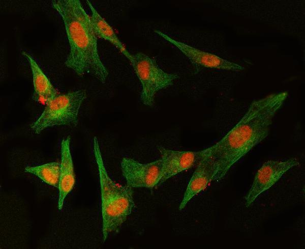
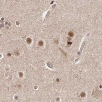


Comprehensive Human Protein Atlas IHC scoring per tissue (reliability: Approved — Medium consistency between antibody staining and RNA expression data. Pending external verification.). Rows are taken directly from the HPA tissue chart — click any row's HPA link to view the source.
| Tissue | Cell type | Level | Evidence | Source |
|---|---|---|---|---|
| Adipose tissue | Adipocytes | Medium | Protein (IHC) | HPA → |
| Adrenal gland | Glandular cells | Medium | Protein (IHC) | HPA → |
| Appendix | Glandular cells | Medium | Protein (IHC) | HPA → |
| Bone marrow | Hematopoietic cells | Medium | Protein (IHC) | HPA → |
| Bronchus | Respiratory epithelial cells | Medium | Protein (IHC) | HPA → |
| Tissue | Cell type | Level | Evidence | Source |
|---|---|---|---|---|
| None in HPA: MED8 is detected in all 45 scored tissues. Use a no-primary (secondary-only) and an isotype control instead. | ||||
For MED8 chromogenic IHC, assess nuclear staining alongside controls for retrieval, background, and compartment specificity (UniProt Q96G25; HPA tissue IHC).
A08094-1 has paraffin-section IHC data in mouse and rat testis (IHC captions) and IF data in U87 cells (IF caption); listed reactivity covers human, mouse and rat (catalog).
A08094-1 has IHC images from paraffin-embedded mouse and rat testis (IHC captions). A08094-1 also has an IF image from U87 cells (IF caption), and the catalog lists IF and ICC applications (catalog).
Which to pick: For tissue IHC, choose A08094-1 for paraffin sections based on its mouse and rat testis images (IHC captions); the fixative is unreported (IHC captions). For IF/ICC, A08094-1 is the listed option, with an IF image from U87 cells (IF caption; catalog: IF, ICC). For work across species, A08094-1 lists human, mouse and rat reactivity (catalog), while its tissue IHC images document mouse and rat samples (IHC captions).