This website uses cookies to ensure you get the best experience on our website.


- Table of Contents
Plan paraffin IHC for MEMO1 around its cytoplasmic tissue staining and high staining in colon glandular cells (HPA tissue IHC). Use consistent fixation and appropriate tissue controls when comparing chromogenic staining across sections.

Expected localisation, validated protocols, controls and antibodies — the at-a-glance facts below, then the full design guide.
| Expected localisation | Cytoplasmic staining in most tissues (HPA tissue IHC) | |
| Staining pattern | Cytoplasmic; high in colon glandular cells (HPA tissue IHC) | |
| Antigen retrieval | Citrate pH 6.0 HIER, 95–98 °C, 20 min (rule: cytoplasmic / membrane antigen) | |
| Positive control | Colon+4 more · see all | |
| Negative control | Adipose tissue+4 more · see all |
| Fixation | Keep fixation conditions consistent across sections. (standard IHC practice; not target-specific) | |
| Caveat | Antibody staining and RNA show low consistency (HPA tissue IHC) | |
| Regulation | Expression regulation not annotated (UniProt) | |
| Isoform / epitope | 3 isoforms; epitope coverage unreported (UniProt) |
The catalog antibody protocol is accompanied by a published chromogenic IHC protocol for mouse dorsal skin (PMC4345637).
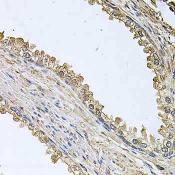
| Sample | Paraffin-embedded human prostate tissue; fixative not specified (datasheet A07852) |
| Fixation | Image fixative and duration unreported (datasheet A07852); verify before use. |
| Sectioning | 4–5 µm sections on charged slides (standard) |
| Deparaffinisation | Xylene, graded ethanol series to water (standard) |
| Antigen retrieval | Heat-induced epitope retrieval in citrate buffer, pH 6.0, 20 min at 95–98 °C (standard rule: cytoplasmic / membrane antigen) |
| Peroxidase block | 3% H2O2, 10 min, room temperature (standard) |
| Blocking | 10% normal serum of the secondary host, 30 min, room temperature (standard) |
| Primary antibody | Rabbit anti-MEMO1, 1:50-1:100 (datasheet A07852) |
| Primary incubation | Overnight at 4 °C (standard) |
| Detection | HRP-polymer secondary, DAB chromogen 5–10 min (standard) |
| Counterstain | Hematoxylin, blue, dehydrate and mount (standard) |
| Expected result | MEMO1-positive staining in glandular cells of colon (HPA tissue IHC: High). HPA tissue profile: Cytoplasmic expression in most tissues. No signal in the no-primary control. |
In paraffin-section IHC, expect mainly cytoplasmic MEMO1 staining, with high staining reported in colon and rectal glandular cells, lung alveolar cells, placental trophoblastic cells, skeletal muscle myocytes, and thyroid glandular cells (HPA: tissue IHC). MEMO1 has no annotated transmembrane segment (UniProt Q9Y316 topology). HPA rates the tissue IHC pattern Approved, while reporting low consistency between antibody staining and RNA expression (HPA: reliability).
| Cytoplasmic staining in colon or rectal glandular cells, or in lung alveolar cells. | This fits the reported high staining in those cells (HPA: tissue IHC). Judge the pattern in the named cells rather than treating all cells in a positive tissue as equivalent; HPA reports cytoplasmic expression across most tissues (HPA: tissue IHC). |
| A dominant nuclear or membrane-outline pattern replaces cytoplasmic staining. | That differs from the tissue IHC pattern (HPA: cytoplasmic expression). Review the antibody and staining controls for an artefact before assigning a new compartment; no transmembrane segment is annotated (UniProt Q9Y316 topology). The IF vesicle call does not establish a nuclear or membrane-outline IHC pattern (HPA: subcellular ICC-IF). |
| Strong colour appears in adipocytes or ovarian stroma cells while expected positive cells are weak. | Those cells are reported as not detected (HPA: adipose tissue and ovary IHC). Consider cross-reactivity or endogenous detection activity (general IHC practice), then compare with a no-primary control. A listed negative is an observed HPA result, not a guarantee for every specimen (HPA: tissue IHC). |
| Colour spreads through extracellular space or appears similarly across unrelated cell types. | Diffuse background obscures the cell-restricted cytoplasmic pattern (HPA: tissue IHC). Assess no-primary staining and wash or blocking conditions (general IHC practice). Broad low-level staining alone is hard to interpret because HPA reports low tissue RNA specificity and low staining–RNA consistency (HPA: tissue IHC reliability). |
| No convincing staining appears in colon glandular cells or another reported high-staining cell population. | This fails an expected positive comparison (HPA: colon and listed high IHC cells), but does not by itself prove MEMO1 absence. Check section quality, retrieval, primary-antibody use, and detection controls (general IHC practice); HPA's Approved rating carries a low staining–RNA consistency caveat (HPA: reliability). |
| Tissue and cell choice | Colon and rectal glandular cells, lung alveolar cells, placental trophoblastic cells, skeletal muscle myocytes, and thyroid glandular cells are reported high (HPA: tissue IHC). Adipocytes and ovarian stroma cells are reported not detected (HPA: tissue IHC); compare named cell populations. |
| Strength of the tissue evidence | The IHC pattern is Approved, but HPA reports low consistency between staining and RNA expression (HPA: reliability). Treat the reported pattern as an interpretation guide and evaluate controls alongside each specimen (general IHC practice). |
| Antibody validation by application | HPA042603 has Approved IHC status, while HPA057952 has Approved ICC status (HPA: antibody validation). An ICC approval does not establish an IHC result for that antibody; use the validation status for the application being interpreted (HPA: antibody validation). |
| Protein topology and variants | MEMO1 has no annotated transmembrane segment, signal peptide, or propeptide, and has three annotated isoforms (UniProt Q9Y316). The supplied record gives no antibody epitope or isoform coverage, so it cannot predict which variants a given stain detects. |
| Modified residue | UniProt annotates a phosphotyrosine at residue 210 (UniProt Q9Y316). Without a supplied antibody epitope or modification-specific validation, this annotation does not justify calling a brighter or weaker IHC signal a phosphorylation change. |
| IF/ICC: what pattern is reported? | HPA reports Approved vesicle localization in ICC-IF, with images from RT-4 and U2OS (HPA: subcellular ICC-IF). This is a separate cell-imaging observation; tissue IHC is summarized as cytoplasmic (HPA: tissue IHC). |
| Situation | Likely cause | Next action |
|---|---|---|
| The reported positive cells have no chromogenic signal. | The stain may have failed, or the selected section may not show the expected cells (HPA: high-staining cell populations; general IHC practice). | Confirm the named cell population on the section, then review retrieval, primary-antibody application, detection reagents, and a concurrent positive control (general IHC practice). Do not assign MEMO1 absence from a failed control. |
| Nuclei dominate the stain. | This conflicts with HPA's cytoplasmic tissue pattern (HPA: tissue IHC); nonspecific staining or detection background is possible (general IHC practice). | Compare the no-primary control, inspect counterstain and chromogen separately, and reassess antibody specificity (general IHC practice). Keep the compartment call tied to the reported cytoplasmic IHC pattern (HPA: tissue IHC). |
| Adipocytes, ovarian stroma cells, or parathyroid glandular cells stain strongly. | HPA reports MEMO1 as not detected in those cell populations (HPA: tissue IHC). Cross-reactivity or endogenous detection activity may explain unexpected colour (general IHC practice). | Check a no-primary control and the detection system's endogenous-activity controls; compare staining with a reported high-staining cell population (general IHC practice; HPA: tissue IHC). |
| The whole section has diffuse colour that hides cell boundaries. | Background may reflect detection, blocking, or washing conditions (general IHC practice), making the expected cytoplasmic cell pattern difficult to score (HPA: tissue IHC). | Inspect no-primary staining and review blocking, washes, and detection exposure (general IHC practice). Score MEMO1 only where cell-associated colour remains distinguishable from background. |
| An ICC-IF image looks punctate, but tissue IHC looks broadly cytoplasmic. | HPA assigns vesicles in ICC-IF and cytoplasmic expression in tissue IHC (HPA: subcellular ICC-IF; tissue IHC). The listed antibodies also have approvals in different applications (HPA: antibody validation). | Interpret each image against its own application and antibody validation (HPA: antibody validation). For paraffin-section IHC, use the tissue cell pattern and staining controls for the call (HPA: tissue IHC; general IHC practice). |
| A specimen disagrees with the reported intensity ranking. | HPA reports low consistency between antibody staining and RNA expression despite Approved IHC reliability (HPA: reliability); specimen or staining variation also requires control review (general IHC practice). | Check cell identity, section quality, and concurrent controls before comparing intensity (general IHC practice). Report the observed cells and compartment, and describe the disagreement with HPA's tissue result (HPA: tissue IHC). |


Comprehensive Human Protein Atlas IHC scoring per tissue (reliability: Approved — Low consistency between antibody staining and RNA expression data.). Rows are taken directly from the HPA tissue chart — click any row's HPA link to view the source.
| Tissue | Cell type | Level | Evidence | Source |
|---|---|---|---|---|
| Colon | Glandular cells | High | Protein (IHC) | HPA → |
| Lung | Alveolar cells | High | Protein (IHC) | HPA → |
| Placenta | Trophoblastic cells | High | Protein (IHC) | HPA → |
| Rectum | Glandular cells | High | Protein (IHC) | HPA → |
| Skeletal muscle | Myocytes | High | Protein (IHC) | HPA → |
| Tissue | Cell type | Level | Evidence | Source |
|---|---|---|---|---|
| Adipose tissue | Adipocytes | Not detected | Protein (IHC) | HPA → |
| Bone marrow | Hematopoietic cells | Not detected | Protein (IHC) | HPA → |
| Cerebellum | Cells in granular layer | Not detected | Protein (IHC) | HPA → |
| Ovary | Ovarian stroma cells | Not detected | Protein (IHC) | HPA → |
| Parathyroid gland | Glandular cells | Not detected | Protein (IHC) | HPA → |
Troubleshoot MEMO1 staining in paraffin sections by checking retrieval, controls, cellular distribution and scoring before interpreting chromogenic signal.
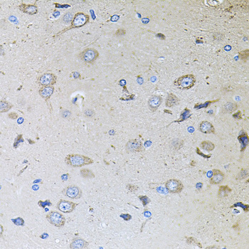
A07852 has IHC images from paraffin-embedded human prostate and rat brain (catalog image captions). Human, Mouse and Rat reactivity is listed (catalog reactivity); IF/ICC validation is unreported (catalog applications).
A07852 will render with an IHC figure from paraffin-embedded human prostate at 1:100 (catalog image caption). Its second caption documents paraffin-embedded rat brain at 1:100 (catalog image caption); Human, Mouse and Rat reactivity is listed (catalog reactivity).
Which to pick: Choose A07852 for paraffin-section IHC, supported by its own human prostate and rat brain captions (catalog image captions); the fixative is unreported (catalog image captions). No SKU here has a listed IF/ICC application or IF image, so IF/ICC validation is unreported (catalog applications; catalog IF images). For cross-species IHC, A07852 lists Human, Mouse and Rat reactivity (catalog reactivity), with images for Human and Rat only (catalog image captions); clonality is unreported (catalog clone field).