This website uses cookies to ensure you get the best experience on our website.


- Table of Contents
Plan METAP2 chromogenic IHC in paraffin sections using its widespread cytoplasmic tissue pattern as a reference (HPA tissue IHC). Start with the catalog antibody’s IHC dilution of 2–5 μg/ml (datasheet A03648-2) and compare sections fixed consistently (standard IHC practice).

Expected localisation, validated protocols, controls and antibodies — the at-a-glance facts below, then the full design guide.
| Expected localisation | Cytoplasmic staining across tissues (HPA tissue IHC) | |
| Staining pattern | Widespread cytoplasmic staining across tissues (HPA tissue IHC) | |
| Antigen retrieval | EDTA pH 8.0 HIER, heat-mediated (datasheet A03648-2) | |
| Positive control | Bone marrow+4 more · see all | |
| Negative control | None in HPA (detected in all 45 tissues); use no-primary + isotype controls |
| Fixation | Keep fixation consistent across sections. (standard IHC practice; not target-specific) | |
| Caveat | Widespread staining limits negative tissue controls (HPA tissue IHC) | |
| Regulation | Low tissue specificity; no regulator reported (HPA tissue IHC; UniProt) | |
| Isoform / epitope | 3 isoforms; epitope effects are unresolved (UniProt) |
The catalog antibody uses heat-mediated EDTA retrieval at pH 8.0 (datasheet A03648-2). The published METAP2 IHC protocols below provide sample-specific methods (PMC3324555; PMC11082409; PMC8137584; PMC7484650).
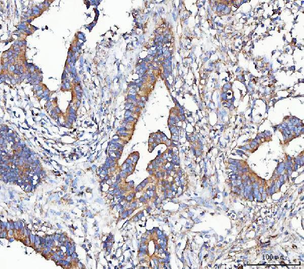
| Sample | Paraffin-embedded human colorectal adenocarcinoma tissue; fixative not specified (datasheet A03648-2) |
| Fixation | Image fixative and duration unreported (datasheet A03648-2); verify before use. |
| Sectioning | 4–5 µm sections on charged slides (standard) |
| Deparaffinisation | Xylene, graded ethanol series to water (standard) |
| Antigen retrieval | Heat retrieval: EDTA pH 8.0 (datasheet A03648-2); 20 min, 95–100 °C (standard) |
| Peroxidase block | 3% H2O2, 10 min, room temperature (standard) |
| Blocking | 10% goat serum (datasheet A03648-2) |
| Primary antibody | Rabbit anti-METAP2, 2-5 μg/ml (datasheet A03648-2) |
| Primary incubation | Overnight at 4 °C (datasheet A03648-2) |
| Detection | HRP-conjugated secondary, DAB chromogen (datasheet A03648-2) |
| Counterstain | Hematoxylin, blue, dehydrate and mount (standard) |
| Expected result | METAP2-positive staining in hematopoietic cells of bone marrow (HPA tissue IHC: High). HPA tissue profile: Ubiquitous cytoplasmic expression in all tissues. No signal in the no-primary control. |
METAP2 should show widespread cytoplasmic staining in paraffin tissue sections, with particularly strong staining in the cell populations listed below (HPA: ubiquitous cytoplasmic tissue IHC; Enhanced reliability). UniProt places METAP2 in the cytoplasm and reports no transmembrane segment (UniProt P50579). HPA calls the tissue IHC reliability Enhanced while describing only medium consistency between antibody staining and RNA expression; interpret intensity differences with that qualification (HPA: tissue IHC reliability).
| Cytoplasmic staining across several tissues, strongest in selected cell populations. | This fits HPA's ubiquitous cytoplasmic pattern. High staining is reported in bone marrow hematopoietic cells, kidney glomerular cells and lung alveolar type I cells (HPA: tissue IHC). Compare cells within each tissue; widespread expression does not imply equal intensity everywhere (HPA: tissue IHC levels). |
| Predominantly nuclear staining, or a crisp membrane rim without cytoplasmic staining. | These are unexpected as the principal chromogenic tissue IHC pattern (HPA: ubiquitous cytoplasmic tissue IHC; UniProt P50579: cytoplasm). Check whether the signal follows tissue edges, pigment or detection background before assigning METAP2. A membrane location is reported separately in ICC-IF and does not establish a membrane-only tissue IHC pattern (HPA: subcellular ICC-IF). |
| Strong staining confined to a cell population reported as low, with weak staining in nearby expected-positive cells. | Consider cross-reactivity or cell-specific detection background before calling this a METAP2-enriched population. HPA reports low staining in cardiomyocytes, skeletal myocytes, smooth muscle cells, fibroblasts and adipocytes; low is not absent (HPA: tissue IHC). |
| Uniform haze over cells and extracellular areas, obscuring cell boundaries. | Treat this as background until cellular staining can be separated from it. Uneven blocking, residual chromogen or endogenous detection activity are general IHC possibilities, not METAP2-specific findings (standard IHC practice). Compare a no-primary control and the expected cytoplasmic cell pattern (HPA: tissue IHC). |
| No signal in a section containing an HPA high-staining population. | An absent result in bone marrow hematopoietic cells or tonsil germinal center cells conflicts with the reported tissue pattern (HPA: High in those cells). First verify section quality, retrieval, antibody application and detection with suitable controls (standard IHC practice); a blank slide alone cannot establish true absence of METAP2. |
| Tissue and cell choice | HPA reports High staining in bone marrow hematopoietic cells, glomerular cells, alveolar type I cells, lymph node and tonsil germinal center cells, seminal vesicle glandular cells, and late spermatids (HPA: tissue IHC). It reports Medium in adrenal glandular cells and Low in several stromal, muscle and adipose populations; these levels guide comparison rather than define negative tissue (HPA: tissue IHC). |
| Location evidence and antibody validation | The tissue reference pattern is cytoplasmic, and both listed antibodies have Enhanced IHC status (HPA: tissue IHC; HPA019095 and CAB013025). Enhanced reflects independent-antibody or orthogonal support, while HPA still reports medium staining–RNA consistency; neither statement validates every unexpected compartment or cell type (HPA: reliability description and antibody validation). |
| Isoforms and epitope coverage | UniProt lists three METAP2 isoforms and a processed chain spanning residues 2–478 (UniProt P50579). The supplied records give no epitope location or isoform-specific IHC result, so a difference between antibodies cannot be assigned to a particular isoform from this evidence. |
| Detection chemistry | In chromogenic IHC, endogenous enzyme activity or tissue pigment can resemble antibody-derived colour (standard IHC practice). A no-primary control and review of cellular localisation help distinguish these from the expected cytoplasmic signal (HPA: tissue IHC pattern). This is a general workflow consideration, not a reported METAP2 tissue property. |
| IF/ICC Q&A | Q: Should IF/ICC look identical to paraffin IHC? A: HPA reports cytosol as supported and plasma membrane as approved in ICC-IF, with images from A-431, U-251MG and U2OS (HPA: subcellular ICC-IF). Interpret those cellular images on their own terms; the tissue IHC reference remains cytoplasmic (HPA: tissue IHC). |
| Situation | Likely cause | Next action |
|---|---|---|
| Expected-positive tissue is blank. | The run may have lost antigen detection or the selected section may lack the relevant cells; absence in HPA high-staining cells conflicts with the reference pattern (HPA: tissue IHC). | Confirm the expected cell population is present. Review retrieval, primary-antibody application and detection controls as general IHC checks; assess a known-positive section in the same run (standard IHC practice). |
| Signal is faint everywhere, including in reported high-staining cells. | Weak staining may reflect a general assay sensitivity problem; the supplied sources give no METAP2-specific fixation effect or required antibody dilution (HPA: tissue IHC; supplied source scope). | Check the catalog antibody's own IHC-P instructions, reagent performance and retrieval settings before changing interpretation. Compare high and low HPA populations under the same staining conditions (standard IHC practice; HPA: tissue IHC levels). |
| Strong nuclear-only or membrane-only chromogen dominates. | The principal tissue IHC pattern is cytoplasmic (HPA: tissue IHC; UniProt P50579). HPA's plasma-membrane observation comes from ICC-IF, so it does not by itself explain membrane-only paraffin IHC (HPA: subcellular ICC-IF). | Inspect cell outlines and counterstain, compare a no-primary control, and repeat with the IHC-validated antibody or an independently validated antibody if available (standard IHC practice; HPA: Enhanced IHC antibodies). |
| Diffuse colour covers cells and spaces between them. | Nonspecific reagent binding, endogenous detection activity or pigment can produce background in chromogenic IHC (standard IHC practice). Such haze cannot be scored as METAP2's cellular cytoplasmic pattern (HPA: tissue IHC). | Use a no-primary control, check blocking and detection steps, and score only signal that can be assigned to cells with the expected localisation (standard IHC practice; HPA: tissue IHC). |
| A low-staining population appears stronger than an HPA high-staining population. | HPA levels describe observed patterns, not absolute expression cutoffs; a reversal may reflect section composition, scoring or nonspecific staining (HPA: tissue IHC levels; standard IHC interpretation). | Identify the cell types before comparing intensity, then check controls and staining across another suitable section. Do not treat HPA Low populations as negative controls (HPA: tissue IHC). |
| Two IHC antibodies give different cell patterns. | Both listed antibodies have Enhanced IHC status, but the supplied evidence does not identify their epitopes or isoform coverage (HPA: HPA019095 and CAB013025; UniProt P50579: three isoforms). | Compare the same tissue and cell populations with matched detection controls; report the disagreement and avoid attributing it to an isoform or processing site without further evidence (standard IHC practice). |


Comprehensive Human Protein Atlas IHC scoring per tissue (reliability: Enhanced — Medium consistency between antibody staining and RNA expression data.). Rows are taken directly from the HPA tissue chart — click any row's HPA link to view the source.
| Tissue | Cell type | Level | Evidence | Source |
|---|---|---|---|---|
| Bone marrow | Hematopoietic cells | High | Protein (IHC) | HPA → |
| Kidney | Cells in glomeruli | High | Protein (IHC) | HPA → |
| Lung | Alveolar cells type I | High | Protein (IHC) | HPA → |
| Lymph node | Germinal center cells | High | Protein (IHC) | HPA → |
| Seminal vesicle | Glandular cells | High | Protein (IHC) | HPA → |
| Tissue | Cell type | Level | Evidence | Source |
|---|---|---|---|---|
| None in HPA: METAP2 is detected in all 45 scored tissues. Use a no-primary (secondary-only) and an isotype control instead. | ||||
Troubleshoot METAP2 staining in paraffin sections using the catalog antibody’s tissue result, cellular localisation, and appropriate IHC controls.
Anti-METAP2 options include pictured human paraffin-section IHC (A03648-2 image captions) and a listed IF/ICC application without a supplied image (M03648-1 catalog).
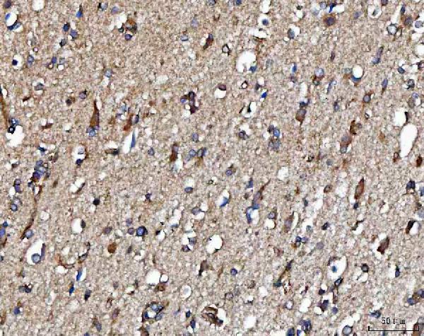
A03648-2 has IHC images from human colorectal adenocarcinoma, brain, and tonsil paraffin sections (A03648-2 image captions). M03648-1 lists IHC and IF/ICC applications and human, mouse, and rat reactivity, but supplies no IHC or IF image (M03648-1 catalog).
Which to pick: For tissue IHC, choose A03648-2: its human paraffin-section captions document EDTA retrieval at pH 8.0 and antibody at 2 μg/ml; the fixative is unreported (A03648-2 image captions). For IF/ICC, M03648-1 is the rabbit monoclonal option, clone 20M17, with IF/ICC listed but no supplied IF image (M03648-1 catalog). For cross-species planning, A03648-2 lists human, mouse, rat, and monkey reactivity, while M03648-1 lists human, mouse, and rat; the pictured IHC results for A03648-2 are human (catalog reactivity; A03648-2 image captions).