MFF / Mitochondrial fission factor · IHC design guide

Design Immunohistochemistry for MFF

Plan chromogenic MFF IHC in paraffin sections using the IHC-validated antibody (datasheet A02563-1). This guide pairs the reported cytoplasmic tissue pattern (HPA tissue IHC) with antibody concentration and controls for interpreting cell-type differences.

Evidence assembled Oct 2026 · For research use; verify linked source records and product datasheet before use
Immunohistochemistry protocol sheet for MFF (IHC for MFF): expected localisation Cytoplasmic IHC; mitochondrial outer membrane expected (HPA tissue IHC; UniProt), antibody A02563-1, validated IHC image, and IHC protocol steps
Printable MFF IHC protocol sheet — expected localisation Cytoplasmic IHC; mitochondrial outer membrane expected (HPA tissue IHC; UniProt), antibody A02563-1, controls and protocol steps. Open the full MFF IHC guide →

MFF Immunohistochemistry Experimental Design Guide

Expected localisation, validated protocols, controls and antibodies — the at-a-glance facts below, then the full design guide.

Must know before staining
Expected localisation Cytoplasmic IHC; mitochondrial outer membrane expected (HPA tissue IHC; UniProt)
Staining pattern Cytoplasmic staining across diverse tissue cell types (HPA tissue IHC)
Antigen retrieval EDTA pH 8.0 HIER, heat-mediated (datasheet A02563-1)
Positive control ⓘ Appendix+4 more · see all
Negative control ⓘ Adipose tissue+4 more · see all
Important caveats
Reasons your staining may differ from the expected pattern.
Fixation Keep fixation conditions consistent across sections (standard IHC practice; not target-specific)
Caveat Heart is high by UniProt, but cardiomyocytes lack detected staining (UniProt; HPA tissue IHC)
Regulation Expression regulation is unspecified (UniProt)
Isoform / epitope 5 isoforms; check epitope coverage across variants (UniProt)
Section 1

Recommended MFF IHC & IF Protocols

The catalog antibody protocol uses heat-mediated EDTA pH 8.0 retrieval (datasheet A02563-1). The published MFF IHC protocols below cover tumor and brain sections (PMC6838406; PMC4558123; PMC13612925; PMC6911621).

Recommended immunohistochemistry (IHC-P) protocol parameters
SampleParaffin-embedded human breast cancer tissue; fixative not specified (datasheet A02563-1)
FixationImage fixative and duration unreported (datasheet A02563-1); verify before use.
Sectioning4–5 µm sections on charged slides (standard)
DeparaffinisationXylene, graded ethanol series to water (standard)
Antigen retrievalHeat retrieval: EDTA pH 8.0 (datasheet A02563-1); 20 min, 95–100 °C (standard)
Peroxidase block3% H2O2, 10 min, room temperature (standard)
Blocking10% goat serum (datasheet A02563-1)
Primary antibodyRabbit anti-MFF, 2-5 μg/ml (datasheet A02563-1)
Primary incubationOvernight at 4 °C (datasheet A02563-1)
DetectionHRP-conjugated secondary, DAB chromogen (datasheet A02563-1)
CounterstainHematoxylin, blue, dehydrate and mount (standard)
Expected resultMFF-positive staining in glandular cells of appendix (HPA tissue IHC: Medium). HPA tissue profile: Cytoplasmic expression in most tissues. No signal in the no-primary control.
💡Decision noteStart with EDTA pH 8.0 heat retrieval for the catalog antibody (datasheet A02563-1); the cited articles do not specify retrieval conditions.
Section 2

What Is the Expected MFF Staining Pattern?

MFF is anchored in the mitochondrial outer membrane, with additional peroxisomal and vesicle localization (UniProt Q9GZY8). In paraffin IHC, expect cytoplasmic staining in documented positive cells, including appendix glandular cells and cerebellar Purkinje cells (HPA tissue IHC). HPA calls the tissue staining reliability Enhanced, while reporting medium consistency with RNA expression (HPA tissue IHC). Its short intermembrane tail follows a cytoplasm-facing region (UniProt Q9GZY8 topology).

What am I looking at on my slide?
Cytoplasmic staining in appendix glandular cells or cerebellar Purkinje cells.These are documented medium-staining populations (HPA tissue IHC). Score the named cells and their cytoplasm; chromogenic IHC alone cannot prove that individual puncta are mitochondria (general IHC practice).
Predominantly nuclear staining, or signal confined to cell borders.That distribution conflicts with MFF’s outer-membrane localization and HPA’s cytoplasmic tissue profile (UniProt Q9GZY8; HPA tissue IHC). Treat it as suspect and compare with control sections and detection controls (general IHC practice).
Strong staining in a population reported as not detected, such as adipocytes or smooth muscle cells.The result differs from the sampled HPA pattern (HPA tissue IHC). Possible causes include cross-reactivity or endogenous detection activity; a single discordant population does not establish which cause applies (general IHC practice).
Uniform color across cells, stroma, and blank areas.This obscures the cell-restricted cytoplasmic pattern reported by HPA (HPA tissue IHC). Check nonspecific antibody binding, wash steps, and endogenous detection activity with appropriate controls (general IHC practice).
No staining in appendix glandular cells or cerebellar Purkinje cells.Both populations have medium HPA staining, so a blank result warrants assay review (HPA tissue IHC). Verify the tissue and antibody, then review retrieval, detection, and control performance (general IHC practice).
💡Expected MFF appearanceA credible positive is cytoplasmic signal of about medium intensity in appendix glandular cells or cerebellar Purkinje cells (HPA tissue IHC); dominant nuclear staining or uniform background is suspect (UniProt Q9GZY8; general IHC practice).
How each factor affects the staining
Membrane topologyMFF has a transmembrane segment at residues 323–340 and a cytoplasm-facing region at 1–322 (UniProt Q9GZY8 topology). Interpret tissue signal as cytoplasmic localization; topology alone does not predict an antibody’s staining intensity.
Choice of reference cellsAppendix glandular, Purkinje, cortical neuronal, and esophageal squamous epithelial cells are reported at medium intensity (HPA tissue IHC). Stomach glandular cells are low, making them a less clear visual benchmark (HPA tissue IHC).
Tissue-expression caveatUniProt reports high expression in heart and muscle, yet HPA reports cardiomyocytes and skeletal myocytes as not detected by tissue IHC (UniProt Q9GZY8; HPA tissue IHC). Do not equate expression summaries with a guaranteed paraffin-section signal.
Isoforms and antibody validationUniProt lists 5 MFF isoforms; the supplied record gives no epitope or isoform coverage for a catalog antibody (UniProt Q9GZY8). HPA010968 has Enhanced IHC validation, whereas the supplied HPA entry gives HPA074625 no IHC status (HPA antibodies).
IF/ICC Q: where should fluorescence appear?A: Mainly at mitochondria; HPA also reports the sperm principal piece (HPA ICC-IF). This is an IF/ICC localization reference, not an IHC-P protocol or proof that every chromogenic cytoplasmic granule is mitochondrial.
Retrieval and detection controlsUse the selected antibody’s IHC-P instructions to choose retrieval; no MFF-specific retrieval response is supplied (general IHC practice). A detection-only control helps assess endogenous color development (general IHC practice).
Why is my staining missing, weak or wrong?
SituationLikely causeNext action
Expected positive cells are blank.The run may have lost antigen signal or detection sensitivity; the record does not identify an MFF-specific fixation effect (general IHC practice).Confirm the sampled cell population against HPA, then check the antibody’s IHC-P instructions, retrieval, detection reagents, and a working positive control (HPA tissue IHC; general IHC practice).
Color is mainly nuclear.Nuclear dominance conflicts with the documented cytoplasmic tissue pattern and membrane topology (HPA tissue IHC; UniProt Q9GZY8).Review morphology and staining controls; repeat with the IHC-validated antibody and compare the same cell populations (HPA antibodies; general IHC practice).
A reported negative cell population stains strongly.The finding is discordant with HPA’s sampled result and could reflect nonspecific binding or endogenous detection activity (HPA tissue IHC; general IHC practice).Check a detection-only control and compare a documented medium-positive population on the same run before assigning the signal to MFF (HPA tissue IHC; general IHC practice).
Background hides cell boundaries.Excess nonspecific color can prevent assessment of HPA’s cell-specific cytoplasmic profile (HPA tissue IHC; general IHC practice).Inspect blank and detection-only controls; review antibody concentration, blocking, washing, and chromogen development under the chosen IHC-P workflow (general IHC practice).
Stomach glandular cells show faint signal.HPA rates this population low, so faint staining can fit the reference pattern (HPA tissue IHC).Judge the run alongside a medium reference population, such as appendix glandular cells, before calling it a failure (HPA tissue IHC).
Heart or skeletal muscle conflicts with an expression summary.UniProt lists high tissue expression, while HPA reports cardiomyocytes and skeletal myocytes as not detected by IHC (UniProt Q9GZY8; HPA tissue IHC).Record the exact cell type and assay result; use documented HPA-positive cells to assess staining performance rather than assuming those muscle cells must stain (HPA tissue IHC).

Sample controls for MFF IHC & IF

🧪Run cerebellum first and look for staining in Purkinje cells (HPA: Medium in cerebellar Purkinje cells). Use adipose tissue as a negative comparator, focusing on adipocytes (HPA: Not detected in adipocytes); on the cerebellar slide, treat unstained non-Purkinje cells as background comparators, since their MFF status is unreported in the supplied HPA rows.
Positive control tissue: Appendix (Glandular cells, HPA Medium)
Negative control tissue: Adipose tissue (HPA Not detected)
ICC-IF cell lines (HPA subcellular resource): HPA ICC-IF images show MFF in HEK293, SiHa, U2OS, Sperm, with annotated localisation: Mitochondria (supported) (HPA subcellular).
Technical controls: Include a no-primary (secondary-only) control, a host-matched rabbit IgG isotype control (caption: rabbit anti-MFF), and a matched MFF knockout specimen or a validated peptide-block control. Quench endogenous peroxidase and inspect pigment before interpreting DAB staining (caption: HRP/DAB detection; standard IHC practice).
⚠️Feasibility: The selected paraffin-section example uses heat-mediated EDTA retrieval at pH 8.0, but does not establish that retrieval is required (caption: heat-mediated EDTA retrieval). Its fixative is unreported, and no target-specific fixation window or fixation effect is reported (caption: fixative not stated). Paraffin IHC has a supplied example; whether frozen sections or IF are easier is unreported, and cerebellar pigment or autofluorescence should be checked when interpreting neuronal staining (standard IHC/IF practice).

HPA tissue IHC evidence for MFF

Comprehensive Human Protein Atlas IHC scoring per tissue (reliability: Enhanced — Medium consistency between antibody staining and RNA expression data.). Rows are taken directly from the HPA tissue chart — click any row's HPA link to view the source.

Positive expression · recommended positive controls

TissueCell typeLevelEvidenceSource
Appendix Glandular cells Medium Protein (IHC) HPA →
Cerebellum Purkinje cells Medium Protein (IHC) HPA →
Cerebral cortex Neuronal cells Medium Protein (IHC) HPA →
Esophagus Squamous epithelial cells Medium Protein (IHC) HPA →
Oral mucosa Squamous epithelial cells Medium Protein (IHC) HPA →

Undetected expression · recommended negative controls

TissueCell typeLevelEvidenceSource
Adipose tissue Adipocytes Not detected Protein (IHC) HPA →
Caudate Glial cells Not detected Protein (IHC) HPA →
Heart muscle Cardiomyocytes Not detected Protein (IHC) HPA →
Liver Cholangiocytes Not detected Protein (IHC) HPA →
Ovary Follicle cells Not detected Protein (IHC) HPA →
Section 3

Advanced MFF IHC Tips

Troubleshoot MFF staining in paraffin sections by checking retrieval, cell type, compartment and detection controls before comparing chromogenic IHC scores.

Which antigen retrieval should I start with for MFF paraffin sections?
Use heat-mediated antigen retrieval in EDTA at pH 8.0 for paraffin sections stained with the catalog antibody (datasheet A02563-1). The documented workflow detected MFF after that retrieval, followed by 2 μg/ml primary antibody overnight at 4°C (datasheet A02563-1). If staining is weak, vary heating time in small steps while keeping the EDTA buffer constant, and compare sections from the same specimen in one staining run (standard IHC optimisation practice). Judge improvement by signal in the expected cytoplasmic compartment and by background on a no-primary control, since tissue IHC shows cytoplasmic expression in most tissues (HPA tissue IHC).
Could fixation explain weak or uneven MFF staining?
The selected tissue caption identifies a paraffin section but does not state its fixative, so MFF-specific sensitivity to fixation is unknown (datasheet A02563-1). Record each specimen’s fixative and fixation time, then compare sections processed together before changing antibody concentration (standard IHC practice). Use the documented EDTA retrieval at pH 8.0 and 2 μg/ml primary antibody as a common starting point when comparing fixation conditions (datasheet A02563-1). If staining varies within a specimen, inspect morphology and compare central and peripheral regions; fixation gradients can produce uneven antigen preservation in paraffin-section IHC (standard IHC practice).
What staining pattern is plausible for MFF in chromogenic IHC?
Expect predominantly cytoplasmic staining at tissue-section resolution, consistent with the reported IHC profile of cytoplasmic expression in most tissues (HPA tissue IHC). MFF is assigned to the mitochondrial outer membrane and peroxisomes, with additional vesicle localisation, so fine cytoplasmic puncta are plausible (UniProt Q9GZY8 subcellular). Its 323–340 transmembrane segment anchors a protein whose 1–322 region faces the cytoplasm (UniProt Q9GZY8 topology). Do not call an individual DAB punctum a mitochondrion from chromogenic IHC alone; compare its distribution with an organelle marker using a suitable imaging method (standard IHC interpretation practice).
How can isoforms or epitope accessibility affect my MFF result?
MFF has 5 annotated isoforms, so confirm which sequence region the catalog antibody recognises before treating staining as a measure of every isoform (UniProt Q9GZY8 isoforms). The record places residues 1–322 on the cytoplasmic side and the membrane-spanning segment at 323–340, but it does not identify this antibody’s epitope (UniProt Q9GZY8 topology; supplied datasheet caption). Phosphorylated residues include 115, 155 and 157; their presence alone does not establish an effect on antibody binding (UniProt Q9GZY8 modified residues). If results conflict across specimens, check the antibody’s epitope documentation and compare with an independently validated reagent or genetic control (standard IHC validation practice).
How should I investigate MFF localisation with multiplex immunofluorescence?
Use the separate IF/ICC guide to establish staining conditions; this page’s selected antibody evidence describes paraffin-section chromogenic IHC (datasheet A02563-1). Multiplex MFF with a marker for the expected cell type, such as a Purkinje-cell marker when examining cerebellum, where Purkinje cells show medium staining (HPA tissue IHC). Choose spectrally separated fluorophores and consider a far-red channel if tissue autofluorescence obscures shorter wavelengths (standard IF practice). For an antibody directed to the cytoplasmic region, titrate permeabilisation to expose residues 1–322 while preserving organelle morphology; the actual epitope is unspecified here (UniProt Q9GZY8 topology; supplied datasheet caption).
How can I reduce diffuse brown background without losing MFF signal?
Start by comparing a no-primary control with the catalog workflow, which used 10% goat serum block and 2 μg/ml primary antibody overnight at 4°C (datasheet A02563-1). The caption used a peroxidase-linked secondary and DAB development, making peroxidase blocking and careful chromogen timing appropriate general controls for this detection chemistry (datasheet A02563-1; standard chromogenic IHC practice). If background persists, titrate primary concentration and washing conditions on matched sections while holding retrieval at EDTA pH 8.0 (datasheet A02563-1; standard IHC optimisation practice). Preserve a discernible cytoplasmic signal; the reported tissue profile is predominantly cytoplasmic (HPA tissue IHC).
What should I measure when comparing MFF staining across sections? ⚠ ANSWER MARKED FOR VERIFICATION
Score MFF within a predefined cell population and cytoplasmic compartment, because the reported tissue IHC profile is cytoplasmic and expression differs among cell types (HPA tissue IHC). An H-score combines the percentage of cells at intensity grades 0–3 into a 0–300 score; alternatively report percentage positive or positive-cell density per mm² (standard IHC scoring practice). Normalise to the number of evaluable cells or viable tissue area in that population, and apply the same threshold and imaging settings across sections (standard IHC quantification practice). Report cell type and compartment alongside the score, since HPA describes only medium consistency between staining and RNA expression (HPA tissue IHC reliability).
How do I distinguish convincing MFF staining from artefact?
A convincing result follows cell-type and cytoplasmic patterns: Purkinje cells show medium staining, whereas cardiomyocytes are listed as not detected in the cited tissue IHC dataset (HPA tissue IHC). Do not read the cardiomyocyte result as proof that MFF is absent from all heart cells or conditions; UniProt reports high expression in heart without assigning that statement to cardiomyocytes (HPA tissue IHC; UniProt Q9GZY8 tissue specificity). Treat isolated nuclear staining, section-edge enhancement and necrotic-area deposits cautiously, and examine no-primary and peroxidase-block controls for detection artefact (standard chromogenic IHC practice). Chromogenic cytoplasmic signal alone cannot establish mitochondrial rather than peroxisomal localisation (UniProt Q9GZY8 subcellular; standard IHC interpretation practice).
Boster reagents

Best MFF / Mitochondrial fission factor IHC Antibodies

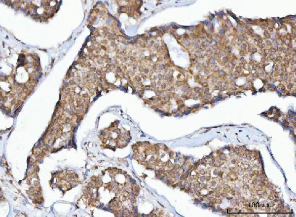
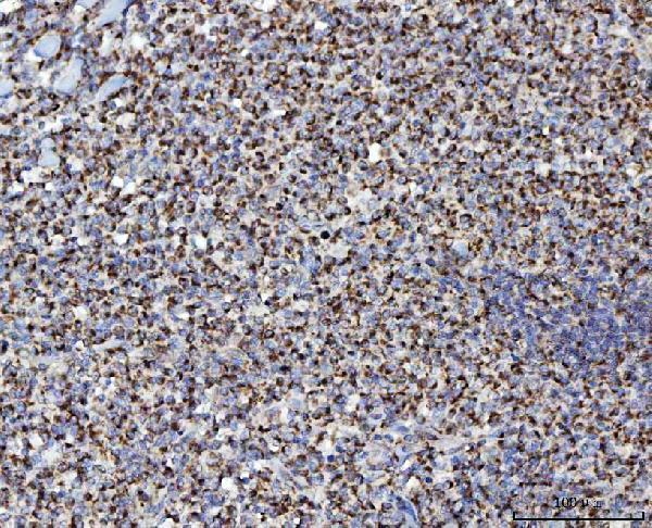
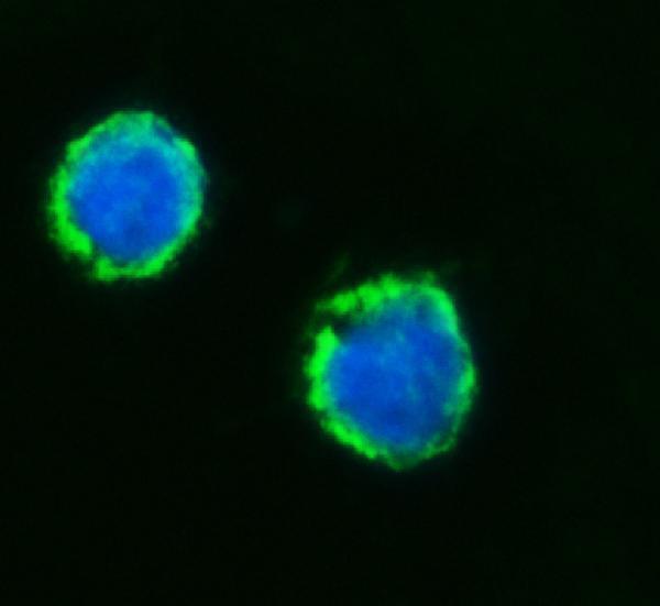
A02563-1 has IHC images from human cancer and lymphoma paraffin sections and mouse brain paraffin sections, plus IF images from SiHa cells and human lymphoma paraffin sections (catalog image captions).

Real IHC data IHC analysis of MFF using anti-MFF antibody (A02563-1). MFF was detected in a paraffin-embedded section of human breast cancer tissue. Heat mediated antigen retrieval was performed in EDTA buffer (pH 8.0, epitope retrieval solution). The tissue section was blocked with 10% goat serum. The tissue section was then incubated with 2 μg/ml rabbit anti-MFF Antibody (A02563-1) overnight at 4°C. Peroxidase Conjugated Goat Anti-rabbit IgG was used as secondary antibody and incubated for 30 minutes at 37°C. The tissue section was developed using HRP Conjugated Rabbit IgG Super Vision Assay Kit (Catalog # SV0002) with DAB as the chromogen.
Anti-MFF Antibody ®
Cat # A02563-1

A02563-1 is listed for IHC, ICC and IF in human, mouse and rat (catalog applications and reactivity). Its IHC captions show human breast cancer, lymphoma and rectal cancer paraffin sections and mouse brain paraffin sections; its IF captions show SiHa cells and human lymphoma paraffin sections (catalog image captions).

Which to pick: For tissue IHC, choose A02563-1 for paraffin sections, the preparation shown in its IHC captions; the fixative is unreported (catalog IHC image captions). For IF/ICC, A02563-1 has images from SiHa cells and human lymphoma paraffin sections; clonality is unreported (catalog IF image captions; catalog clone field). For cross-species work, A02563-1 is listed as human, mouse and rat reactive, with IHC images for human and mouse samples (catalog reactivity; catalog IHC image captions).

Each figure is that product's own IHC / IF validation image from its datasheet.

References

  1. UniProt Consortium. UniProt entry Q9GZY8 (MFF_HUMAN, Mitochondrial fission factor).
  2. Human Protein Atlas. MFF tissue IHC expression (reliability: Enhanced).
  3. Human Protein Atlas. MFF subcellular location (ICC-IF): Mainly localized to the mitochondria. In addition localized to the principal piece..
  4. Human Protein Atlas. MFF antibody validation summary (2 antibodies).
  5. Mitochondrial fission factor is a novel Myc-dependent regulator of mitochondrial permeability in cancer. EBioMedicine 2019 — PMC6838406.
  6. Mitochondrial fission determines cisplatin sensitivity in tongue squamous cell carcinoma through the BRCA1-miR-593-5p-MFF axis. Oncotarget 2015 — PMC4558123.
  7. Mitochondrial alterations in pellagra-associated central chromatolysis: Comparison with ballooned achromatic neurons of other etiologies. Journal of neuropathology and experimental neurology 2026 — PMC13612925.
  8. MFF Regulation of Mitochondrial Cell Death Is a Therapeutic Target in Cancer. Cancer research 2019 — PMC6911621.
  9. PubMed PMID:18353969 — UniProt-cited evidence.
  10. PubMed PMID:14702039 — UniProt-cited evidence.
  11. PubMed PMID:15815621 — UniProt-cited evidence.