MFSD12 / Major facilitator superfamily domain-containing protein 12 · IHC design guide

Design Immunohistochemistry for MFSD12

Plan chromogenic IHC on paraffin sections with the catalog antibody at 2–5 μg/ml (datasheet A16855-1). Compare the reported cytoplasmic tissue staining (HPA tissue IHC) with MFSD12's melanosome and lysosome membrane annotation (UniProt), and treat the tissue profile as pending external verification (HPA tissue IHC).

Evidence assembled Oct 2026 · For research use; verify linked source records and product datasheet before use
Immunohistochemistry protocol sheet for MFSD12 (IHC for MFSD12): expected localisation General cytoplasmic tissue staining (HPA tissue IHC), antibody A16855-1, validated IHC image, and IHC protocol steps
Printable MFSD12 IHC protocol sheet — expected localisation General cytoplasmic tissue staining (HPA tissue IHC), antibody A16855-1, controls and protocol steps. Open the full MFSD12 IHC guide →

MFSD12 Immunohistochemistry Experimental Design Guide

Expected localisation, validated protocols, controls and antibodies — the at-a-glance facts below, then the full design guide.

Must know before staining
Expected localisation General cytoplasmic tissue staining (HPA tissue IHC)
Staining pattern Cytoplasmic in glial and seminiferous-duct cells (HPA tissue IHC)
Antigen retrieval EDTA pH 8.0 HIER, heat-mediated (datasheet A16855-1)
Positive control ⓘ Cerebral cortex+4 more · see all
Negative control ⓘ Adipose tissue+4 more · see all
Important caveats
Reasons your staining may differ from the expected pattern.
Fixation Matched tissue-IHC evidence does not establish the fixation claim. Validate the specimen-specific method before use. (selected-SKU IHC image A16855-1)
Caveat Tissue IHC profile awaits external verification (HPA tissue IHC)
Regulation High in primary melanocytes (UniProt)
Isoform / epitope 4 isoforms; cytoplasmic vs lumenal epitope map unknown (UniProt)
Section 1

Recommended MFSD12 IHC & IF Protocols

The catalog antibody’s IHC-P protocol (datasheet: A16855-1) and a published melanoma tissue microarray protocol (PMC6462865) provide starting conditions for chromogenic MFSD12 staining.

Recommended immunohistochemistry (IHC-P) protocol parameters
SampleParaffin-embedded human testicular seminoma tissue; fixative not specified (datasheet A16855-1)
FixationImage fixative and duration unreported (datasheet A16855-1); verify before use.
Sectioning4–5 µm sections on charged slides (standard)
DeparaffinisationXylene, graded ethanol series to water (standard)
Antigen retrievalHeat retrieval: EDTA pH 8.0 (datasheet A16855-1); 20 min, 95–100 °C (standard)
Peroxidase block3% H2O2, 10 min, room temperature (standard)
Blocking10% goat serum (datasheet A16855-1)
Primary antibodyRabbit anti-MFSD12, 2-5 μg/ml (datasheet A16855-1)
Primary incubationOvernight at 4 °C (datasheet A16855-1)
DetectionHRP-conjugated secondary, DAB chromogen (datasheet A16855-1)
CounterstainHematoxylin, blue, dehydrate and mount (standard)
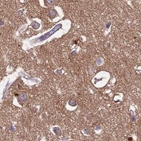
Expected resultMFSD12-positive staining in glial cells of cerebral cortex (HPA tissue IHC: High). HPA tissue profile: General cytoplasmic expression. No signal in the no-primary control.
💡Decision noteStart with heat-mediated EDTA retrieval at pH 8.0 (datasheet: A16855-1); the published protocol reports antigen retrieval without specifying conditions (PMC6462865).
Section 2

What Is the Expected MFSD12 Staining Pattern?

MFSD12 is a 12-pass protein of melanosome and lysosome membranes (UniProt Q6NUT3 topology and subcellular location). In paraffin-section IHC, expect cytoplasmic staining in positive cells: HPA reports high staining in cerebral-cortex glia, seminiferous-duct cells and tonsil germinal-center cells (HPA tissue IHC). Treat the pattern as provisional because HPA rates its tissue-IHC reliability Uncertain, pending external verification (HPA tissue IHC).

What am I looking at on my slide?
Cytoplasmic staining in cerebral-cortex glia, seminiferous-duct cells or tonsil germinal-center cells (HPA tissue IHC).This matches HPA's reported high-staining cell populations. MFSD12 resides on intracellular organelle membranes, although chromogenic IHC may not resolve individual membranes (UniProt Q6NUT3 subcellular location; HPA tissue IHC). Judge the pattern alongside controls because HPA rates the tissue evidence Uncertain (HPA tissue IHC).
Predominantly nuclear staining, with little cytoplasmic staining in an expected-positive cell population (HPA tissue IHC).A nuclear-dominant pattern conflicts with the reported cytoplasmic tissue profile and melanosome/lysosome membrane assignment (HPA tissue IHC; UniProt Q6NUT3 subcellular location). Investigate nonspecific detection or tissue artefact before assigning the nuclear signal to MFSD12; the available evidence does not support nuclear localisation.
Strong staining in adipocytes or heart cardiomyocytes (HPA: Not detected in these cells).This conflicts with those HPA cell-specific observations, but does not by itself prove cross-reactivity: the HPA IHC assessment is Uncertain (HPA tissue IHC). Consider nonspecific antibody binding and endogenous detection activity; compare with a no-primary control using the same chromogen (general IHC practice).
Diffuse color across cells and extracellular areas, without a discernible cell-specific pattern.Uniform background is harder to reconcile with HPA's cell-specific tissue observations (HPA tissue IHC). In chromogenic IHC, inadequate blocking, excessive antibody or detection-reagent background can obscure localisation (general IHC practice). Interpret only signal that separates from the matched no-primary control (general IHC practice).
No staining in cerebral-cortex glia, seminiferous-duct cells or tonsil germinal-center cells (HPA: High in each).A missing expected signal calls for review of tissue identity, retrieval, antibody use and detection performance (general IHC practice). HPA's Uncertain reliability means these are candidate positive controls, not guaranteed positives for every antibody or preparation (HPA tissue IHC).
💡Expected MFSD12 appearanceA plausible positive is cell-associated cytoplasmic chromogen in HPA-reported high-staining glia, seminiferous-duct cells or germinal-center cells (HPA tissue IHC), consistent with intracellular organelle membranes (UniProt Q6NUT3); diffuse field-wide or nuclear-dominant color is suspect, subject to controls (general IHC practice).
How each factor affects the staining
Intracellular membrane topologyMFSD12 has 12 transmembrane segments and is assigned to melanosome and lysosome membranes (UniProt Q6NUT3 topology and subcellular location). Score cytoplasmic cell-associated signal; ordinary chromogenic sections may not distinguish those two organelles (general IHC practice).
Cell-specific tissue distributionHPA reports high staining in cerebral-cortex glia, seminiferous-duct cells and tonsil germinal-center cells, but no detection in adipocytes or cardiomyocytes (HPA tissue IHC). Compare the specified cell populations rather than calling an entire tissue positive or negative.
Antibody validation limitHPA labels tissue-IHC reliability Uncertain, pending external verification; both listed antibodies have Uncertain IHC status (HPA tissue IHC; HPA antibodies). A visually plausible pattern therefore needs matched controls and should not be treated as independently verified.
Epitope and isoform informationUniProt lists 4 isoforms and maps cytoplasmic and lumenal regions across MFSD12's membrane topology (UniProt Q6NUT3 isoforms and topology). No antibody epitope is supplied, so these records do not establish isoform coverage or predict which retrieval condition will work.
Why is my staining missing, weak or wrong?
SituationLikely causeNext action
Expected-positive cells show no chromogen (HPA: High in the specified cell populations).The run may have a retrieval, primary-antibody or detection problem; HPA's Uncertain rating also limits confidence that every preparation will reproduce its pattern (general IHC practice; HPA tissue IHC).Check tissue and cell identity, then verify the detection system with an appropriate control and review the antibody's IHC-P instructions and retrieval condition (general IHC practice). Do not infer MFSD12 absence from one failed slide.
The entire section has weak, diffuse color rather than a cellular pattern.Background from primary antibody or detection reagents may obscure the cell-specific contrast described by HPA (general IHC practice; HPA tissue IHC).Inspect a no-primary control, confirm blocking and washes, and adjust antibody concentration only within the validated IHC-P workflow (general IHC practice). Reassess whether specific cells remain distinguishable after background is reduced.
Nuclei dominate the staining in an expected-positive field.This does not fit MFSD12's reported intracellular membrane location or HPA's general cytoplasmic tissue profile (UniProt Q6NUT3 subcellular location; HPA tissue IHC).Compare nuclear color with a no-primary control and review counterstain and detection artefacts (general IHC practice). Report the pattern as unconfirmed unless cell-associated cytoplasmic staining is independently supported.
Adipocytes or cardiomyocytes appear strongly positive (HPA: Not detected).Nonspecific binding or endogenous chromogenic activity is possible; HPA's Uncertain reliability prevents treating its negative calls as absolute (general IHC practice; HPA tissue IHC).Compare a no-primary slide, inspect background in the same cell type, and verify endogenous-enzyme blocking if using enzyme-based detection (general IHC practice). Interpret discordance as unresolved until controls support specificity.
Brown granules in pigmented cells resemble positive chromogen.Native melanin can resemble a brown IHC reaction product (general IHC practice). UniProt reports high MFSD12 expression in primary melanocytes, so pigment and plausible target-bearing cells may coincide (UniProt Q6NUT3 tissue specificity).Inspect an adjacent no-primary section and compare pigment distribution with chromogen before scoring (general IHC practice). Require antibody-dependent signal in the expected cellular compartment.
Can an IF/ICC membrane pattern confirm this IHC result?HPA summarizes MFSD12 as membrane-associated in its subcellular record, but provides no main-location assignment or ICC-IF image-bearing cell lines (HPA subcellular). UniProt assigns melanosome and lysosome membranes (UniProt Q6NUT3 subcellular location).Treat any IF/ICC comparison as supplementary and evaluate it with its own controls (general IF practice). The supplied HPA record cannot establish a reference IF image pattern; use the separate IF/ICC guide for that application.

Sample controls for MFSD12 IHC & IF

🧪Run testis first and look for staining in cells in seminiferous ducts (HPA: High in cells in seminiferous ducts). Run adipose tissue as the negative and expect adipocytes to lack specific staining (HPA: Not detected in adipocytes); on the testis slide, cells without specific signal should show counterstain without brown DAB deposit (standard IHC practice).
Positive control tissue: Cerebral cortex (Glial cells, HPA High)
Negative control tissue: Adipose tissue (HPA Not detected)
ICC-IF cell lines (HPA subcellular resource): HPA carries no ICC-IF cell line for MFSD12; derive a cell-line control from the positive tissue's cell type (Glial cells) and confirm it by RNA or western blot first.
Technical controls: Include a no-primary, secondary-only control; an isotype control matched to the rabbit primary antibody, with clonality matched if known; and MFSD12 knockout tissue as a biological negative where available (caption: rabbit anti-MFSD12; standard IHC practice). Quench endogenous peroxidase in the testis section before HRP/DAB detection (caption: HRP/DAB; standard IHC practice).
⚠️Feasibility: A target-specific fixation window or fixation effect is unreported, and the selected A16855-1 tissue-IHC caption does not state a fixative (selected A16855-1 tissue-IHC caption: fixative not stated). The paraffin-section example used heat retrieval in EDTA at pH 8.0, but it does not establish that retrieval is required; whether frozen sections or IF are easier is unreported (selected A16855-1 tissue-IHC caption: EDTA retrieval; HPA subcellular: no ICC-IF images listed). Score normal testis by seminiferous-duct cell type because the illustrated specimen is testicular seminoma, whereas the HPA positive row identifies cells in seminiferous ducts (selected A16855-1 tissue-IHC caption: testicular seminoma; HPA: High in cells in seminiferous ducts).

HPA tissue IHC evidence for MFSD12

Comprehensive Human Protein Atlas IHC scoring per tissue (reliability: Uncertain — Pending external verification.). Rows are taken directly from the HPA tissue chart — click any row's HPA link to view the source.

Positive expression · recommended positive controls

TissueCell typeLevelEvidenceSource
Cerebral cortex Glial cells High Protein (IHC) HPA →
Testis Cells in seminiferous ducts High Protein (IHC) HPA →
Tonsil Germinal center cells High Protein (IHC) HPA →
Adrenal gland Glandular cells Medium Protein (IHC) HPA →
Appendix Glandular cells Medium Protein (IHC) HPA →

Undetected expression · recommended negative controls

TissueCell typeLevelEvidenceSource
Adipose tissue Adipocytes Not detected Protein (IHC) HPA →
Bone marrow Hematopoietic cells Not detected Protein (IHC) HPA →
Caudate Glial cells Not detected Protein (IHC) HPA →
Heart muscle Cardiomyocytes Not detected Protein (IHC) HPA →
Liver Cholangiocytes Not detected Protein (IHC) HPA →
Section 3

Advanced MFSD12 IHC Tips

Troubleshoot MFSD12 chromogenic IHC in paraffin sections by checking retrieval, compartment, cell identity, and assay controls before interpreting staining intensity.

Which retrieval condition should I try first for MFSD12 staining in paraffin sections?
Start with heat-mediated antigen retrieval in EDTA pH 8.0 (datasheet A16855-1). The selected tissue image used this retrieval before 2 μg/ml primary antibody overnight at 4°C, providing a reference condition for optimization (datasheet A16855-1). Keep section thickness, heating, and detection consistent while comparing retrieval durations on adjacent sections (standard IHC practice). If staining remains weak, test citrate pH 6.0 as a fallback alongside the documented EDTA condition, and judge both against tissue morphology and background (standard IHC practice). The caption identifies the specimen as paraffin embedded but does not report its fixative, which limits conclusions about retrieval performance across differently processed samples (datasheet A16855-1).
Could fixation explain weak or inconsistent MFSD12 IHC staining?
MFSD12 specific fixation sensitivity is unknown because the selected paraffin section caption does not state the fixative (datasheet A16855-1). Record fixation conditions for each specimen and compare sections processed together using the documented EDTA pH 8.0 retrieval condition (datasheet A16855-1; standard IHC practice). When staining varies, inspect preservation and morphology before changing primary antibody concentration, since tissue processing can affect antigen accessibility and interpretation (standard IHC practice). Include a specimen with reproducible staining in each run and compare it with a primary antibody omission control (standard IHC practice). Do not attribute a difference specifically to fixation without a controlled comparison of specimens whose fixation histories are known (standard IHC practice).
What staining pattern is plausible for MFSD12 in chromogenic IHC?
MFSD12 is annotated at melanosome and lysosome membranes, with 12 transmembrane segments (UniProt Q6NUT3 localisation and topology). In tissue sections, assess intracellular cytoplasmic staining and its distribution rather than expecting a crisp plasma membrane outline; the tissue atlas describes a general cytoplasmic pattern (HPA: general cytoplasmic expression; UniProt Q6NUT3 localisation). Apparent granules may be compatible with organelle localisation, but DAB alone cannot establish which organelle contains the signal (UniProt Q6NUT3 localisation; standard IHC interpretation). Compare stained cells with neighbouring cells and a primary antibody omission control under the same detection conditions (standard IHC practice). Treat a predominantly nuclear pattern as discordant with the annotated locations and investigate background or antibody specificity (UniProt Q6NUT3 localisation; standard IHC interpretation).
How should isoforms and membrane topology affect interpretation of a negative section?
MFSD12 has 4 annotated isoforms and 12 transmembrane segments, so epitope accessibility may depend on the antibody's binding region (UniProt Q6NUT3 isoforms and topology; standard IHC practice). The supplied caption does not identify that region or establish which isoforms the catalog antibody detects (datasheet A16855-1). Check the available immunogen or epitope documentation before interpreting absent staining as absent MFSD12, especially when comparing specimens (standard IHC interpretation). UniProt places residues 240–279 on the cytoplasmic side and annotates phosphorylation at residue 254, but neither fact establishes an effect on this antibody without epitope evidence (UniProt Q6NUT3 topology and modified residues). Retain a known staining control during retrieval optimization (standard IHC practice).
How can IF help assess an ambiguous MFSD12 IHC pattern?
Use IF as a complementary localisation check, while keeping chromogenic IHC scoring tied to the paraffin section assay (standard IHC/IF practice). Multiplex MFSD12 with a validated marker for the expected cell type in the specimen, then evaluate whether their cell assignments agree (standard IF practice). Choose fluorophores after checking unstained tissue autofluorescence, and include single stain controls to assess signal spillover (standard IF practice). Match permeabilisation to the documented antibody epitope: a cytoplasmic epitope and a lumen facing epitope can require different access conditions across MFSD12's 12 transmembrane segments (UniProt Q6NUT3 topology; standard IF practice). Because the supplied evidence does not map this antibody's epitope or report an IF validation, optimize access empirically rather than assuming an IHC result transfers to IF (datasheet A16855-1; standard IF practice).
What should I check when MFSD12 DAB staining appears widespread?
First compare the stained section with a primary antibody omission control and inspect whether signal follows tissue edges, damaged regions, or endogenous pigment (standard IHC practice). The documented assay used peroxidase conjugated secondary detection and DAB, so include a peroxidase blocking step and check the detection system for nonspecific color (datasheet A16855-1; standard IHC practice). The selected image used 10% goat serum blocking and 2 μg/ml primary antibody overnight at 4°C, which provide documented starting conditions for a controlled dilution series (datasheet A16855-1). Evaluate any reduction in background alongside retention of interpretable cellular staining and preserved morphology (standard IHC practice). Pigment and DAB can overlap visually in pigmented specimens, so inspect unstained and control sections before assigning brown deposits to MFSD12 (UniProt Q6NUT3 function; standard IHC practice).
How should I quantify MFSD12 staining across tissue sections? ⚠ ANSWER MARKED FOR VERIFICATION
Define the cell population and tissue region before scoring, then apply the same threshold and imaging conditions across sections (standard IHC quantification practice). Report the percentage of positive cells and an H-score from 0–300 when intensity grading is reproducible (standard IHC quantification practice). For sparse positive cells, density per mm² of viable tissue can complement the percentage; exclude folds, necrosis, and section edges consistently (standard IHC quantification practice). Normalize counts to the number of eligible cells or the viable area within the same annotated compartment, and document any staining run used for comparison (standard IHC quantification practice). Keep cell types separate because the tissue atlas reports differing levels across cell populations and labels its IHC reliability uncertain (HPA: tissue profile and uncertain reliability).
How can I distinguish convincing MFSD12 staining from artefact?
Look for reproducible intracellular staining in morphologically intact cells, consistent with the annotated melanosome and lysosome membrane locations (UniProt Q6NUT3 localisation; standard IHC interpretation). Compare the expected cell population with a primary antibody omission control, and scrutinize nuclear color, edge effects, and necrotic areas before calling cells positive (standard IHC practice). Check peroxidase dependent color with appropriate detection controls because the documented assay used peroxidase and DAB (datasheet A16855-1; standard IHC practice). The tissue atlas reports high staining in testicular seminiferous duct cells but rates the IHC evidence uncertain, so use that pattern as context rather than independent validation (HPA: testis high; HPA: uncertain reliability). Require concordant cell identity, compartment, morphology, and controls before interpreting intensity differences biologically (standard IHC interpretation).
Boster reagents

Best MFSD12 / Major facilitator superfamily domain-containing protein 12 IHC Antibodies

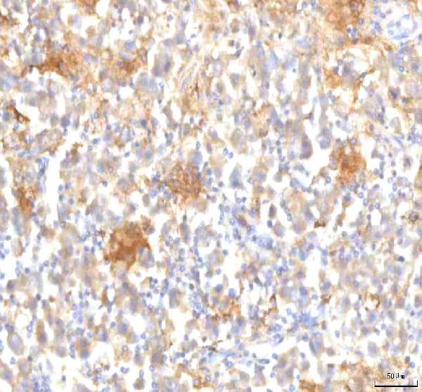
The catalog includes one human-reactive anti-MFSD12 antibody with paraffin-section IHC images from testicular seminoma and colon adenocarcinoma (catalog: A16855-1 image captions); no IF image is supplied (catalog: IF image list).

Real IHC data IHC analysis of MFSD12 using anti-MFSD12 antibody (A16855-1). MFSD12 was detected in a paraffin-embedded section of human testicular seminoma tissue. Heat mediated antigen retrieval was performed in EDTA buffer (pH 8.0, epitope retrieval solution). The tissue section was blocked with 10% goat serum. The tissue section was then incubated with 2 μg/ml rabbit anti-MFSD12 Antibody (A16855-1) overnight at 4°C. Peroxidase Conjugated Goat Anti-rabbit IgG was used as secondary antibody and incubated for 30 minutes at 37°C. The tissue section was developed using HRP Conjugated Rabbit IgG Super Vision Assay Kit (Catalog # SV0002) with DAB as the chromogen.
Anti-MFSD12 Antibody ®
Cat # A16855-1

A16855-1 is listed for human IHC (catalog: applications and reactivity). Its IHC captions show paraffin sections of human testicular seminoma and colon adenocarcinoma (catalog: A16855-1 image captions).

Which to pick: Choose A16855-1 for human paraffin-section IHC: its rendered figure shows testicular seminoma stained after EDTA retrieval, using 2 μg/ml primary antibody and DAB detection (A16855-1 figure caption); the fixative is unreported (A16855-1 figure caption). The antibody is rabbit polyclonal, but IF/ICC is absent from its tested application list and no IF image is supplied (catalog: A16855-1 host, clonality, applications and IF image list). No cross-species option is documented because A16855-1 lists only human reactivity (catalog: A16855-1 reactivity).

Each figure is that product's own IHC / IF validation image from its datasheet.

References

  1. UniProt Consortium. UniProt entry Q6NUT3 (MFS12_HUMAN, Major facilitator superfamily domain-containing protein 12).
  2. Human Protein Atlas. MFSD12 tissue IHC expression (reliability: Uncertain).
  3. Human Protein Atlas. MFSD12 subcellular location (ICC-IF): Membrane.
  4. Human Protein Atlas. MFSD12 antibody validation summary (2 antibodies).
  5. MFSD12 promotes proliferation, metastasis and invasion of hepatocellular carcinoma cells and its potential correlation with HAVCR2/LGALS9 immune checkpoint axis. Frontiers in immunology 2025 — PMC12582970.
  6. Bioinformatics-based analysis reveals elevated MFSD12 as a key promoter of cell proliferation and a potential therapeutic target in melanoma. Oncogene 2019 — PMC6462865.
  7. PubMed PMID:15057824 — UniProt-cited evidence.
  8. PubMed PMID:15489334 — UniProt-cited evidence.
  9. PubMed PMID:22814378 — UniProt-cited evidence.