This website uses cookies to ensure you get the best experience on our website.


- Table of Contents
Plan MGAM chromogenic IHC in paraffin sections around apical microvillar staining in small intestine and kidney, with cytoplasmic staining in subsets of leukocytes (HPA tissue IHC). This guide covers fixation, controls and scoring for those patterns.

Expected localisation, validated protocols, controls and antibodies — the at-a-glance facts below, then the full design guide.
| Expected localisation | Apical microvilli; leukocyte cytoplasm (HPA tissue IHC) | |
| Staining pattern | Membranous in intestinal and kidney microvilli; cytoplasmic in some leukocytes (HPA tissue IHC) | |
| Antigen retrieval | EDTA pH 8.0 HIER, heat-mediated (datasheet A07347) | |
| Positive control | Duodenum+4 more · see all | |
| Negative control | Adipose tissue+4 more · see all |
| Fixation | Keep fixation consistent across paraffin sections. (standard IHC practice; not target-specific) | |
| Caveat | Presumed off-target staining was observed and disregarded (HPA tissue IHC) | |
| Regulation | High expression in small intestine (UniProt) | |
| Isoform / epitope | 2 isoforms; check epitopes against cytoplasmic 1–13 and luminal 35–2753 regions (UniProt) |
The catalog antibody’s IHC-P protocol is accompanied by published MGAM protocols for duodenal tissue (PMC3520432) and colonic biopsies (PMC8155054).
| Sample | Paraffin-embedded rat colon tissue; fixative not specified (datasheet A07347) |
| Fixation | Image fixative and duration unreported (datasheet A07347); verify before use. |
| Sectioning | 4–5 µm sections on charged slides (standard) |
| Deparaffinisation | Xylene, graded ethanol series to water (standard) |
| Antigen retrieval | Heat retrieval: EDTA pH 8.0 (datasheet A07347); 20 min, 95–100 °C (standard) |
| Peroxidase block | 3% H2O2, 10 min, room temperature (standard) |
| Blocking | 10% goat serum (datasheet A07347) |
| Primary antibody | Rabbit anti-MGAM, 2-5 μg/ml (datasheet A07347) |
| Primary incubation | Overnight at 4 °C (datasheet A07347) |
| Detection | HRP-conjugated secondary, DAB chromogen (datasheet A07347) |
| Counterstain | Hematoxylin, blue, dehydrate and mount (standard) |
| Expected result | MGAM-positive staining in enterocytes - Microvilli of duodenum (HPA tissue IHC: High). HPA tissue profile: Distinct membranous expression in small intestine and kidney as well as cytoplasmic expression in subsets of leukocytes. No signal in the no-primary control. |
MGAM is an apical brush-border membrane protein with residues 35–2753 on the lumenal side (UniProt O43451 topology). In IHC, expect strong microvillar staining in small-intestinal and duodenal enterocytes and kidney proximal tubules (HPA: High). HPA also reports high staining in epididymal glandular cells and cytoplasmic staining in subsets of leukocytes (HPA: tissue IHC). Its tissue IHC reliability is Enhanced, although presumed off-target binding was observed and disregarded (HPA: reliability).
| A crisp apical rim lines intestinal enterocytes or kidney proximal tubules. | This matches the high microvillar staining reported in both sites (HPA: High in enterocyte and proximal-tubule microvilli) and MGAM's brush-border location (UniProt O43451 subcellular location). Judge the rim against cell boundaries, rather than overall brown intensity. |
| Enterocytes show predominantly nuclear, basal, or uniform cytoplasmic staining without an apical rim. | That distribution does not match the expected enterocyte pattern (HPA: distinct membranous expression in small intestine; UniProt O43451: apical brush border). Treat it as a localisation discrepancy and review morphology and staining controls before calling it MGAM. |
| Strong staining appears in an unexpected cell population. | Check the cell identity before calling it cross-reactivity: HPA reports cytoplasmic staining in subsets of leukocytes and high staining in epididymal glandular cells (HPA: tissue IHC). Staining confined to adipocytes, adrenal glandular cells, or bronchial respiratory epithelium conflicts with their reported 'Not detected' results (HPA: tissue IHC). |
| Brown signal spreads across stroma, lumina, and unrelated cells. | A diffuse pattern obscures the compartment needed for interpretation (HPA: distinct membranous expression in small intestine and kidney). In chromogenic IHC, nonspecific detection or endogenous enzyme activity can contribute to background (general IHC practice); compare detection controls before assigning target staining. |
| No apical signal appears in an intact small-intestinal positive control. | This disagrees with high enterocyte-microvillar staining (HPA: High in small intestine and duodenum). Check tissue preservation and section orientation, then the antibody, retrieval, and detection steps (general IHC practice). One failed section does not establish absent MGAM expression. |
| Which compartment should anchor scoring? | MGAM has one transmembrane segment at residues 14–34 and a lumenal region at 35–2753 (UniProt O43451 topology). Score epithelial apical staining in its tissue context (HPA: enterocyte and proximal-tubule microvilli). |
| How broad is the positive-tissue range? | HPA reports High staining in duodenum, small intestine, kidney, and epididymis; Medium in bone marrow, parathyroid gland, and splenic red-pulp cells (HPA: tissue IHC). The reported compartment and cell population differ by site, so use a site-specific expectation. |
| How strong is the IHC pattern evidence? | The tissue profile has Enhanced reliability and reports presumed off-target binding that was disregarded (HPA: reliability). HPA002270 has Enhanced IHC validation (HPA: antibody record). These ratings support the reported pattern but do not validate every stain in a new specimen. |
| Can sequence features predict retrieval conditions? | UniProt lists 20 glycosylation sites and two isoforms (UniProt O43451). Neither record identifies the catalog antibody's epitope or a suitable antigen-retrieval condition; select retrieval from the antibody's validated IHC-P instructions, then assess the positive control (general IHC practice). |
| Q: Does this establish an IF/ICC staining pattern? | A: HPA summarises MGAM as Membrane but provides no main location or cell-line ICC-IF images (HPA: subcellular record); HPA002270 has no ICC validation listed (HPA: antibody record). The IHC tissue pattern cannot by itself validate an IF/ICC result. |
| Situation | Likely cause | Next action |
|---|---|---|
| The small-intestinal positive control has no apical staining. | The result conflicts with High enterocyte-microvillar staining (HPA: small intestine). Tissue orientation or an IHC detection step may be limiting interpretation (general IHC practice). | Confirm that intact enterocyte apices are present; check the IHC-validated antibody's instructions, retrieval, primary-antibody step, and detection controls (general IHC practice). |
| Kidney tubules stain, but no microvillar rim is discernible. | HPA reports High staining in proximal-tubule microvilli (HPA: kidney); a broad tubular signal cannot resolve that location. | Identify proximal tubules morphologically and inspect the luminal edge at suitable magnification; reduce chromogen development if the signal is saturated (general IHC practice). |
| Most of the section has diffuse brown background. | Background can arise from nonspecific antibody or detection binding, or endogenous enzyme activity in chromogenic IHC (general IHC practice). It is not the distinct membranous pattern reported for intestine and kidney (HPA: tissue IHC). | Review the no-primary and detection controls; optimise blocking, washes, antibody concentration, and endogenous-enzyme blocking as appropriate (general IHC practice). |
| Enterocytes stain mainly in the nucleus or basal cytoplasm. | That compartment conflicts with MGAM's apical brush-border assignment (UniProt O43451) and HPA's enterocyte-microvillar staining (HPA: High). | Recheck cell boundaries and the apical surface; compare a known-positive section and staining controls before scoring the displaced signal as MGAM (general IHC practice). |
| Unexpected cells stain in a nominally negative tissue. | HPA's 'Not detected' calls apply to specified cell types, including adipocytes and bronchial respiratory epithelium, rather than every cell in those tissues (HPA: tissue IHC). | Identify the stained cells first; if staining remains in a reported negative population, assess no-primary controls and antibody specificity before interpreting it (general IHC practice). |
| Cytoplasmic leukocyte staining accompanies epithelial staining. | HPA reports cytoplasmic MGAM staining in subsets of leukocytes, while UniProt reports expression in granulocytes (HPA: tissue IHC; UniProt O43451: tissue specificity). These sources do not identify every stained leukocyte subtype. | Score leukocytes separately from epithelial apical staining and confirm their identity morphologically; do not treat their cytoplasmic signal alone as proof of epithelial mislocalisation (HPA: tissue IHC). |


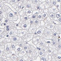
Comprehensive Human Protein Atlas IHC scoring per tissue (reliability: Enhanced — High consistency between antibody staining and RNA expression data. Presumed off target binding observed and disregarded.). Rows are taken directly from the HPA tissue chart — click any row's HPA link to view the source.
| Tissue | Cell type | Level | Evidence | Source |
|---|---|---|---|---|
| Duodenum | Enterocytes - Microvilli | High | Protein (IHC) | HPA → |
| Epididymis | Glandular cells | High | Protein (IHC) | HPA → |
| Kidney | Proximal tubules (microvilli) | High | Protein (IHC) | HPA → |
| Small intestine | Enterocytes - Microvilli | High | Protein (IHC) | HPA → |
| Bone marrow | Hematopoietic cells | Medium | Protein (IHC) | HPA → |
| Tissue | Cell type | Level | Evidence | Source |
|---|---|---|---|---|
| Adipose tissue | Adipocytes | Not detected | Protein (IHC) | HPA → |
| Adrenal gland | Glandular cells | Not detected | Protein (IHC) | HPA → |
| Appendix | Endocrine cells | Not detected | Protein (IHC) | HPA → |
| Breast | Adipocytes | Not detected | Protein (IHC) | HPA → |
| Bronchus | Respiratory epithelial cells | Not detected | Protein (IHC) | HPA → |
Troubleshoot MGAM chromogenic IHC by checking retrieval, apical localisation, staining controls and compartment-specific scoring (UniProt O43451; HPA tissue IHC).
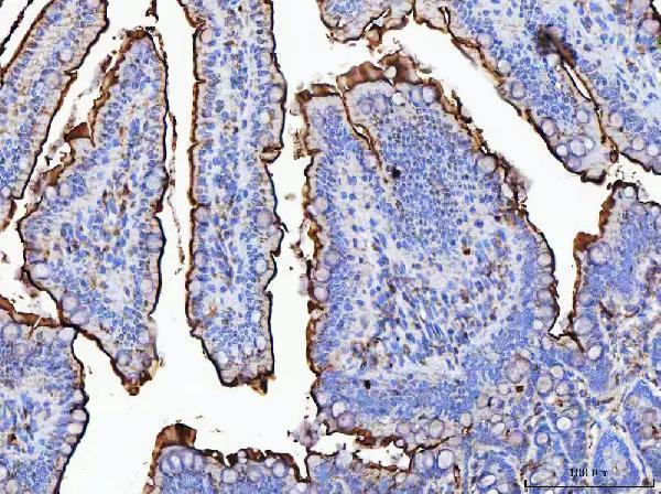
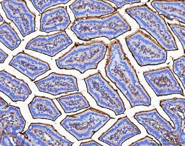
A07347 lists IHC and human, mouse and rat reactivity (catalog applications/reactivity). Its IHC images show mouse and rat colon paraffin sections (catalog image captions); no IF image is supplied (catalog IF images).
Only A07347 will render; it lists IHC and human, mouse and rat reactivity (catalog applications/reactivity). Its IHC images show mouse and rat colon paraffin sections stained with 2 μg/ml primary antibody (catalog image captions).
Which to pick: For tissue IHC, A07347 is the listed option: its mouse and rat paraffin-section captions describe EDTA retrieval at pH 8.0 (catalog image captions), but the fixative is unreported and the captions name a different antibody identifier (catalog image captions; catalog SKU). For cross-species work, A07347 lists human, mouse and rat reactivity, while the shown IHC images cover mouse and rat only; clone type is unreported (catalog reactivity; catalog image captions; catalog clone field). No listed SKU has an IF/ICC application or IF image, so this payload supports no IF/ICC pick (catalog applications; catalog IF images).