This website uses cookies to ensure you get the best experience on our website.


- Table of Contents
Plan chromogenic MGST1 IHC in paraffin sections using liver hepatocytes as a high-staining reference (HPA tissue IHC). This guide covers granular cytoplasmic scoring (HPA tissue IHC) and the 2 µg/mL catalog antibody condition (datasheet: PB9723).

Expected localisation, validated protocols, controls and antibodies — the at-a-glance facts below, then the full design guide.
| Expected localisation | Granular cytoplasm in tissue sections (HPA tissue IHC) | |
| Staining pattern | Granular cytoplasmic staining in most tissues (HPA tissue IHC) | |
| Antigen retrieval | EDTA pH 8.0 HIER, heat-mediated (datasheet PB9723) | |
| Positive control | Adrenal gland+4 more · see all | |
| Negative control | Bone marrow+4 more · see all |
| Fixation | Keep paraffin-section fixation consistent (standard IHC practice; not target-specific) | |
| Caveat | Staining and RNA show medium consistency (HPA tissue IHC) | |
| Regulation | High expression in liver (UniProt) | |
| Isoform / epitope | 2 isoforms; map epitopes to cytoplasmic or lumenal loops (UniProt) |
The catalog antibody’s IHC-P protocol (datasheet PB9723) is paired with a published MGST1 protocol for paraffin-embedded aortic tissue (PMC9339483).
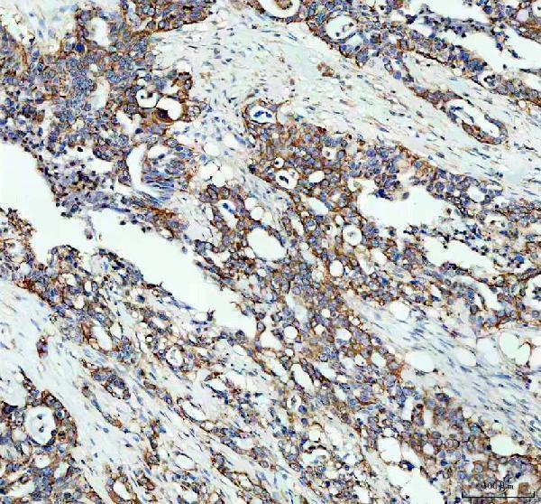
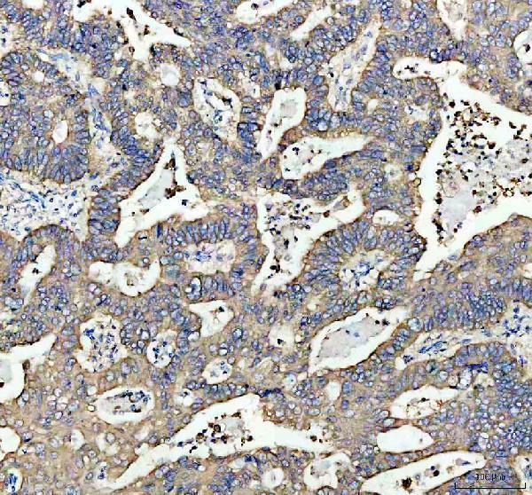
| Sample | Paraffin-embedded human pancreas cancer tissue; fixative not specified (datasheet PB9723) |
| Fixation | Image fixative and duration unreported (datasheet PB9723); verify before use. |
| Sectioning | 4–5 µm sections on charged slides (standard) |
| Deparaffinisation | Xylene, graded ethanol series to water (standard) |
| Antigen retrieval | Heat retrieval: EDTA pH 8.0 (datasheet PB9723); 20 min, 95–100 °C (standard) |
| Peroxidase block | 3% H2O2, 10 min, room temperature (standard) |
| Blocking | 10% goat serum (datasheet PB9723) |
| Primary antibody | Rabbit anti-MGST1, 2-5μg/ml (datasheet PB9723) |
| Primary incubation | Overnight at 4 °C (datasheet PB9723) |
| Detection | HRP-conjugated secondary, DAB chromogen (datasheet PB9723) |
| Counterstain | Hematoxylin, blue, dehydrate and mount (standard) |
| Expected result | MGST1-positive staining in glandular cells of adrenal gland (HPA tissue IHC: High). HPA tissue profile: Cytoplasmic expression with a granular pattern in most tissues. No signal in the no-primary control. |
MGST1 should appear mainly as granular cytoplasmic staining in IHC sections (HPA: tissue IHC profile). Look for strong staining in liver hepatocytes and kidney collecting ducts, among other HPA-positive cells (HPA: High). Its membrane localization in the endoplasmic reticulum and mitochondrial outer membrane supports an extranuclear pattern (UniProt P10620 topology). HPA rates the tissue IHC profile Enhanced, while reporting medium consistency between staining and RNA data (HPA: tissue IHC reliability).
| Granular cytoplasmic staining in hepatocytes, with little nuclear staining (HPA: liver hepatocytes High; tissue IHC profile). | This fits the reported liver pattern and membrane localization (HPA: tissue IHC profile; UniProt P10620 subcellular location). Judge the signal in hepatocytes rather than by whole-section darkness: adjacent cell populations need not share the same staining level (HPA: cell-specific tissue IHC levels). |
| Predominantly nuclear staining, or a pattern with no discernible cytoplasmic component (HPA: expected tissue IHC profile). | Treat this as a compartment mismatch requiring investigation, since the reported IHC pattern is cytoplasmic and the annotated locations are membrane-bound (HPA: tissue IHC profile; UniProt P10620 subcellular location). Check morphology, counterstain and detection controls before calling it specific (general IHC practice). |
| Strong staining in cells reported as not detected, such as skeletal-muscle myocytes (HPA: skeletal muscle myocytes Not detected). | Unexpected signal may reflect cross-reactivity or endogenous detection activity (general IHC practice). Compare a no-primary control and an HPA-positive tissue processed in the same run; a single discordant section cannot establish new MGST1 expression (general IHC practice; HPA: tissue IHC levels). |
| Uniform brown haze across cells and extracellular spaces, obscuring granular detail (HPA: expected tissue IHC profile). | This is background rather than the reported cell-associated granular pattern (HPA: tissue IHC profile). Review blocking, washing and chromogen development, and inspect a no-primary control for detection-system background (general chromogenic IHC practice). |
| No detectable hepatocyte staining in an otherwise interpretable liver section (HPA: liver hepatocytes High). | A failed positive reference is inconclusive for MGST1 absence (general IHC practice; HPA: liver hepatocytes High). Verify tissue preservation and the staining run, then optimize retrieval or antibody conditions according to the antibody's IHC-P instructions (general IHC practice). |
| Compartment and topology (UniProt P10620 topology). | MGST1 has four transmembrane segments and is assigned to ER and mitochondrial outer membranes (UniProt P10620 topology; subcellular location). Paraffin-section IHC reports their combined granular cytoplasmic appearance; it does not by itself resolve which organelle contributes each granule (HPA: tissue IHC profile; general IHC interpretation). |
| Choice of positive and low-signal tissue (HPA: tissue IHC levels). | Hepatocytes and kidney collecting ducts are High; adipocytes are Medium, while skeletal-muscle myocytes are Not detected (HPA: tissue IHC levels). Use a High population to assess whether the run worked. A weak result in a lower-signal population is a less decisive technical check (general IHC practice). |
| Evidence and antibody scope (HPA: tissue IHC reliability; antibody validation). | HPA labels the tissue IHC profile Enhanced but describes only medium consistency with RNA expression data (HPA: tissue IHC reliability). The listed antibody HPA044840 has Enhanced IHC validation (HPA: antibody validation). These labels support a reference pattern; they do not validate every antibody or explain each discordant sample (general interpretation). |
| Epitope and isoform uncertainty (UniProt P10620 topology; isoforms). | Two isoforms are listed, but the supplied record does not map the IHC antibody epitope or establish isoform-specific staining (UniProt P10620 isoforms; HPA: antibody record). Topology alone cannot predict retrieval requirements or fixation sensitivity (UniProt P10620 topology). Treat changes in those conditions as general IHC optimization, not a documented MGST1 effect. |
| IF/ICC Q&A: What localization should an IF image show? (HPA: subcellular ICC-IF). | HPA reports mainly mitochondrial signal with additional ER localization in ICC-IF (HPA: approved subcellular locations). That supports the organelle interpretation of cytoplasmic IHC staining, but IHC sections need not resolve mitochondria from ER (HPA: tissue IHC profile; general microscopy interpretation). IF/ICC conditions belong in its separate guide. |
| Situation | Likely cause | Next action |
|---|---|---|
| Liver positive reference is blank or very faint (HPA: liver hepatocytes High). | The run may have insufficient antigen exposure or detection sensitivity; a blank positive reference cannot distinguish technical failure from biology (general IHC practice). | Confirm hepatocyte morphology and reagent performance, then optimize retrieval and antibody conditions within the antibody's IHC-P workflow (general IHC practice). |
| Nuclear staining dominates the image (HPA: cytoplasmic tissue IHC profile). | The observed compartment conflicts with the annotated ER and mitochondrial outer-membrane locations (UniProt P10620 subcellular location). Nonspecific staining or interpretation of counterstain is possible (general IHC practice). | Review the counterstain and no-primary control; require a distinct cell-associated cytoplasmic pattern in a positive reference before scoring MGST1 (HPA: tissue IHC profile; general IHC practice). |
| A reported Not detected cell population stains strongly (HPA: tissue IHC levels). | Cross-reactivity or endogenous detection activity could explain unexpected signal (general IHC practice); HPA's cell-level reference alone cannot identify the mechanism (HPA: tissue IHC levels). | Compare with a no-primary control and a High reference on the same run; assess the suspect cell type separately from neighboring positive cells (general IHC practice; HPA: cell-specific tissue IHC levels). |
| Diffuse brown haze masks cell boundaries (HPA: granular cytoplasmic tissue IHC profile). | Background from detection reagents, incomplete blocking or excessive development is possible (general chromogenic IHC practice). | Inspect a no-primary control, review blocking and wash steps, and shorten development if controls show excess background (general chromogenic IHC practice). |
| Different tissue regions appear to disagree (HPA: cell-specific tissue IHC levels). | The sampled regions may contain different cell populations with different reported MGST1 levels (HPA: tissue IHC levels); section quality or uneven staining is also possible (general IHC practice). | Score identifiable cells with their morphology and counterstain, and compare equivalent regions on a positive reference before changing the interpretation (general IHC practice). |
| IHC looks granular, but its pattern does not identify a single organelle (HPA: tissue IHC profile). | MGST1 is assigned to both ER and mitochondrial outer membranes (UniProt P10620 subcellular location), while HPA's IHC description is cytoplasmic and granular (HPA: tissue IHC profile). | Report the IHC result as granular cytoplasmic staining. Use the separate IF/ICC localization evidence when an organelle-level comparison is needed (HPA: approved subcellular ICC-IF locations; general IHC interpretation). |


Comprehensive Human Protein Atlas IHC scoring per tissue (reliability: Enhanced — Medium consistency between antibody staining and RNA expression data.). Rows are taken directly from the HPA tissue chart — click any row's HPA link to view the source.
| Tissue | Cell type | Level | Evidence | Source |
|---|---|---|---|---|
| Adrenal gland | Glandular cells | High | Protein (IHC) | HPA → |
| Appendix | Enterocytes | High | Protein (IHC) | HPA → |
| Endometrium | Ciliated epithelial cells | High | Protein (IHC) | HPA → |
| Kidney | Collecting ducts | High | Protein (IHC) | HPA → |
| Liver | Hepatocytes | High | Protein (IHC) | HPA → |
| Tissue | Cell type | Level | Evidence | Source |
|---|---|---|---|---|
| Bone marrow | Hematopoietic cells | Not detected | Protein (IHC) | HPA → |
| Epididymis | Glandular cells | Not detected | Protein (IHC) | HPA → |
| Oral mucosa | Squamous epithelial cells | Not detected | Protein (IHC) | HPA → |
| Skeletal muscle | Myocytes | Not detected | Protein (IHC) | HPA → |
| Smooth muscle | Smooth muscle cells | Not detected | Protein (IHC) | HPA → |
Use the catalog antibody’s paraffin-section IHC conditions as the starting point, then check whether staining matches MGST1’s expected cells and compartments.
PB9723 has IHC images from human pancreas cancer, stomach cancer, and liver paraffin sections (PB9723 image captions). M04703 lists human, mouse, and rat reactivity for IHC and IF/ICC, without image evidence (M04703 catalog).
PB9723 is the only card that will render; its IHC images show human pancreas cancer, stomach cancer, and liver paraffin sections (PB9723 image captions). Its catalog lists human, mouse, and rat reactivity, but reports IHC-specific testing in human samples (PB9723 catalog).
Which to pick: Choose PB9723 for human paraffin-section IHC: its captions document EDTA pH 8.0 retrieval and 2 μg/ml primary antibody; the fixative is unreported (PB9723 image captions). For IF/ICC, M04703 lists those applications and is a rabbit monoclonal, but has no IF image in the payload (M04703 catalog). For mouse or rat work, both SKUs list reactivity with those species; confirm IHC performance in your samples because PB9723’s IHC-specific testing and images are human, while M04703 has no IHC images here (PB9723 catalog and image captions; M04703 catalog).