This website uses cookies to ensure you get the best experience on our website.


- Table of Contents
Plan MLST8 chromogenic IHC in paraffin sections using the catalog antibody’s human IHC examples (datasheet A05357-2). Assess cytoplasmic staining by cell type and intensity, allowing for low consistency between antibody staining and RNA expression (HPA tissue IHC).

Expected localisation, validated protocols, controls and antibodies — the at-a-glance facts below, then the full design guide.
| Expected localisation | Cytoplasmic in tissue (HPA tissue IHC); lysosomal membrane location (UniProt) | |
| Staining pattern | Cytoplasmic staining in most tissues (HPA tissue IHC) | |
| Antigen retrieval | EDTA pH 8.0 HIER, heat-mediated (datasheet A05357-2) | |
| Positive control | Adrenal gland+4 more · see all | |
| Negative control | Adipose tissue |
| Fixation | Keep fixation consistent across sections. (standard IHC practice; not target-specific) | |
| Caveat | Low consistency between antibody staining and RNA expression (HPA tissue IHC) | |
| Regulation | No expression regulator specified (UniProt) | |
| Isoform / epitope | 4 isoforms; no transmembrane segment; epitope coverage unknown (UniProt) |
The catalog antibody’s IHC-P protocol (datasheet A05357-2) is accompanied by one published MLST8 immunoperoxidase protocol (PMC4408021).
| Sample | Paraffin-embedded human esophageal squamous carcinoma tissue; fixative not specified (datasheet A05357-2) |
| Fixation | Image fixative and duration unreported (datasheet A05357-2); verify before use. |
| Sectioning | 4–5 µm sections on charged slides (standard) |
| Deparaffinisation | Xylene, graded ethanol series to water (standard) |
| Antigen retrieval | Heat retrieval: EDTA pH 8.0 (datasheet A05357-2); 20 min, 95–100 °C (standard) |
| Peroxidase block | 3% H2O2, 10 min, room temperature (standard) |
| Blocking | 10% goat serum (datasheet A05357-2) |
| Primary antibody | Rabbit anti-MLST8, 2-5 μg/ml (datasheet A05357-2) |
| Primary incubation | Overnight at 4 °C (datasheet A05357-2) |
| Detection | HRP-conjugated secondary, DAB chromogen (datasheet A05357-2) |
| Counterstain | Hematoxylin, blue, dehydrate and mount (standard) |
| Expected result | MLST8-positive staining in glandular cells of adrenal gland (HPA tissue IHC: Medium). HPA tissue profile: Cytoplasmic expression in most tissues. No signal in the no-primary control. |
MLST8 should usually appear cytoplasmic in tissue IHC, including medium staining in glandular, respiratory epithelial, hematopoietic and selected glial cells (HPA tissue IHC). UniProt also places MLST8 in the cytoplasm and at lysosome membranes, with no transmembrane segment (UniProt Q9BVC4). Treat this as a working pattern: HPA rates its tissue IHC evidence Approved but reports low consistency between antibody staining and RNA expression (HPA tissue IHC).
| Cytoplasmic staining in appendix glandular cells or bone marrow hematopoietic cells. | This matches the reported medium signal in those cells (HPA tissue IHC). Judge the cell type and compartment together; intensity alone cannot establish specificity, particularly given the reported low agreement with RNA expression (HPA tissue IHC). |
| Strong, nucleus-only staining in tissue sections, with little cytoplasmic signal. | This departs from the predominant cytoplasmic tissue pattern and warrants an artefact check (HPA tissue IHC). Nuclear signal is not automatically false: HPA reports approved nucleoplasmic localization in ICC-IF, a different application (HPA subcellular ICC-IF). |
| Prominent signal in adipocytes while expected glandular or hematopoietic cells are weak. | HPA reports adipocytes as not detected, versus medium signal in several glandular populations and bone marrow hematopoietic cells (HPA tissue IHC). Consider cross-reactivity or endogenous detection activity; neither cause is established by the pattern alone (standard IHC practice). |
| Diffuse color across cells and surrounding tissue, without clear cytoplasmic boundaries. | This is hard to score as cellular MLST8 staining (standard IHC practice). Check background from the detection workflow before interpreting intensity; HPA describes cytoplasmic expression in most tissues, not a diffuse tissue-wide deposit (HPA tissue IHC). |
| No signal in appendix glandular cells despite interpretable tissue morphology. | HPA reports medium staining in these cells, so first check the IHC run and whether the intended cell population is present (HPA tissue IHC; standard IHC practice). A single blank section cannot establish MLST8 absence because HPA reports limited staining–RNA consistency (HPA tissue IHC). |
| Tissue and cell population | MLST8 is broadly expressed (UniProt Q9BVC4); HPA reports cytoplasmic staining in most tissues, medium signal in several named cell populations, low signal in hippocampal glia and ovarian stroma, and no detected signal in adipocytes (HPA tissue IHC). Score the relevant cell population rather than the section as a whole. |
| IF/ICC Q: Should its pattern match tissue IHC? | HPA's approved ICC-IF localization is mainly nucleoplasmic, with additional Golgi apparatus and cell junction signal (HPA subcellular ICC-IF). This differs from HPA's mainly cytoplasmic tissue IHC profile (HPA tissue IHC). Evaluate each application against its own evidence; the ICC-IF observation does not establish a nuclear tissue IHC expectation. |
| Nutrient-dependent location | UniProt describes recruitment of mTORC1 to lysosome membranes according to amino acid availability and lists MLST8 in the cytoplasm and at lysosome membranes (UniProt Q9BVC4). The supplied tissue IHC profile does not specify a punctate staining requirement (HPA tissue IHC); avoid scoring puncta alone as proof of MLST8. |
| Validation and isoforms | Three listed antibodies are Approved for IHC, while HPA reports low consistency between tissue staining and RNA expression (HPA antibodies; HPA tissue IHC). UniProt lists four MLST8 isoforms, but the supplied evidence gives no epitope coverage for an antibody (UniProt Q9BVC4); do not infer which isoforms a stain detects. |
| Situation | Likely cause | Next action |
|---|---|---|
| A reported medium-positive population is blank or unexpectedly faint. | The result conflicts with the reported tissue pattern, but could reflect an IHC workflow problem or sample variation (HPA tissue IHC; standard IHC practice). | Verify the named cells are present, review the catalog antibody's IHC-P procedure, and check retrieval, detection and counterstain against a working run (standard IHC practice). Do not assign an MLST8-specific fixation effect: none is supplied. |
| Adipocytes stain prominently. | HPA reports adipocytes as not detected, making this pattern discordant; nonspecific antibody binding or endogenous detection activity are possibilities (HPA tissue IHC; standard IHC practice). | Inspect a matched detection control and staining distribution, then compare with a reported positive cell population (standard IHC practice; HPA tissue IHC). Treat the adipocyte observation as a specificity question, not proof of a particular mechanism. |
| The tissue section shows predominantly nuclear staining. | This differs from HPA's cytoplasmic tissue IHC profile, although HPA separately reports approved nucleoplasmic ICC-IF localization (HPA tissue IHC; HPA subcellular ICC-IF). | Check cellular boundaries and background, then compare the tissue pattern with the catalog antibody's IHC evidence (standard IHC practice). Record the application difference before calling the nuclear signal an artefact. |
| Broad, hazy chromogen obscures cell boundaries. | Background from blocking, washing or detection may prevent compartment scoring (standard IHC practice); the HPA tissue description requires an interpretable cytoplasmic pattern (HPA tissue IHC). | Review blocking and washes, inspect a detection control, and reassess only cells with clear boundaries (standard IHC practice). Do not count uniform haze as medium cytoplasmic staining. |
| Only fine puncta are visible, with no convincing cellular pattern. | Lysosome membrane association is biologically plausible under amino acid-dependent conditions (UniProt Q9BVC4), but the supplied tissue IHC evidence does not validate puncta as a stand-alone readout (HPA tissue IHC). | Check whether puncta lie within the expected stained cells, assess background and compare the overall pattern with HPA tissue IHC (standard IHC practice; HPA tissue IHC). Report puncta descriptively without assigning them to lysosomes from chromogenic IHC alone. |
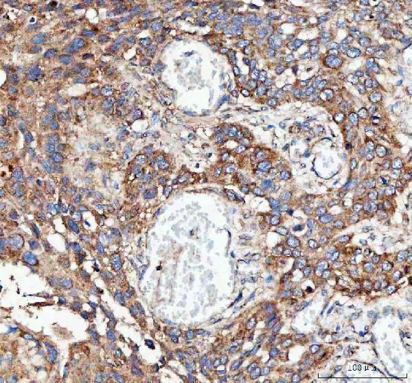
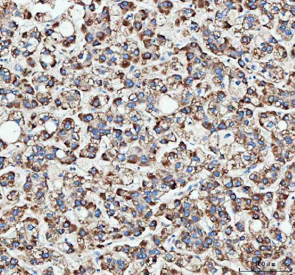
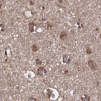


Comprehensive Human Protein Atlas IHC scoring per tissue (reliability: Approved — Low consistency between antibody staining and RNA expression data.). Rows are taken directly from the HPA tissue chart — click any row's HPA link to view the source.
| Tissue | Cell type | Level | Evidence | Source |
|---|---|---|---|---|
| Adrenal gland | Glandular cells | Medium | Protein (IHC) | HPA → |
| Appendix | Glandular cells | Medium | Protein (IHC) | HPA → |
| Bone marrow | Hematopoietic cells | Medium | Protein (IHC) | HPA → |
| Breast | Glandular cells | Medium | Protein (IHC) | HPA → |
| Bronchus | Respiratory epithelial cells | Medium | Protein (IHC) | HPA → |
| Tissue | Cell type | Level | Evidence | Source |
|---|---|---|---|---|
| Adipose tissue | Adipocytes | Not detected | Protein (IHC) | HPA → |
Troubleshoot MLST8 staining in paraffin sections by checking retrieval, compartment, controls and scoring before interpreting changes in signal.
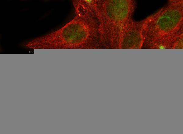
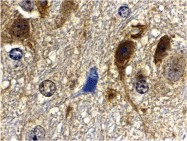
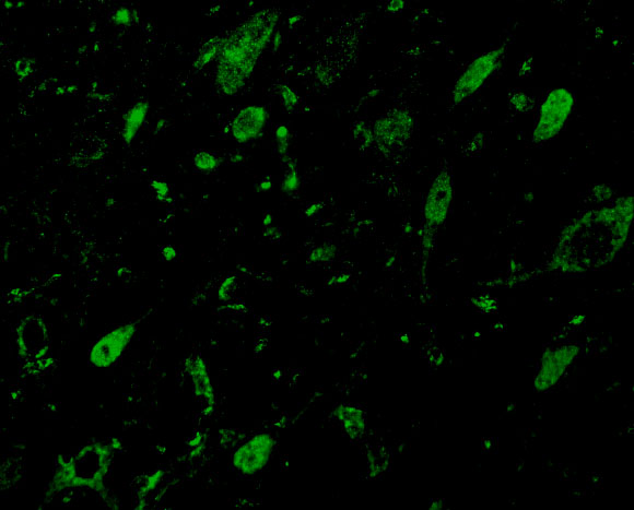
Two anti-MLST8 antibodies have IHC images from human paraffin carcinoma sections and mouse brain tissue, plus IF images from human cells and sections and mouse brain cells (catalog: image captions).
A05357-2 shows IHC in paraffin sections of human esophageal, liver, lung, and rectal cancers, and IF in U2OS cells and human cancer sections (catalog: A05357-2 image captions). A05357-1 shows IHC in mouse brain tissue and IF in mouse brain cells (catalog: A05357-1 image captions).
Which to pick: Choose A05357-2 for human paraffin-section IHC and A05357-1 for mouse brain IHC, matching each antibody’s own image (catalog: IHC captions). For IF/ICC, A05357-2 lists both applications and has human cell and section images; A05357-1 lists IF and has a mouse brain cell image (catalog: applications and IF captions). Both list Human, Mouse, and Rat reactivity, while clonality and the fixative used for the paraffin-section IHC images are unreported (catalog: reactivity, clone fields, IHC captions).