This website uses cookies to ensure you get the best experience on our website.


- Table of Contents
Plan MRPL51 chromogenic IHC around cytoplasmic staining in most tissues (HPA tissue IHC) and mitochondrial localization (UniProt). The guide highlights high hepatocyte staining (HPA tissue IHC), an IHC dilution range of 1:100–1:300 (datasheet), and low consistency between staining and RNA expression (HPA tissue IHC).

Expected localisation, validated protocols, controls and antibodies — the at-a-glance facts below, then the full design guide.
| Expected localisation | Cytoplasmic staining (HPA tissue IHC); mitochondrial location (UniProt) | |
| Staining pattern | Cytoplasmic staining in most tissues (HPA tissue IHC) | |
| Antigen retrieval | Citrate pH 6.0 HIER, 95–98 °C, 20 min (rule: cytoplasmic / membrane antigen) | |
| Positive control | Appendix+4 more · see all | |
| Negative control | Bone marrow+4 more · see all |
| Fixation | Keep fixation consistent across sections (standard IHC practice; not target-specific) | |
| Caveat | Staining and RNA expression show low consistency (HPA tissue IHC) | |
| Regulation | No expression regulator annotated (UniProt) | |
| Isoform / epitope | 0 isoforms annotated; mature chain aa 32–128 (UniProt) |
Compare the catalog antibody’s IHC-P protocol with one published MRPL51 IHC protocol using human lung adenocarcinoma tissue (PMC12557611).
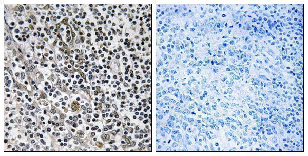
| Sample | Paraffin-embedded human tonsil tissue; fixative not specified (datasheet A14477) |
| Fixation | Image fixative and duration unreported (datasheet A14477); verify before use. |
| Sectioning | 4–5 µm sections on charged slides (standard) |
| Deparaffinisation | Xylene, graded ethanol series to water (standard) |
| Antigen retrieval | Heat-induced epitope retrieval in citrate buffer, pH 6.0, 20 min at 95–98 °C (standard rule: cytoplasmic / membrane antigen) |
| Peroxidase block | 3% H2O2, 10 min, room temperature (standard) |
| Blocking | 10% normal serum of the secondary host, 30 min, room temperature (standard) |
| Primary antibody | Rabbit anti-MRPL51, 1:100-1:300 (datasheet A14477) |
| Primary incubation | Overnight at 4 °C (standard) |
| Detection | HRP-polymer secondary, DAB chromogen 5–10 min (standard) |
| Counterstain | Hematoxylin, blue, dehydrate and mount (standard) |
| Expected result | MRPL51-positive staining in glandular cells of appendix (HPA tissue IHC: High). HPA tissue profile: Cytoplasmic expression in most tissues. No signal in the no-primary control. |
MRPL51 is a mitochondrial protein without a transmembrane segment (UniProt Q4U2R6 topology). In paraffin IHC, expect cytoplasmic staining in many tissues, including strong staining in liver hepatocytes and intestinal glandular cells (HPA: cytoplasmic expression in most tissues; High in hepatocytes and intestinal glandular cells). Treat the pattern as provisional: HPA rates the tissue IHC antibody Approved, but reports low agreement with RNA expression and pending external verification (HPA: tissue IHC reliability).
| Cytoplasmic staining in hepatocytes or intestinal glandular cells. | This fits HPA's High tissue IHC observations and UniProt's mitochondrial location (HPA: High in liver hepatocytes and intestinal glandular cells; UniProt Q4U2R6 location). Chromogenic IHC can establish a cytoplasmic pattern without resolving individual mitochondria; judge the stained cell population and background together (general IHC practice). |
| Predominantly nuclear or nucleolar staining, with little cytoplasmic signal. | A nuclear-dominant IHC result conflicts with the primary mitochondrial assignment (UniProt Q4U2R6 location; HPA: mitochondria supported by ICC-IF). HPA also reports an uncertain nucleolar ICC-IF location, so a nucleolar component alone is inconclusive; check controls and repeat before calling the pattern specific (HPA: nucleoli uncertain; general IHC practice). |
| Strong staining in bone marrow hematopoietic cells or smooth muscle cells. | These populations were Not detected in HPA tissue IHC, making strong staining a reason to investigate cross-reactivity or endogenous detection activity (HPA: Not detected in bone marrow hematopoietic cells and smooth muscle cells; general IHC practice). Their HPA results are comparison points, not proof that every specimen must be negative (HPA: Approved; pending external verification). |
| Broad, even color over cells, stroma, or slide background. | A distribution that ignores cell boundaries and the expected cytoplasmic pattern is difficult to score as MRPL51 (HPA: cytoplasmic expression in most tissues; general IHC practice). Compare a no-primary control, inspect washing and blocking, and assess the detection system before assigning cellular staining (general IHC practice). |
| No staining in liver hepatocytes despite a readable section. | HPA reports High hepatocyte staining, so absence warrants checking assay performance (HPA: High in liver hepatocytes). Inspect the positive control, antibody and detection reagents, retrieval conditions, and section integrity; one failed run cannot establish absent MRPL51 (general IHC practice). HPA's low antibody–RNA agreement also limits certainty about tissue expectations (HPA: reliability description). |
| Cell and tissue choice | HPA reports High staining in appendix, colon, duodenum, gallbladder, parathyroid and small-intestinal glandular cells, plus liver hepatocytes and breast adipocytes (HPA: tissue IHC). These are candidate positive comparators; HPA also describes low tissue RNA specificity and uncertain agreement between staining and RNA (HPA: RNA specificity; reliability description). |
| Antibody evidence | The listed antibody, HPA039923, is Approved for IHC, while the tissue profile remains pending external verification (HPA: antibody validation; tissue IHC reliability). Do not treat Approved as proof that every observed cell or compartment is specific (HPA: low agreement with RNA expression). |
| Topology and processing | UniProt places MRPL51 in mitochondria, annotates no transmembrane segment, and identifies residues 32–128 as the mature chain (UniProt Q4U2R6 location, topology and processing). These annotations support intracellular interpretation; they do not establish an epitope, retrieval requirement, or fixation sensitivity (UniProt Q4U2R6 annotations). |
| IF/ICC: where should signal appear? | Mainly in mitochondria; HPA marks that ICC-IF location supported and an additional nucleolar location uncertain (HPA: subcellular summary). This answers the localization question only; the paraffin IHC result must still be interpreted against its own tissue controls (HPA: tissue IHC; general IHC practice). |
| Situation | Likely cause | Next action |
|---|---|---|
| Positive tissue stains weakly throughout. | The run may have inadequate detection or unsuitable retrieval; HPA does not report an MRPL51-specific fixation effect (general IHC practice; HPA: tissue IHC scope). | Verify the positive control and reagent sequence, then optimize retrieval and antibody concentration within the assay's validated range (general IHC practice). |
| No signal in the positive control or test section. | A missing or ineffective primary, detection reagent, or chromogen can produce an assay-wide blank result (general IHC practice). | Check reagent identity, expiration and application; repeat with a documented positive tissue such as liver hepatocytes (general IHC practice; HPA: High in liver hepatocytes). |
| Color appears in the no-primary control. | Endogenous detection activity or nonspecific detection chemistry can create signal without primary antibody (general IHC practice). | Review the detection method and use the appropriate endogenous-activity block and no-primary comparison before scoring cells (general IHC practice). |
| High staining appears in an HPA Not detected cell population. | Cross-reactivity, endogenous activity or specimen variation may explain the discrepancy; HPA tissue IHC is pending external verification (general IHC practice; HPA: reliability description). | Compare morphology and controls, assess another antibody if available, and report the disagreement rather than treating one HPA negative as absolute (general IHC practice; HPA: selected cells Not detected). |
| Nuclear staining dominates the section. | That distribution differs from the supported mitochondrial location; HPA's nucleolar ICC-IF finding is uncertain (UniProt Q4U2R6 location; HPA: subcellular locations). | Check no-primary staining and assess whether a reproducible cytoplasmic component tracks the expected positive cells before interpreting nuclear color (general IHC practice; HPA: High in selected cells). |
| Diffuse background obscures cell boundaries. | Nonspecific binding, incomplete washing or excessive detection signal can conceal cellular distribution (general IHC practice). | Review blocking, washes and detection strength, then score only sections where cytoplasmic signal is distinguishable from background (general IHC practice; HPA: cytoplasmic expression). |
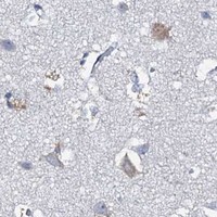


Comprehensive Human Protein Atlas IHC scoring per tissue (reliability: Approved — Low consistency between antibody staining and RNA expression data. Pending external verification.). Rows are taken directly from the HPA tissue chart — click any row's HPA link to view the source.
| Tissue | Cell type | Level | Evidence | Source |
|---|---|---|---|---|
| Appendix | Glandular cells | High | Protein (IHC) | HPA → |
| Breast | Adipocytes | High | Protein (IHC) | HPA → |
| Colon | Glandular cells | High | Protein (IHC) | HPA → |
| Duodenum | Glandular cells | High | Protein (IHC) | HPA → |
| Gallbladder | Glandular cells | High | Protein (IHC) | HPA → |
| Tissue | Cell type | Level | Evidence | Source |
|---|---|---|---|---|
| Bone marrow | Hematopoietic cells | Not detected | Protein (IHC) | HPA → |
| Cerebral cortex | Endothelial cells | Not detected | Protein (IHC) | HPA → |
| Hippocampus | Glial cells | Not detected | Protein (IHC) | HPA → |
| Ovary | Ovarian stroma cells | Not detected | Protein (IHC) | HPA → |
| Placenta | Decidual cells | Not detected | Protein (IHC) | HPA → |
Troubleshoot MRPL51 staining in paraffin-section IHC by checking retrieval, cellular pattern, controls, and scoring; use IF as a separate localisation check.
A14477 has a human paraffin-section IHC image with peptide-blocked comparison (catalog image caption); IF is listed without an IF image (catalog applications; catalog IF images). Human and mouse reactivity is listed (catalog reactivity).
A14477 is the only card and shows IHC on paraffin-embedded human tonsil tissue with a peptide-blocked comparison (catalog image caption). IF is listed as an application, and human and mouse reactivity is listed; no IF image is supplied (catalog applications; catalog reactivity; catalog IF images).
Which to pick: Choose A14477 for tissue IHC because its own image shows a paraffin-embedded human tonsil section; the fixative is unreported (catalog image caption). For IF/ICC, A14477 lists IF and a 1:50 IF dilution, but supplies no IF image or ICC validation (catalog applications; catalog IF dilution; catalog IF images). It is a rabbit polyclonal antibody with listed human and mouse reactivity, so mouse use should be verified in the intended assay (catalog host; catalog dilution_raw; catalog reactivity).