This website uses cookies to ensure you get the best experience on our website.


- Table of Contents
Source-linked MYO1C Western blot protocol options, expected-band and isoform facts, troubleshooting for weak or shifted signal, and recommended anti-MYO1C WB antibodies. Everything you need to plan the experiment before you commit precious samples.

Expected bands, source-linked protocol options, controls and antibodies — the at-a-glance facts below, then the full design guide.
| Expected band | ~121.7 kDa | |
| Gel | 8–10% (standard starting point) | |
| Positive control | Appendix (IHC candidate; verify WB) +4 more | |
| Negative control | Bone marrow (IHC candidate; verify WB) |
| PTM | Phosphorylated + Acetylated | |
| Caveat | Phosphorylation-state controls | |
| Gene-set association | MSigDB Hallmark membership | |
| Isoform | 3 isoform(s) |
The A03399 protocol combines labelled catalog values with standard starting conditions. Published comparisons retain their own sample, reagent and detection scope.
| Sample / lysate | extracts of various cell lines, (catalog A03399) |
| Gel % | 8–10% (standard starting point) |
| Load | 20–30 µg total protein per lane; optimize for abundance (standard starting point) |
| Transfer | Wet/tank transfer; optimize duration (standard starting point) |
| Membrane | 0.45 µm PVDF (standard starting point) |
| Blocking | 5% milk or 5% BSA in TBST (standard starting point) |
| Primary antibody | A03399; use the WB datasheet starting dilution (standard starting point) |
| Primary incubation | Overnight at 4 °C (standard starting point) |
| Secondary antibody | Species-matched HRP conjugate at validated dilution (standard starting point) |
| Secondary incubation | 1 h at room temperature (standard starting point) |
| Wash | 3 × 5 min in TBST (standard starting point) |
| Detection | ECL; bracket exposures to avoid saturation (standard starting point) |
MYO1C has a predicted mass of 121.7 kDa; three isoforms could affect migration, but no distinct band sizes are demonstrated.
| Band near 121.7 kDa | consistent with the predicted MYO1C mass; confirm identity with an antibody control |
| Several bands near the expected region | could reflect isoforms 1, 2, and 3 if their migration differs |
| Single band despite three isoforms | the isoforms may not resolve as separate bands |
| Weak band in a soluble fraction | MYO1C may be retained in membrane or cell cortex fractions |
| UniProt predicted mass | places the reference band near 121.7 kDa |
| Splice isoform 1 | its individual mass and migration relative to the other isoforms are not supplied |
| Splice isoform 2 | may differ in size from the other isoforms, but its mass is not supplied |
| Splice isoform 3 | may differ in size from the other isoforms, but its mass is not supplied |
| Situation | Likely cause | Next action |
|---|---|---|
| No band in lysate | MYO1C extraction may be incomplete from membrane or cell cortex fractions | check the insoluble fraction and a positive control lysate |
| Band higher than expected | band identity or migration is uncertain | compare with a positive control and verify antibody specificity |
| Band lower than expected | an isoform or unrelated band is possible; individual isoform masses are unavailable | check antibody specificity and compare isoform expression |
| Multiple bands | isoforms 1, 2, and 3 may contribute if they migrate differently | verify bands with isoform-aware controls |
| Weak or no signal | MYO1C abundance or extraction may be low in the sampled fraction | check loading and examine membrane-associated fractions |
Comprehensive Human Protein Atlas IHC scoring per tissue. Rows are taken directly from the HPA tissue chart — click any row's HPA link to view the source.
| Tissue | Cell type | Level | Evidence | Source |
|---|---|---|---|---|
| Appendix | glandular cells | High | Protein (IHC) | HPA → |
| Colon | glandular cells | High | Protein (IHC) | HPA → |
| Duodenum | glandular cells | High | Protein (IHC) | HPA → |
| Gallbladder | glandular cells | High | Protein (IHC) | HPA → |
| Lung | endothelial cells | High | Protein (IHC) | HPA → |
| Tissue | Cell type | Level | Evidence | Source |
|---|---|---|---|---|
| Bone marrow | hematopoietic cells | Not detected | Protein (IHC) | HPA → |
| Skeletal muscle | myocytes | Not detected | Protein (IHC) | HPA → |
| Heart muscle | cardiomyocytes | Low | Protein (IHC) | HPA → |
| Hippocampus | glial cells | Low | Protein (IHC) | HPA → |
| Spleen | cells in red pulp | Low | Protein (IHC) | HPA → |
Deeper troubleshooting and optimisation questions for MYO1C, answered from its protein features.
Catalog antibodies with Western blot application and product-specific WB images. Evaluate suitability with the reported sample, controls and experimental conditions.
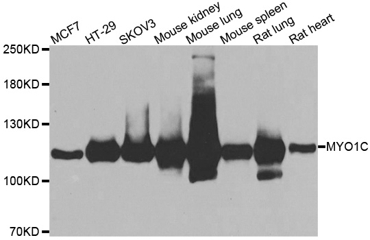
The catalog reports anti-MYO1C antibody A03399 for Western blot, with reported Human, Mouse, and Rat reactivity. A WB image is available for extracts from various cell lines; the supplied caption does not identify the lines or establish validation across all listed species.
Which to pick: A03399 is the only listed MYO1C antibody and has a WB image. Check that its reported Human, Mouse, or Rat reactivity fits your sample; the supplied caption does not name the cell lines tested.