This website uses cookies to ensure you get the best experience on our website.
- Table of Contents
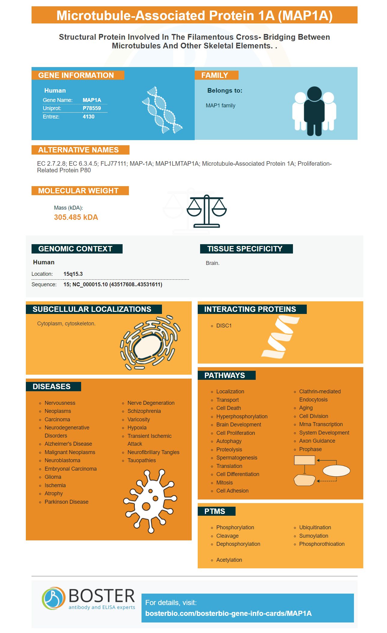
Facts about Microtubule-associated protein 1A.
Human | |
---|---|
Gene Name: | MAP1A |
Uniprot: | P78559 |
Entrez: | 4130 |
Belongs to: |
---|
MAP1 family |
EC 2.7.2.8; EC 6.3.4.5; FLJ77111; MAP-1A; MAP1LMTAP1A; microtubule-associated protein 1A; Proliferation-related protein p80
Mass (kDA):
305.485 kDA
Human | |
---|---|
Location: | 15q15.3 |
Sequence: | 15; NC_000015.10 (43517608..43531611) |
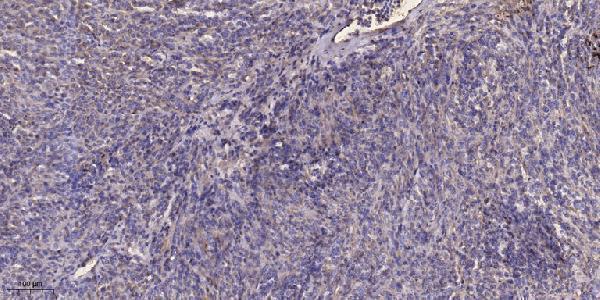
Brain.
Cytoplasm, cytoskeleton.
Scientists can use MAP1A markers to determine the molecular arrangement of microtubules within a variety if species. Boster scientists may submit results for species-specific applications or samples. The JNK protein phosphorylates the MAP1A. Boster can receive results from scientists all over the globe.
MAPs bind to tubulin dimers to regulate their dynamics. They also stabilize their structures. These proteins promote their polymerization. MARK is a protein that can phosphorylate MAPs. MAPs that are phosphorylated will detach from microtubules, and then fall apart. This can disrupt the growth of neurons and affect other cell processes.
Both MAP1A & MAP1B are known to increase microtubule elongation as well as decrease tubulin concentrations, which is critical for axon cell growth. However, MAP1B exhibits weaker activity than MAP1A, which may be due to differential phosphorylation or inhibition of the light chain by the heavy chain. Despite being less active than MAP2A or MAP2C it is still a key protein in neuronal microtubule regulation and stability.
MAP1A is a member the AAA family. It plays a role in the disassembly microtubules, binding alpha/beta tubulin dimers to inhibit their polymerization and thereby binding them. It also acts a scaffold for interaction between Aurora-kinases/septins. The Aurora B Protein is also found in the brain. It regulates SEP7 and MAP1A's interaction.
The light chain of LC1 or LC2 is responsible for triggering assembly of microtubules. However, the presence of NH2-terminal domains in LC1 and LC2 results in a lag phase in which little or no microtubule polymerization takes place. This suggests that light chain proteins play a major part in microtubule creation.
MAP1A, which is a microtubulite stabiliser, has been implicated as preventing the formation and maintenance of spindle structures. However, it remains to be determined if septins play a role in microtubule organization as well as dynamics. MAP1A is thought to be one of the mechanisms that regulate intracellular cargo spatial transport. This discovery is exciting, but more research needs to be done.
It is not known what the heavy chain of MAP1A looks like. However, its replacement with a light chain during development has been reported. This is consistent with the model of light and heavy-chain duties in the MAP1 complex. This further highlights the importance and value of light chains. This protein is also involved in the formation synapses, which are responsible for controlling cell division. But what is its function?
MAP1A is a high-molecular mass microtubule-associated protein expressed in the nervous system. It is found in neurons in the early stages of neuronal developmental development and is downregulated by the adult brain. But MAP1B has additional microtubule-binding sites. MAP1A & MAP1B both have basic repeats KKE. However, the sequences flanking KKE can bind microtubules by themselves. However, these sequences have not yet been mapped. All members of this family are cleaved in close proximity to their carboxyl terminus.
MAP1A and MAP1B both have a similar structure. However, they differ in the heavy chains. Positively charged, the heavy chains of MAP1A have heavy chains. In addition, the two proteins contain an amino acid motif that is repeated 11 times in MAP1A and 21 times in MAP1B. They also both have a projection domain. However, they are not understood to play any role in the brain.
MAP1B is a high-molecular-weight protein with a calculated molecular mass of 256 kDa. It is translated into a precursor protein and then cleaved to two distinct proteins, the heavy and light chains (MAP1BHC and MAP1BLC1). It is believed to play a role in the differentiation and movement of neuronal cells, as well as in axon regeneration and the expression signaling molecules in brain. GSK3ss is a kinase that regulates GSK3-dependent enzymes. It regulates the phosphorylation, dephosphorylation and expression of signaling molecules in the brain.
Recent studies suggest that FMRP regulates MAP1B translation. However, it is not connected to a comprehensive model MAP1B regulation. It is not known what the biochemical consequences of misregulated MAP1B gene translation will be. Further research is required to determine the precise role played by FMRP in regulating MAP1B gene expression. The study's findings have important implications for understanding the mechanisms underlying synaptogenesis and neuronal network development.
MAP1B is enriched in the dense MT cytoskeleton of cell bodies and primary processes. Its interaction of actin raises interesting possibilities for coordinated organization of microtubules, microfilaments. This could impact synaptic and growth cone dynamics. It may also regulate the activity and function of microtubule binders proteins in brain.
MAPs play a key role in axon growth, as well as regulating the growth and development of neurons. MAP1B and MAP1B are not the only signals proteins involved in regulating synapse development at neuromuscular junctions. The signaling protein ringer, which is microtubule-based, also regulates synapse creation. Mutating futsch in mice led to futsch mRNA levels decreasing and futsch protein levels decreasing. Futsch had a more dramatic impact on axon renewal. It may also contribute to additional signaling that is independent of the ringer.
MAP1B regulates cell-cycle progression. It is involved the regulation of rice's blooming time. It regulates the expression of other proteins within the plant. It also controls RNA binding. It is a critical component of the signaling pathway involved in oligodendrocyte cell development. It is an essential gene that plays an important role during flowering.
It is unclear what role MAP1A or MAP1B play in neuromorphogenesis. While both MAP1B (and MAP1A) are involved in signaling pathways it is not clear how they function. This study examined the function of MAP1A (and MAP1B) in this study. Both MAP1 proteins get phosphorylated via JNK.
Although MAP1A is phosphorylated both by JNK, the heavy chain of these proteins is related. The ratio of MAP1A/MAP1B has not been determined. These two proteins are very similar, but their functions are different. Both MAP1A as well as MAP1B have heavy chains which preferentially interacts with LC2, whereas LC3 in Boster Bio is phosphorylated using JNK.
AP-1 signaling relies on JNK to phosphorylate MAP1A. The IKK complex (AKT/IKKa), activates the AP-1 proteins in response to lipopolysaccharide. This activation of MAP1A is dependent on phosphorylation histone H3 within the c-fos enhancer. This experiment shows that JNK may be necessary for phosphorylation MAP1A to function properly.
In the Caco-2 cell strain, extensive research was done to understand the role played by MAP kinase when renal cells induce IL-6. Pharmacological inhibitors have demonstrated that all three MAP kinases respond to IL-1 as well as IL-6. The MAP kinase pathway is also required for the induction IL-6.
MAP1A phosphorylation by JNK has a number of consequences for the immune system. It activates a host of inflammatory genes such as NFkB. MAP kinases MAP1A (and IKKa) are key factors in activating NFkB. The resulting phosphorylation may cause inflammation and cell growth.
The inhibitory effect of dnAKT upon IL-6 promoter activation was reversed by the overexpression of IKKaWT. JNK and MAP1A share a common pathway to transcription of the IL-6 gene as a response to IL-1. Both kinases can also affect NFkB. The transcription of IL-6 can be stimulated when the two kinases phosphorylate MAP1A.
LY294002 was a PI 3-kinase inhibitor that prevented IKKa phosphorylation from Ser473. It also inhibited AKT activation (Ser473), as well as GSK3b. LY294002 also inhibited phosphorylation. The PI 3-kinase inhibitor also inhibited the phosphorylation of JNK on Ser473 (Fig. 2).
In a study involving the NFkB pathway, MAP1A's Ser473 is phosphorylated. For the synthesis IL-6 to occur, IKKaT23 must have phosphorylation. MAP1A phosphorylation at phospho-Ser473 inhibits canonical NFkB activation. Boster bio also phosphorylates MAP1A.
Mutation at the NFkB site reduced IL-6 activation but not pIL-1 promoter activated. LY294002/wortmannin do not inhibit NFkB. However, it does have an effect on the pIL-6 promor. Both mNFkB as well as pIL-6 luc651 phosphorylated with JNK inhibit IL-1 activity.
PMID: 8812494 by Fink J.K., et al. Human microtubule-associated protein 1a (MAP1A) gene: genomic organization, cDNA sequence, and developmental- and tissue-specific expression.
PMID: 7629894 by Fukuyama R., et al. Brain-specific expression of human microtubule-associated protein 1A (MAP1A) gene and its assignment to human chromosome 15.