This website uses cookies to ensure you get the best experience on our website.


- Table of Contents
Plan paraffin-section NAMPT IHC using the catalog antibody’s documented workflow (datasheet PB10009). Compare glandular-cell staining with HPA tissue observations and account for secreted NAMPT when interpreting signal (HPA tissue IHC; UniProt).

Expected localisation, validated protocols, controls and antibodies — the at-a-glance facts below, then the full design guide.
| Expected localisation | Predominantly cytoplasmic tissue staining (HPA tissue IHC) | |
| Staining pattern | Cytoplasmic glandular-cell staining in colon and duodenum (HPA tissue IHC) | |
| Antigen retrieval | EDTA pH 8.0 HIER, heat-mediated (datasheet PB10009) | |
| Positive control | Adrenal gland+4 more · see all | |
| Negative control | Adipose tissue+4 more · see all |
| Fixation | Keep fixation consistent across sections (standard IHC practice; not target-specific); assess its effects empirically. | |
| Caveat | Secreted NAMPT may complicate tissue staining interpretation (UniProt) | |
| Regulation | IL-1β stimulates endothelial secretion (UniProt) | |
| Isoform / epitope | No annotated isoforms; one 1–491 chain (UniProt) |
The catalog antibody’s IHC-P protocol (datasheet PB10009) is accompanied by four published NAMPT IHC protocols (PMC7771344; PMC7771308; PMC7771238; PMC6411120).
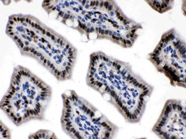
| Sample | Paraffin-embedded mouse intestine tissue; fixative not specified (datasheet PB10009) |
| Fixation | Image fixative and duration unreported (datasheet PB10009); verify before use. |
| Sectioning | 4–5 µm sections on charged slides (standard) |
| Deparaffinisation | Xylene, graded ethanol series to water (standard) |
| Antigen retrieval | Heat retrieval: EDTA pH 8.0 (datasheet PB10009); 20 min, 95–100 °C (standard) |
| Peroxidase block | 3% H2O2, 10 min, room temperature (standard) |
| Blocking | 10% goat serum (datasheet PB10009) |
| Primary antibody | Rabbit anti-NAMPT, 0.5-1μg/ml (datasheet PB10009) |
| Primary incubation | Overnight at 4 °C (datasheet PB10009) |
| Detection | Streptavidin-biotin complex (SABC), DAB chromogen (datasheet PB10009) |
| Counterstain | Hematoxylin, blue, dehydrate and mount (standard) |
| Expected result | NAMPT-positive staining in glandular cells of adrenal gland (HPA tissue IHC: Medium). HPA tissue profile: General cytoplasmic expression, highest abundance in lymphoid tissues and gastrointestinal tract. Positivity in plasma. No signal in the no-primary control. |
In paraffin-section IHC, expect mainly cytoplasmic NAMPT staining, especially in lymphoid and gastrointestinal tissues; HPA also reports plasma positivity (HPA tissue IHC: Approved, pending external verification). Nuclear staining can be biologically plausible because NAMPT is reported in the nucleus and cytoplasm (UniProt P43490: subcellular location). NAMPT has no transmembrane segment, so a membrane-only pattern needs scrutiny (UniProt P43490: topology).
| Cytoplasmic staining in identifiable tissue cells, with stronger overall staining in lymphoid or gastrointestinal tissue. | This fits the reported IHC profile, but intensity should be judged by cell population: HPA describes general cytoplasmic expression and the highest abundance in lymphoid tissues and gastrointestinal tract (HPA tissue IHC: profile). |
| Nuclear staining accompanies the cytoplasmic pattern, or staining appears confined to cell borders. | Do not dismiss nuclear signal solely because the IHC profile is cytoplasmic: granular nuclear NAMPT is reported under non-inflammatory conditions (UniProt P43490: subcellular location). An isolated border pattern needs review because NAMPT has no transmembrane segment (UniProt P43490: topology). |
| Strong signal appears in adipocytes, cardiomyocytes or skeletal myocytes while nearby expected cells are unstained. | These cell populations were reported as not detected in HPA tissue IHC (HPA tissue IHC: adipocytes, cardiomyocytes, skeletal myocytes). Check morphology and controls for cross-reactivity or endogenous detection activity (general IHC practice); staining alone cannot identify which caused the signal. |
| Color is diffuse across tissue, plasma-rich spaces or the negative control, without clear cellular boundaries. | Plasma positivity is reported and NAMPT can be secreted (HPA tissue IHC: profile; UniProt P43490: subcellular location). Diffuse color therefore cannot establish cellular localisation; assess the negative control and detection background (general IHC practice). |
| No staining is visible in a selected positive-control tissue or its expected cell population. | First confirm the chosen population: HPA reports medium glandular-cell staining in appendix and colon, while its overall IHC assessment remains pending external verification (HPA tissue IHC: positive tissues, reliability). A blank control calls for a workflow check (general IHC practice). |
| Tissue and cell selection | HPA reports its highest overall abundance in lymphoid tissues and gastrointestinal tract, but the listed glandular cells in appendix and colon are medium (HPA tissue IHC: profile, positive tissues). Score the named cell population rather than assigning one intensity to every cell. |
| Secreted protein | NAMPT is also reported as secreted, and HPA notes plasma positivity and potentially complex RNA–protein agreement for secreted variants (UniProt P43490: subcellular location; HPA tissue IHC: profile, reliability). Extracellular color needs cellular context before interpretation. |
| Compartment and topology | UniProt lists nucleus and cytoplasm and describes predominantly granular nuclear staining under non-inflammatory conditions; it lists no transmembrane segment (UniProt P43490: subcellular location, topology). Treat nuclear signal as plausible and a membrane-only result as a reason to inspect controls. |
| Evidence strength | The supplied antibodies HPA047776 and CAB034349 are IHC Approved; the supplied record does not mark either IHC Enhanced (HPA antibodies: IHC status). HPA calls tissue IHC Approved while noting pending external verification (HPA tissue IHC: reliability). |
| IF/ICC Q&A: Should the IHC pattern match IF/ICC? | No direct match is established: HPA reports nuclear speckles as the main ICC-IF location and cell junctions as an additional location, whereas its tissue IHC profile is generally cytoplasmic (HPA subcellular: approved locations; HPA tissue IHC: profile). Compare each result within its assay. |
| Tissue-level discrepancy | UniProt reports NAMPT expression in muscle and heart, while HPA reports no detected staining in skeletal myocytes and cardiomyocytes (UniProt P43490: tissue specificity; HPA tissue IHC: negative tissues). Record the assayed cell type and method when comparing these observations. |
| Situation | Likely cause | Next action |
|---|---|---|
| Expected cytoplasmic staining is absent from an appendix or colon control. | The selected cells may be misidentified, or the IHC workflow may have failed (general IHC practice); HPA reports medium staining specifically in glandular cells there (HPA tissue IHC: positive tissues). | Locate glandular cells on the section, then review the IHC antibody, antigen retrieval and detection controls using the validated workflow (general IHC practice). |
| The entire section has weak or uneven color. | Section handling or uneven reagent coverage can affect an IHC run (general IHC practice). This payload provides no NAMPT-specific fixation or retrieval sensitivity. | Inspect section integrity and reagent coverage; compare with a concurrently processed control and the selected antibody's validated IHC conditions (general IHC practice). |
| Brown color appears in the negative control as well as the NAMPT slide. | Residual endogenous enzyme activity or nonspecific detection can produce color in chromogenic IHC (general IHC practice). | Review the negative control, enzyme-blocking and detection steps; interpret the NAMPT slide only after the background source is resolved (general IHC practice). |
| Signal appears only along cell borders, with no convincing cytoplasmic staining. | A border-only pattern does not fit HPA's general cytoplasmic tissue profile or UniProt's lack of a transmembrane segment (HPA tissue IHC: profile; UniProt P43490: topology). | Check morphology, adjacent cells and negative controls; repeat with an IHC Approved antibody if needed (general IHC practice; HPA antibodies: IHC status). |
| Adipocytes or myocytes stain strongly, contrary to the selected HPA reference. | HPA reports no detection in adipocytes, cardiomyocytes or skeletal myocytes (HPA tissue IHC: negative tissues). Cross-reactivity or endogenous detection activity remains possible (general IHC practice). | Confirm cell identity, inspect the negative control and compare an independently validated IHC antibody before assigning the signal to NAMPT (general IHC practice; HPA antibodies: IHC status). |
| Plasma-rich spaces stain, but individual positive cells are hard to identify. | HPA reports plasma positivity and UniProt lists a secreted form of NAMPT (HPA tissue IHC: profile; UniProt P43490: subcellular location). | Score identifiable cells separately from extracellular color; use morphology and a negative control to judge whether a cellular staining pattern is present (general IHC practice). |


Comprehensive Human Protein Atlas IHC scoring per tissue (reliability: Approved — At least one protein variant secreted, tissue location of RNA and protein might differ and correlation is complex. Pending external verification.). Rows are taken directly from the HPA tissue chart — click any row's HPA link to view the source.
| Tissue | Cell type | Level | Evidence | Source |
|---|---|---|---|---|
| Adrenal gland | Glandular cells | Medium | Protein (IHC) | HPA → |
| Appendix | Glandular cells | Medium | Protein (IHC) | HPA → |
| Breast | Glandular cells | Medium | Protein (IHC) | HPA → |
| Cervix | Glandular cells | Medium | Protein (IHC) | HPA → |
| Colon | Glandular cells | Medium | Protein (IHC) | HPA → |
| Tissue | Cell type | Level | Evidence | Source |
|---|---|---|---|---|
| Adipose tissue | Adipocytes | Not detected | Protein (IHC) | HPA → |
| Caudate | Glial cells | Not detected | Protein (IHC) | HPA → |
| Cerebellum | Cells in granular layer | Not detected | Protein (IHC) | HPA → |
| Heart muscle | Cardiomyocytes | Not detected | Protein (IHC) | HPA → |
| Hippocampus | Glial cells | Not detected | Protein (IHC) | HPA → |
Troubleshoot NAMPT staining in paraffin sections by checking retrieval, compartment, tissue context, controls and scoring before interpreting chromogenic signal.
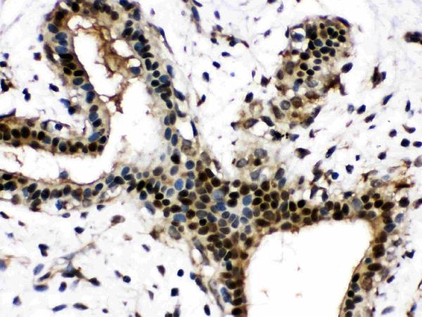
PB10009 has paraffin-section IHC images from human mammary cancer and mouse and rat intestine (IHC image captions), plus an IF/ICC image from A549 cells (IF image caption).
PB10009 is listed for IHC and IF/ICC in human, mouse and rat (catalog applications; catalog reactivity). Its IHC captions show human mammary cancer and mouse and rat intestine paraffin sections; its IF caption shows A549 cells (IHC and IF image captions).
Which to pick: For tissue IHC, choose PB10009: its own images document staining in paraffin sections, and the fixative is unreported (IHC image captions). For IF/ICC, the same SKU is listed for both applications and has an A549-cell IF image (catalog applications; IF image caption). For work across species, PB10009 lists human, mouse and rat reactivity and has a paraffin-section IHC image for each; clonality is unreported (catalog reactivity; IHC image captions; catalog clone field).