NAT2 / Arylamine N-acetyltransferase 2 · IHC design guide

Design Immunohistochemistry for NAT2

Plan NAT2 staining in paraffin sections using cytoplasmic tissue staining as the expected pattern (HPA tissue IHC). Compare positive tissues with controls and interpret staining cautiously because antibody results show low agreement with RNA expression (HPA tissue IHC).

Evidence assembled Oct 2026 · For research use; verify linked source records and product datasheet before use
Immunohistochemistry protocol sheet for NAT2 (IHC for NAT2): expected localisation Cytoplasmic staining in several tissues (HPA tissue IHC), antibody A00966, validated IHC image, and IHC protocol steps
Printable NAT2 IHC protocol sheet — expected localisation Cytoplasmic staining in several tissues (HPA tissue IHC), antibody A00966, controls and protocol steps. Open the full NAT2 IHC guide →

NAT2 Immunohistochemistry Experimental Design Guide

Expected localisation, validated protocols, controls and antibodies — the at-a-glance facts below, then the full design guide.

Must know before staining
Expected localisation Cytoplasmic staining in several tissues (HPA tissue IHC)
Staining pattern Cytoplasmic staining in hepatocytes and intestinal glands (HPA tissue IHC)
Antigen retrieval Citrate pH 6.0 HIER, 95–98 °C, 20 min (rule: cytoplasmic / membrane antigen)
Positive control ⓘ Breast+4 more · see all
Negative control ⓘ Adipose tissue+4 more · see all
Important caveats
Reasons your staining may differ from the expected pattern.
Fixation Keep fixation consistent across sections. (standard IHC practice; not target-specific)
Caveat Antibody staining may reflect more than one gene product (HPA tissue IHC)
Regulation Expression regulation is unreported (UniProt)
Isoform / epitope No annotated isoforms; one unprocessed 1–290 chain (UniProt)
Section 1

Recommended NAT2 IHC & IF Protocols

Compare the catalog antibody’s IHC-P protocol with 3 published NAT2/Nat2 IHC protocols (PMC2430772; PMC8635240; PMC7947712).

Recommended immunohistochemistry (IHC-P) protocol parameters
SampleParaffin-embedded rat lung tissue; fixative not specified (datasheet A00966)
FixationImage fixative and duration unreported (datasheet A00966); verify before use.
Sectioning4–5 µm sections on charged slides (standard)
DeparaffinisationXylene, graded ethanol series to water (standard)
Antigen retrievalHeat-induced epitope retrieval in citrate buffer, pH 6.0, 20 min at 95–98 °C (standard rule: cytoplasmic / membrane antigen)
Peroxidase block3% H2O2, 10 min, room temperature (standard)
Blocking10% normal serum of the secondary host, 30 min, room temperature (standard)
Primary antibodyRabbit anti-NAT2, 1:50-1:200 (datasheet A00966)
Primary incubationOvernight at 4 °C (standard)
DetectionHRP-polymer secondary, DAB chromogen 5–10 min (standard)
CounterstainHematoxylin, blue, dehydrate and mount (standard)
Expected resultNAT2-positive staining in glandular cells of breast (HPA tissue IHC: High). HPA tissue profile: Cytoplasmic expression in several different tissue types. No signal in the no-primary control.
💡Decision noteStart with citrate pH 6.0 HIER at 95–98 °C for 20 min (page antigen retrieval); NAT2 is cytoplasmic (UniProt P11245).
Section 2

What Is the Expected NAT2 Staining Pattern?

NAT2 is a cytoplasmic protein with no transmembrane segment (UniProt P11245). In tissue IHC, expect cytoplasmic staining in hepatocytes, intestinal glandular cells, kidney tubule cells and lung macrophages, among other reported cells (HPA: High in these cells). Treat this as a provisional pattern: HPA rates tissue IHC reliability Uncertain because antibody staining and RNA expression have low consistency, and warns that the antibody may recognize protein from more than one gene (HPA: reliability Uncertain).

What am I looking at on my slide?
Cytoplasmic signal in hepatocytes, duodenal or small-intestinal glandular cells, kidney tubule cells, or lung macrophages.This matches reported high-staining cell populations (HPA: High in each). Score the named cells and their cytoplasm, while retaining the antibody-specific uncertainty in the interpretation (HPA: reliability Uncertain).
Predominantly nuclear or membrane-rim signal, with little cytoplasmic staining.The compartment conflicts with the reported cytoplasmic pattern and a protein lacking a transmembrane segment (HPA: cytoplasmic profile; UniProt P11245 topology). Suspect artefact or nonspecific staining; check controls before assigning it to NAT2 (general IHC practice).
Strong signal in cells reported as not detected, such as adipocytes or adrenal glandular cells.The result conflicts with those HPA cell-level observations (HPA: Not detected in adipocytes and adrenal glandular cells). Cross-reactivity or endogenous detection activity is possible (HPA: antibody may recognize more than one gene; general IHC practice).
Widespread haze obscures cell boundaries and persists across unrelated tissue compartments.This cannot establish the reported cell-specific cytoplasmic pattern (HPA: tissue IHC profile). Review blocking, washes and detection controls for nonspecific background or endogenous activity (general IHC practice).
No interpretable signal in hepatocytes or kidney tubule cells on a test section.These cells were reported as High, so an absent result merits a technical check (HPA: High in both). It does not alone disprove NAT2 expression: the HPA antibody's IHC status is Uncertain (HPA: HPA043704 IHC Uncertain).
💡Expected NAT2 appearanceA provisional positive is predominantly cytoplasmic signal in a reported High cell population, such as hepatocytes or kidney tubule cells; isolated nuclear, membrane-rim or diffuse signal is suspect (HPA: tissue IHC profile and reliability Uncertain; UniProt P11245 topology).
How each factor affects the staining
CompartmentNAT2 is annotated in the cytoplasm and has no transmembrane segment (UniProt P11245). Interpret staining against a cytoplasmic pattern; membrane-only localization needs independent validation (general IHC practice).
Cell populationHPA reports High staining in several distinct cells, including breast and prostate glandular cells and late spermatids, while adipocytes and adrenal glandular cells are Not detected (HPA: tissue IHC). Judge the relevant cells within each section.
Antibody validationHPA043704 has an Uncertain IHC rating; HPA reports low staining–RNA consistency and possible recognition of protein from more than one gene (HPA: antibody status and tissue reliability). A matching image alone cannot establish specificity.
Protein processingUniProt lists one chain spanning residues 1–290, with no signal peptide, propeptide, annotated glycosylation sites or modified residues (UniProt P11245). These annotations provide no basis for predicting a processed or shed staining pattern.
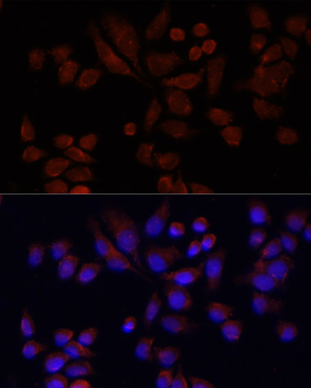
IF/ICC evidenceHPA supplies no NAT2 ICC-IF image-bearing cell lines or main subcellular location, despite a reported RT-4 RNA value of 1.0 nTPM (HPA: subcellular record). Tissue IHC cannot validate an IF/ICC pattern by itself.
Why is my staining missing, weak or wrong?
SituationLikely causeNext action
How should a High liver or kidney signal be reported?High is an observed staining level, while HPA rates the tissue IHC evidence Uncertain (HPA: liver and kidney High; reliability Uncertain).Report the stained cell type and cytoplasmic compartment, then qualify the finding as compatible with NAT2 rather than confirmed target-specific staining (HPA: tissue profile and reliability Uncertain).
Nuclear or membrane-rim staining dominates.That distribution disagrees with the cytoplasmic annotation and lack of a transmembrane segment (HPA: cytoplasmic profile; UniProt P11245 topology).Inspect the negative control and detection background, and reassess localization in reported High cells before scoring (general IHC practice; HPA: High cell populations).
Cells reported as Not detected stain strongly.An off-pattern result may reflect nonspecific binding or endogenous detection activity; HPA also flags possible recognition of another gene product (general IHC practice; HPA: reliability description).Compare the same detection workflow with an appropriate negative control and review which cells stain; do not treat the signal as NAT2-specific without corroboration (general IHC practice; HPA: reliability Uncertain).
Background obscures cytoplasmic detail.Nonspecific binding, insufficient washing or endogenous enzyme activity can obscure chromogenic IHC (general IHC practice).Review blocking and washing; if using an enzyme-based detector, check its endogenous-activity control, then score only cells with interpretable boundaries (general IHC practice).
A reported High population shows no signal.The section or staining run may have failed, but HPA's High observation does not establish a guaranteed result for this antibody (general IHC practice; HPA: tissue levels and IHC Uncertain).Check section integrity, retrieval and detection controls, and the catalog antibody's IHC-P instructions before interpreting absence of staining (general IHC practice; HPA: antibody IHC status Uncertain).
Can this tissue pattern define the expected IF/ICC result?HPA provides no NAT2 ICC-IF images or assigned main subcellular location (HPA: subcellular record).Use cytoplasmic localization only as a hypothesis for IF/ICC and evaluate that application with its own controls; do not infer IF/ICC validation from tissue IHC (UniProt P11245; HPA: subcellular record).

Sample controls for NAT2 IHC & IF

🧪Run lung first: macrophages should show NAT2 staining (HPA: High in lung macrophages). Run adipose tissue as the negative: adipocytes should lack specific staining (HPA: Not detected in adipocytes); on the lung slide, morphologically distinct cells without cytoplasmic staining can serve as internal background comparators, but their NAT2 status needs independent confirmation (UniProt P11245: cytoplasm; HPA: lung macrophages High).
Positive control tissue: Breast (Glandular cells, HPA High)
Negative control tissue: Adipose tissue (HPA Not detected)
ICC-IF cell lines (HPA subcellular resource): HPA carries no ICC-IF cell line for NAT2; derive a cell-line control from the positive tissue's cell type (Glandular cells) and confirm it by RNA or western blot first.
Technical controls: Include a no-primary, secondary-only control; a control immunoglobulin matched to the primary antibody’s host species, class, and clonality as applicable; and NAT2 knockout tissue or an immunizing-peptide competition control if the peptide is available (standard IHC practice). Check endogenous peroxidase and pigment-related autofluorescence in lung macrophages, especially when interpreting chromogenic IHC or IF signal (standard IHC/IF practice).
⚠️Feasibility: A NAT2-specific fixation window or fixation effect is unreported in the supplied evidence, and the selected A00966 paraffin-section caption does not state the fixative (selected tissue-IHC caption). That rat-lung caption uses microwave retrieval in 10 mM PBS, pH 7.2, with the antibody at 1:100; it does not establish that retrieval is required for other specimens (selected tissue-IHC caption). Neither frozen sections nor IF can be identified as easier from the supplied evidence; lung macrophage pigment and autofluorescence warrant particular attention if IF is attempted (HPA: High in lung macrophages; standard IF practice).

HPA tissue IHC evidence for NAT2

Comprehensive Human Protein Atlas IHC scoring per tissue (reliability: Uncertain — Low consistency between antibody staining and RNA expression data. Caution, targets protein from more than one gene.). Rows are taken directly from the HPA tissue chart — click any row's HPA link to view the source.

Positive expression · recommended positive controls

TissueCell typeLevelEvidenceSource
Breast Glandular cells High Protein (IHC) HPA →
Duodenum Glandular cells High Protein (IHC) HPA →
Kidney Cells in tubules High Protein (IHC) HPA →
Liver Hepatocytes High Protein (IHC) HPA →
Lung Macrophages High Protein (IHC) HPA →

Undetected expression · recommended negative controls

TissueCell typeLevelEvidenceSource
Adipose tissue Adipocytes Not detected Protein (IHC) HPA →
Adrenal gland Glandular cells Not detected Protein (IHC) HPA →
Bone marrow Hematopoietic cells Not detected Protein (IHC) HPA →
Caudate Glial cells Not detected Protein (IHC) HPA →
Cerebral cortex Endothelial cells Not detected Protein (IHC) HPA →
Section 3

Advanced NAT2 IHC Tips

Troubleshoot NAT2 staining in paraffin sections by checking cytoplasmic signal, cell identity, controls and scoring consistency (UniProt P11245; HPA tissue IHC).

Which retrieval conditions should I try first for weak NAT2 staining in paraffin sections?
Start with citrate buffer at pH 6.0, heated to 95–98 °C for 20 min (page retrieval setting). Let sections cool consistently, then compare staining with a known positive section in the same run; unequal retrieval can obscure a cytoplasmic signal (UniProt P11245 localisation; standard IHC practice). If staining remains weak, test microwave retrieval in 10 mM PBS at pH 7.2 on adjacent sections; that condition accompanied the catalog antibody’s rat lung paraffin image (A00966 caption). Keep antibody dilution and detection conditions matched during this comparison so a retrieval effect can be judged (standard IHC practice).
How should I investigate variable NAT2 staining across paraffin blocks?
NAT2-specific fixation sensitivity is unknown: the selected paraffin image does not report a fixative, so it cannot establish a preferred fixation condition (A00966 caption). Compare blocks with documented fixation and processing histories, using sections of similar thickness and a shared staining run to reduce technical variation (standard IHC practice). Include a positive control section with each run and record whether loss of signal is widespread or confined to block edges or damaged regions (standard IHC practice). Avoid attributing differences to NAT2 biology until retrieval, antibody incubation and chromogen development have been held constant (standard IHC practice).
Where should convincing NAT2 staining appear within positive cells?
Expect predominantly cytoplasmic chromogenic signal because NAT2 is annotated in the cytoplasm and has no transmembrane segment (UniProt P11245 localisation and topology). Examine the whole cell against its counterstained nucleus, since dense nuclear chromogen alone would conflict with that annotation (UniProt P11245 localisation; standard IHC practice). In liver, hepatocytes are a useful cell type to examine; in lung, assess macrophages specifically rather than treating every stained cell as equivalent (HPA tissue IHC: high in hepatocytes and lung macrophages). Compare the distribution with a negative reagent control and inspect nearby unstained cells before assigning a compartment or cell type (standard IHC practice).
Could an isoform or epitope difference explain discordant NAT2 IHC results?
The supplied record annotates 0 isoforms and one chain spanning residues 1–290, so it supplies no alternative NAT2 isoform to explain discordant staining (UniProt P11245 processing and isoforms). It also lists no signal peptide, propeptide, glycosylation sites or modified residues; those annotations do not identify the catalog antibody’s epitope (UniProt P11245 processing and PTMs; A00966 caption). Ask for an experimentally mapped epitope before interpreting a negative section as selective loss of part of NAT2 (standard IHC practice). Because the tissue atlas cautions that its staining may recognize protein from more than one gene, corroborate any isoform or specificity claim independently (HPA tissue IHC reliability description).
How should I assess NAT2 by IF alongside the paraffin IHC result?
Treat IF as a separate assay and multiplex NAT2 with a validated marker for the cell population being assessed, such as a macrophage marker when examining lung cells (HPA tissue IHC: high in lung macrophages; standard IF practice). Choose fluorophores after inspecting an unstained section for tissue autofluorescence, and include single-label controls to check channel bleed-through (standard IF practice). NAT2 is cytoplasmic with no transmembrane segment, so permeabilise sufficiently to let antibody reach an intracellular epitope, while optimising that step for the IF specimen (UniProt P11245 localisation and topology; standard IF practice). The paraffin IHC caption reports no IF fixation or permeabilisation conditions and provides no IF validation (A00966 caption).
What should I check when NAT2 DAB staining is widespread or granular?
Run a section without primary antibody and inspect where chromogen develops; signal there points to the detection workflow rather than specific primary binding (standard chromogenic IHC practice). Apply a peroxidase block before DAB detection, and assess whether endogenous pigment, trapped reagent or precipitated chromogen resembles the observed granules (standard chromogenic IHC practice). Titrate the primary antibody and shorten development systematically if diffuse staining obscures cytoplasm; the rat lung paraffin image used 1:100, which is a documented image condition rather than a universal optimum (A00966 caption; standard IHC practice). Judge remaining signal against the expected cytoplasmic pattern and a matched positive section (UniProt P11245 localisation; standard IHC practice).
How should I quantify NAT2 staining across comparable paraffin sections? ⚠ ANSWER MARKED FOR VERIFICATION
Define the cell population and analysis region before scoring, then record the percentage of positive cells and cytoplasmic intensity using the same criteria in every section (standard IHC practice; UniProt P11245 localisation). An H-score can combine intensity categories with their positive-cell percentages; report the categories and calculation alongside the result (standard IHC practice). If counting discrete positive cells, express density per mm² of viable analysed tissue and exclude folds, necrosis and empty space consistently (standard IHC practice). Normalise comparisons to the same cell type and sampled tissue area, since the HPA profile spans several tissues and cell populations (HPA tissue IHC profile).
How can I distinguish a true NAT2 positive from staining artefact?
A plausible positive has cellular cytoplasmic staining in an appropriate population, such as hepatocytes or lung macrophages, and is reproducible across comparable sections (UniProt P11245 localisation; HPA tissue IHC: high in hepatocytes and lung macrophages; standard IHC practice). Isolated nuclear staining or signal assigned to the wrong cell type deserves re-examination against morphology and controls (UniProt P11245 localisation; standard IHC practice). Discount staining concentrated at section edges, necrotic areas or sites that remain positive without primary antibody; assess endogenous peroxidase when using DAB (standard chromogenic IHC practice). Interpret even convincing staining cautiously because the HPA tissue assay is rated uncertain and may detect protein from more than one gene (HPA tissue IHC reliability description).
Boster reagents

Best NAT2 / Arylamine N-acetyltransferase 2 IHC Antibodies

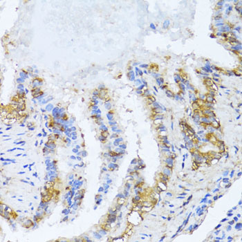
The catalog lists one anti-NAT2 antibody for IHC in human, mouse, and rat (A00966 catalog: applications and reactivity), with IHC images from paraffin-embedded rat lung and heart (A00966 image captions).

Real IHC data Immunohistochemistry of paraffin-embedded rat lung using NAT2 antibody at dilution of 1:100 .Perform microwave antigen retrieval with 10 mM PBS buffer pH 7.2 before commencing with IHC staining protocol.
Anti-NAT2 Antibody
Cat # A00966

A00966 is listed for IHC and reacts with human, mouse, and rat (A00966 catalog: applications and reactivity). Its IHC images show paraffin-embedded rat lung and heart stained at 1:100 after microwave retrieval in 10 mM PBS, pH 7.2 (A00966 image captions).

Which to pick: Choose A00966 for paraffin-section tissue IHC based on its rat lung and heart images; the captions do not report a fixative (A00966 image captions). There is no catalog-supported IF/ICC choice here: A00966 lists IHC and WB, with no IF dilution or IF images (A00966 catalog: applications, IF dilution and IF images). For cross-species planning, A00966 lists human, mouse, and rat reactivity, but its IHC images show rat tissues only; it is rabbit-derived, and clonality is unreported (A00966 catalog: reactivity, host and clone; A00966 image captions).

Each figure is that product's own IHC / IF validation image from its datasheet.

References

  1. UniProt Consortium. UniProt entry P11245 (ARY2_HUMAN, Arylamine N-acetyltransferase 2).
  2. Human Protein Atlas. NAT2 tissue IHC expression (reliability: Uncertain).
  3. Human Protein Atlas. NAT2 subcellular location (ICC-IF): Highest expression in RT-4: 1.0 nTPM.
  4. Human Protein Atlas. NAT2 antibody validation summary (1 antibodies).
  5. Mouse arylamine N-acetyltransferase 2 (Nat2) expression during embryogenesis: a potential marker for the developing neuroendocrine system. Biomarkers : biochemical indicators of exposure, response, and susceptibility to chemicals 2008 — PMC2430772.
  6. VDR Signaling via the Enzyme NAT2 Inhibits Colorectal Cancer Progression. Frontiers in pharmacology 2021 — PMC8635240.
  7. Mining of RNA Methylation-Related Genes and Elucidation of Their Molecular Biology in Gallbladder Carcinoma. Frontiers in oncology 2021 — PMC7947712.
  8. PubMed PMID:2734109 — UniProt-cited evidence.
  9. PubMed PMID:2340091 — UniProt-cited evidence.
  10. PubMed PMID:1968463 — UniProt-cited evidence.