NAT8 / N-acetyltransferase 8 · IHC design guide

Design Immunohistochemistry for NAT8

Plan NAT8 chromogenic IHC on paraffin sections using renal proximal tubules as a high-staining reference (HPA tissue IHC). This guide covers fixation consistency, controls, and interpretation of cytoplasmic tubular staining (HPA tissue IHC).

Evidence assembled Oct 2026 · For research use; verify linked source records and product datasheet before use
Immunohistochemistry protocol sheet for NAT8 (IHC for NAT8): expected localisation Renal tubular cytoplasm (HPA tissue IHC); ER membrane (UniProt), antibody A08414, validated IHC image, and IHC protocol steps
Printable NAT8 IHC protocol sheet — expected localisation Renal tubular cytoplasm (HPA tissue IHC); ER membrane (UniProt), antibody A08414, controls and protocol steps. Open the full NAT8 IHC guide →

NAT8 Immunohistochemistry Experimental Design Guide

Expected localisation, validated protocols, controls and antibodies — the at-a-glance facts below, then the full design guide.

Must know before staining
Expected localisation Renal tubular cytoplasm (HPA tissue IHC); ER membrane (UniProt)
Staining pattern High cytoplasmic staining in proximal tubule cell bodies (HPA tissue IHC)
Antigen retrieval Citrate pH 6.0 HIER, 95–98 °C, 20 min (rule: cytoplasmic / membrane antigen)
Positive control ⓘ Duodenum+2 more · see all
Negative control ⓘ Adipose tissue+4 more · see all
Important caveats
Reasons your staining may differ from the expected pattern.
Fixation Keep fixation consistent across sections (standard IHC practice; not target-specific); Matched tissue-IHC evidence does not establish the fixation claim. Validate the specimen-specific method before use. (selected-SKU IHC image A08414)
Caveat The HPA antibody may detect protein from more than one gene (HPA tissue IHC)
Regulation Kidney-enriched RNA expression (HPA RNA specificity)
Isoform / epitope No isoforms; one 1–227 chain with cytoplasmic 1–42 and luminal 64–227 regions (UniProt)
Section 1

Recommended NAT8 IHC & IF Protocols

The catalog antibody’s IHC-P protocol is followed by one published NAT8 immunohistochemistry protocol (PMC4109999).

Recommended immunohistochemistry (IHC-P) protocol parameters
SampleParaffin-embedded mouse brain tissue; fixative not specified (datasheet A08414)
FixationImage fixative and duration unreported (datasheet A08414); verify before use.
Sectioning4–5 µm sections on charged slides (standard)
DeparaffinisationXylene, graded ethanol series to water (standard)
Antigen retrievalHeat-induced epitope retrieval in citrate buffer, pH 6.0, 20 min at 95–98 °C (standard rule: cytoplasmic / membrane antigen)
Peroxidase block3% H2O2, 10 min, room temperature (standard)
Blocking10% normal serum of the secondary host, 30 min, room temperature (standard)
Primary antibodyRabbit anti-NAT8, 1:50-1:100 (datasheet A08414)
Primary incubationOvernight at 4 °C (standard)
DetectionHRP-polymer secondary, DAB chromogen 5–10 min (standard)
CounterstainHematoxylin, blue, dehydrate and mount (standard)
Expected resultNAT8-positive staining in paneth cells of duodenum (HPA tissue IHC: High). HPA tissue profile: Cytoplasmic expression in renal tubules. No signal in the no-primary control.
💡Decision noteStart with citrate pH 6.0 retrieval at 95–98 °C for 20 min (page retrieval rule; UniProt Q9UHE5 topology).
Section 2

What Is the Expected NAT8 Staining Pattern?

NAT8 is an ER and ER–Golgi intermediate compartment membrane protein with a short cytoplasmic N terminus and a luminal C terminus (UniProt Q9UHE5 topology). In IHC, expect cytoplasmic staining in renal tubules, especially proximal tubule cell bodies, where staining is high (HPA tissue IHC). HPA rates the tissue pattern Supported, while cautioning that its staining data may reflect proteins from more than one gene (HPA tissue IHC).

What am I looking at on my slide?
Strong cytoplasmic staining in kidney proximal tubule cell bodies.This matches the reported high proximal tubule signal and renal tubule cytoplasmic profile (HPA tissue IHC). Score the stained cell population and compartment together; a strong stain elsewhere does not establish the same interpretation.
Predominantly nuclear staining, or a sharply outlined cell surface without cytoplasmic signal.These patterns do not fit the reported renal cytoplasmic profile (HPA tissue IHC) or ER membrane localisation (UniProt Q9UHE5). Check morphology and controls before treating the signal as NAT8.
Strong staining in adipocytes or respiratory epithelial cells.Both cell populations are listed as not detected (HPA tissue IHC). Consider antibody cross-reactivity or endogenous chromogenic activity (general IHC practice); the HPA multi-gene caution makes antibody specificity particularly relevant (HPA tissue IHC).
Uniform colour across cells, stroma, or the entire section.A section-wide haze obscures the cell-restricted pattern reported for renal tubules (HPA tissue IHC). Uneven blocking, excess antibody, or detection background are possible general IHC causes (general IHC practice).
No signal in kidney proximal tubules.This conflicts with the reported high staining in proximal tubule cell bodies (HPA tissue IHC). First establish that the expected cells are present, then assess staining and detection controls (general IHC practice).
💡Expected NAT8 appearanceCall a positive result when proximal tubule cell bodies show high cytoplasmic staining (HPA tissue IHC); isolated nuclear, cell surface, or section-wide colour is inconsistent with that pattern (HPA tissue IHC; UniProt Q9UHE5 localisation).
How each factor affects the staining
Compartment and topologyNAT8 spans the membrane at residues 43–63; residues 1–42 are cytoplasmic and 64–227 luminal (UniProt Q9UHE5 topology). The record does not locate this antibody’s epitope, so topology alone cannot predict its staining sensitivity.
Tissue and cell selectionKidney proximal tubules and duodenal Paneth cells stain high; small intestinal endocrine cells stain medium, while hepatocytes stain low (HPA tissue IHC). Use the named cells when comparing intensity, rather than treating every cell in a tissue as equivalent.
Antibody validation scopeThe tissue IHC profile is Supported, and HPA067855 is IHC Supported (HPA tissue IHC; HPA antibodies). HPA cautions that the tissue signal may include proteins from more than one gene (HPA tissue IHC); a matching pattern alone cannot prove NAT8 specificity.
Chromogenic detectionEndogenous enzyme activity and nonspecific detection can add colour independently of primary antibody binding (general IHC practice). Interpret suspicious staining alongside an appropriate primary-omission control and the expected cell distribution (general IHC practice; HPA tissue IHC).
Why is my staining missing, weak or wrong?
SituationLikely causeNext action
No colour in proximal tubules despite adequate tissue morphology.The expected high-staining cell population is present, but the primary or detection workflow may have failed (HPA tissue IHC; general IHC practice).Confirm the IHC-validated antibody and detection reagents, then review retrieval, antibody concentration, and incubation against the applicable IHC-P protocol (general IHC practice). Do not infer NAT8-specific fixation sensitivity from this result.
Weak kidney signal, with a visible low-level liver signal.Hepatocytes are reported low while proximal tubule cell bodies are high (HPA tissue IHC); the two tissues do not provide interchangeable intensity benchmarks.Compare proximal tubules with the validated kidney reference and check whether the same staining run behaved as expected (HPA tissue IHC; general IHC practice).
Unexpected staining in a cell population listed as not detected.Cross-reactivity or detection background is possible (general IHC practice). HPA explicitly cautions that its tissue data may reflect proteins from more than one gene (HPA tissue IHC).Review cell identity and the primary-omission control, then seek an independent specificity check before assigning the stain to NAT8 (general IHC practice; HPA tissue IHC).
Broad brown haze makes tubules hard to distinguish.Section-wide colour is inconsistent with the reported cell-restricted renal profile (HPA tissue IHC); excessive primary signal or detection background can produce haze (general IHC practice).Inspect the primary-omission control, blocking, washes, and detection exposure; adjust the workflow within its validated range (general IHC practice). Reassess the tubule pattern only after background is controlled.
Nuclear or cell-surface colour dominates an otherwise clean section.That distribution conflicts with cytoplasmic renal staining (HPA tissue IHC) and NAT8’s ER membrane annotation (UniProt Q9UHE5 localisation).Check the counterstain and tissue morphology, compare the primary-omission control, and reassess antibody specificity before scoring the colour as positive (general IHC practice).
What should a corresponding IF/ICC image show?HPA reports a supported ER location in ICC-IF images, while warning that the antibodies may target proteins from multiple genes (HPA subcellular ICC-IF).Look for an intracellular ER-associated pattern rather than predominant nuclear or cell-surface signal (HPA subcellular ICC-IF; UniProt Q9UHE5 localisation). Use the separate IF/ICC guide for that application.

Sample controls for NAT8 IHC & IF

🧪Run kidney first: proximal tubules (cell body) should stain (HPA: High in kidney proximal tubules). Use adipose tissue as the negative tissue, where adipocytes should show no specific staining (HPA: Not detected in adipocytes); on the kidney slide, assess cells outside the annotated proximal tubules as internal background controls without assuming they are NAT8-negative.
Positive control tissue: Duodenum (Paneth cells, HPA High)
Negative control tissue: Adipose tissue (HPA Not detected)
ICC-IF cell lines (HPA subcellular resource): HPA ICC-IF images show NAT8 in CACO-2, Hep-G2, with annotated localisation: Endoplasmic reticulum (supported) (HPA subcellular).
Technical controls: Include no-primary (secondary-only) and host-species- and isotype-matched controls; use a NAT8 knockout specimen or validated peptide block as a biological specificity control (standard IHC practice). For chromogenic kidney IHC, quench endogenous peroxidase and block endogenous biotin if using avidin–biotin detection (standard IHC practice).
⚠️Feasibility: A target-specific fixation window or fixation effect is unreported, and the selected A08414 paraffin-section caption does not state its fixative (selected-SKU tissue-IHC caption). That caption specifies microwave retrieval in 10 mM PBS, pH 7.2 for mouse brain; retrieval dependence in kidney and whether frozen sections or IF are easier are unreported (selected-SKU tissue-IHC caption). For IF/ICC, an ER pattern is reported, but the subcellular annotation warns that it is based on antibodies targeting proteins from multiple genes (HPA subcellular); kidney chromogenic staining also warrants attention to endogenous peroxidase and, with avidin–biotin detection, endogenous biotin (standard IHC practice).

HPA tissue IHC evidence for NAT8

Comprehensive Human Protein Atlas IHC scoring per tissue (reliability: Supported — High consistency between antibody staining and RNA expression data. Caution, targets protein from more than one gene.). Rows are taken directly from the HPA tissue chart — click any row's HPA link to view the source.

Positive expression · recommended positive controls

TissueCell typeLevelEvidenceSource
Duodenum Paneth cells High Protein (IHC) HPA →
Kidney Proximal tubules (cell body) High Protein (IHC) HPA →
Small intestine Endocrine cells Medium Protein (IHC) HPA →

Undetected expression · recommended negative controls

TissueCell typeLevelEvidenceSource
Adipose tissue Adipocytes Not detected Protein (IHC) HPA →
Adrenal gland Glandular cells Not detected Protein (IHC) HPA →
Appendix Endocrine cells Not detected Protein (IHC) HPA →
Bone marrow Hematopoietic cells Not detected Protein (IHC) HPA →
Breast Adipocytes Not detected Protein (IHC) HPA →
Section 3

Advanced NAT8 IHC Tips

Troubleshoot NAT8 staining by checking retrieval, compartment, cell type and controls before comparing signal intensity across sections.

Which retrieval conditions should I try when NAT8 staining is weak in paraffin sections?
Start with citrate buffer at pH 6.0, heated to 95–98 °C for 20 min (page retrieval setting). If staining remains weak, compare it with microwave retrieval in 10 mM PBS at pH 7.2, the condition reported for the catalog antibody in paraffin-embedded mouse brain (A08414 tissue-IHC caption). Keep section thickness, detection chemistry and exposure to chromogen consistent across the comparison (standard IHC practice). Include a positive tissue control, such as kidney proximal tubules, and a no-primary control on each run (HPA tissue IHC; standard IHC practice). Judge improvement by cellular staining and background together, because stronger chromogen alone does not establish greater specificity (standard IHC practice).
How should I assess whether fixation is masking NAT8 in paraffin sections?
Target-specific fixation sensitivity is unknown from the supplied evidence; the mouse brain caption identifies paraffin embedding but does not state its fixative (A08414 tissue-IHC caption). Record the actual fixative and fixation duration for each specimen before attributing weak staining to antigen masking (standard IHC practice). Compare sections prepared under known, matched conditions using the page’s citrate retrieval at pH 6.0, 95–98 °C for 20 min (page retrieval setting). Include kidney proximal tubules as a tissue reference and a no-primary control to separate lost signal from background (HPA tissue IHC; standard IHC practice). Treat any observed fixation effect as specific to the tested preparation until independently reproduced (standard IHC practice).
What staining pattern is plausible for NAT8, and when should I question it?
Expect intracellular staining compatible with an ER-associated membrane protein, rather than a sharply nuclear pattern (UniProt Q9UHE5 subcellular annotation; standard IHC interpretation). NAT8 has one transmembrane segment at residues 43–63, with residues 1–42 cytoplasmic and 64–227 lumenal (UniProt Q9UHE5 topology). In tissue IHC, HPA describes cytoplasmic expression in renal tubules and high staining in proximal tubule cell bodies (HPA tissue IHC). Compare the distribution within cells and across neighboring cell types under the same chromogen development conditions (standard IHC practice). Diffuse extracellular deposits or predominantly nuclear staining warrant review against no-primary and tissue controls before being scored as NAT8 (UniProt Q9UHE5 subcellular annotation; standard IHC practice).
How can I assess epitope accessibility without knowing where this antibody binds?
The supplied record lists no NAT8 isoforms, glycosylation sites or modified residues, so these annotations do not explain a staining difference (UniProt Q9UHE5 record). Antibody epitope position is not supplied; determine whether it lies within cytoplasmic residues 1–42 or lumenal residues 64–227 before making a topology-based accessibility claim (UniProt Q9UHE5 topology). Keep antigen retrieval at the page setting of citrate pH 6.0, 95–98 °C for 20 min while comparing controls (page retrieval setting; standard IHC practice). If an epitope-mapped antibody becomes available, compare its cellular pattern on adjacent sections using the same detection conditions (standard IHC practice). Agreement with the expected renal tubular distribution supports interpretation but does not independently prove antibody specificity (HPA tissue IHC; standard IHC practice).
How should I design an IF check alongside chromogenic NAT8 IHC?
Use IF as a separate application and multiplex NAT8 with a validated marker of the expected cell type, such as a proximal tubule marker in kidney (HPA tissue IHC; standard IF practice). Choose fluorophores after inspecting unstained tissue in each channel, so tissue autofluorescence can be distinguished from antibody signal (standard IF practice). Because NAT8 spans the membrane at residues 43–63, with cytoplasmic 1–42 and lumenal 64–227, select and test permeabilisation only after establishing which side contains the antibody epitope (UniProt Q9UHE5 topology; standard IF practice). Include single-stain and no-primary controls when assessing channel bleed-through and background (standard IF practice). Do not assume the catalog antibody’s paraffin-section retrieval condition establishes its IF performance (A08414 tissue-IHC caption; standard IF practice).
What should I change when NAT8 chromogenic staining is widespread or granular?
First compare the stained section with a no-primary control and inspect whether signal follows cells or tissue edges (standard IHC practice). Block endogenous peroxidase before peroxidase-based chromogenic detection, and assess whether the secondary reagent contributes background (standard IHC practice). Keep chromogen development consistent while adjusting blocking and washing conditions one variable at a time (standard IHC practice). NAT8 is annotated at ER and ER–Golgi intermediate compartment membranes, while HPA reports cytoplasmic renal tubular staining (UniProt Q9UHE5 subcellular annotation; HPA tissue IHC). Widespread extracellular precipitate, edge-only staining or signal in the no-primary control should be investigated before assigning the pattern to NAT8 (standard IHC practice).
How should I score NAT8 staining across kidney sections? ⚠ ANSWER MARKED FOR VERIFICATION
Define the compartment and cell population before scoring: HPA reports high staining in proximal tubule cell bodies and a cytoplasmic renal tubular profile (HPA tissue IHC). For chromogenic IHC, record the percentage of positive proximal tubule cells and an intensity-based H-score, or measure positive area per mm² when boundaries are reliable (standard IHC practice). Normalize each measure to the number of eligible proximal tubule cells or the analyzed proximal tubule area, rather than total section area (standard IHC practice). Apply the same threshold, chromogen development and sampling rules across specimens (standard IHC practice). Report background and excluded damaged regions separately, because either can distort apparent NAT8 abundance (standard IHC practice).
When does a positive NAT8 stain warrant further validation?
A credible result should show intracellular staining in an expected cell population, such as kidney proximal tubules, with limited signal in the no-primary control (HPA tissue IHC; standard IHC practice). The expected compartment is consistent with ER-associated membranes and the renal tubular cytoplasmic pattern reported by HPA (UniProt Q9UHE5 subcellular annotation; HPA tissue IHC). Investigate predominantly nuclear staining, signal concentrated at section edges or necrotic areas, and staining attributable to endogenous peroxidase (UniProt Q9UHE5 subcellular annotation; standard IHC practice). HPA rates tissue staining as supported but cautions that the antibodies target proteins from more than one gene (HPA tissue IHC). Therefore, cellular pattern and controls strengthen interpretation without establishing NAT8-specific binding on their own (HPA tissue IHC; standard IHC practice).
Boster reagents

Best NAT8 / N-acetyltransferase 8 IHC Antibodies

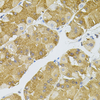
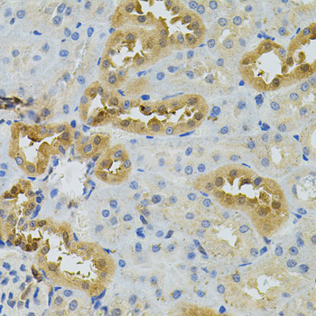
A08414 has real NAT8 IHC images from paraffin sections of mouse brain, rat kidney, and human stomach (catalog: A08414 IHC image captions). No IF data are supplied (catalog: A08414 IF images).

Real IHC data Immunohistochemistry of paraffin-embedded mouse brain using NAT8 antibody .Perform microwave antigen retrieval with 10 mM PBS buffer pH 7.2 before commencing with IHC staining protocol.
Anti-N-acetyltransferase 8 NAT8 Antibody
Cat # A08414

A08414 is listed for IHC in human, mouse, and rat samples (catalog: A08414 applications and reactivity). Its IHC images show paraffin sections of mouse brain, rat kidney, and human stomach (catalog: A08414 IHC image captions).

Which to pick: Choose A08414 for tissue IHC across human, mouse, and rat samples; its own captions document paraffin-section staining in all three species (catalog: A08414 reactivity and IHC image captions). No IF/ICC-validated SKU or IF dilution is supplied, and A08414’s clone is unreported (catalog: A08414 applications, IF dilution, and clone). Its captions specify microwave retrieval in 10 mM PBS at pH 7.2, but do not report the fixative (catalog: A08414 IHC image captions).

Each figure is that product's own IHC / IF validation image from its datasheet.

References

  1. UniProt Consortium. UniProt entry Q9UHE5 (NAT8_HUMAN, N-acetyltransferase 8).
  2. Human Protein Atlas. NAT8 tissue IHC expression (reliability: Supported).
  3. Human Protein Atlas. NAT8 subcellular location (ICC-IF): Localized to the endoplasmic reticulum. Caution: Based on antibodies targeting proteins from multiple genes..
  4. Human Protein Atlas. NAT8 antibody validation summary (2 antibodies).
  5. The role of N-acetyltransferase 8 in mesenchymal stem cell-based therapy for liver ischemia/reperfusion injury in rats. PloS one 2014 — PMC4109999.
  6. Squalene synthase predicts poor prognosis in stage I-III colon adenocarcinoma and synergizes squalene epoxidase to promote tumor progression. Cancer science 2022 — PMC8898704.
  7. PubMed PMID:9852678 — UniProt-cited evidence.
  8. PubMed PMID:11397015 — UniProt-cited evidence.
  9. PubMed PMID:15489334 — UniProt-cited evidence.