This website uses cookies to ensure you get the best experience on our website.


- Table of Contents
Plan chromogenic NCAPG IHC on paraffin sections using the catalog antibody at 2–5 μg/ml (datasheet A05501-1). Compare staining with the reported cytoplasmic tissue pattern, while accounting for its uncertain reliability (HPA tissue IHC).

Expected localisation, validated protocols, controls and antibodies — the at-a-glance facts below, then the full design guide.
| Expected localisation | Cytoplasmic staining in tissues (HPA tissue IHC) | |
| Staining pattern | Cytoplasmic staining across several tissue cell types (HPA tissue IHC) | |
| Antigen retrieval | EDTA pH 8.0 HIER, heat-mediated (datasheet A05501-1) | |
| Positive control | Cerebellum+4 more · see all | |
| Negative control | Adipose tissue+4 more · see all |
| Fixation | Keep paraffin-section fixation consistent (standard IHC practice; not target-specific); Matched tissue-IHC evidence does not establish the fixation claim. Validate the specimen-specific method before use. (selected-SKU IHC image A05501-1) | |
| Caveat | Presumed off-target staining; verification pending (HPA tissue IHC) | |
| Regulation | Mitotic chromatin association (UniProt) | |
| Isoform / epitope | No annotated isoforms; one 1–1015 chain (UniProt) |
The catalog antibody uses EDTA pH 8.0 heat retrieval (datasheet A05501-1). Published LUAD and glioma IHC methods provide two additional protocols (PMC8380402; PMC8906777).
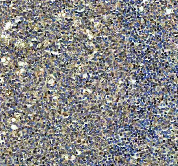
| Sample | Paraffin-embedded human tonsil tissue; fixative not specified (datasheet A05501-1) |
| Fixation | Image fixative and duration unreported (datasheet A05501-1); verify before use. |
| Sectioning | 4–5 µm sections on charged slides (standard) |
| Deparaffinisation | Xylene, graded ethanol series to water (standard) |
| Antigen retrieval | Heat retrieval: EDTA pH 8.0 (datasheet A05501-1); 20 min, 95–100 °C (standard) |
| Peroxidase block | 3% H2O2, 10 min, room temperature (standard) |
| Blocking | 10% goat serum (datasheet A05501-1) |
| Primary antibody | Rabbit anti-NCAPG, 2-5 μg/ml (datasheet A05501-1) |
| Primary incubation | Overnight at 4 °C (datasheet A05501-1) |
| Detection | HRP-conjugated secondary, DAB chromogen (datasheet A05501-1) |
| Counterstain | Hematoxylin, blue, dehydrate and mount (standard) |
| Expected result | NCAPG-positive staining in purkinje cells of cerebellum (HPA tissue IHC: High). HPA tissue profile: Cytoplasmic expression in several different tissues. No signal in the no-primary control. |
NCAPG is found in the cytoplasm, nucleus and chromosomes, with chromosome association increasing during mitosis (UniProt Q9BPX3). In tissue IHC, HPA reports cytoplasmic staining across several tissues, including high staining in esophageal squamous epithelial cells and lymph node germinal center cells (HPA tissue IHC). NCAPG has no transmembrane segment (UniProt Q9BPX3 topology). Treat the tissue pattern cautiously: HPA rates its IHC evidence Uncertain because presumed off-target binding was observed (HPA tissue IHC).
| Cell-associated cytoplasmic stain in esophageal squamous epithelial cells or lymph node germinal center cells (HPA tissue IHC). | These are reported high-staining populations, suitable for assessing whether the slide shows the reported tissue pattern (HPA tissue IHC). Cytoplasmic signal is also consistent with the predominantly cytoplasmic condensin complex in interphase cells (UniProt Q9BPX3). Neither match establishes antibody specificity, given HPA’s Uncertain IHC rating (HPA tissue IHC). |
| Stain concentrates on condensed chromosomes in cells with recognizable mitotic morphology (UniProt Q9BPX3). | Chromosome association is plausible during mitosis, when most condensin complex associates with chromatin; association falls in late telophase (UniProt Q9BPX3). Compare stained structures with the nuclear counterstain and cell morphology (general IHC practice). A predominantly cytoplasmic pattern in adjacent interphase cells can still fit the same protein’s cell-cycle behavior (UniProt Q9BPX3). |
| Sharp membrane-only staining or extracellular deposits dominate the section (UniProt Q9BPX3 topology). | This distribution does not fit the reported cytoplasmic, nuclear and chromosome locations or the absence of a transmembrane segment (UniProt Q9BPX3). Consider nonspecific staining or detection artifact before calling it NCAPG; HPA has already flagged presumed off-target binding (HPA tissue IHC). Check whether the signal follows cell boundaries, tissue edges or deposits (general IHC practice). |
| Strong staining appears in adipocytes or skeletal muscle myocytes (HPA tissue IHC). | HPA reports NCAPG as not detected in these cell populations, so strong signal conflicts with its reference IHC pattern (HPA tissue IHC). Cross-reactivity or endogenous detection activity is possible (general IHC practice). Because HPA’s own NCAPG IHC evidence is Uncertain, use this discrepancy as a prompt to check controls, not as proof of the cause (HPA tissue IHC). |
| Diffuse color covers tissue, empty spaces or the slide outside identifiable cells (general IHC practice). | This differs from the cell-associated cytoplasmic expression described by HPA (HPA tissue IHC). Review the no-primary and detection controls for background from the staining system, and assess blocking, washing and reagent concentration (general IHC practice). Diffuse cytoplasm within identifiable cells alone is not a failure criterion: cytoplasmic localization is reported for NCAPG (HPA tissue IHC; UniProt Q9BPX3). |
| Cell-cycle state (UniProt Q9BPX3). | Interphase condensin is mostly cytoplasmic, with a minority associated with chromatin or chromosome foci; during mitosis, most associates with chromatin (UniProt Q9BPX3). Interpret compartment alongside cell morphology rather than requiring every cell to show the same distribution (general IHC practice). |
| Tissue reference pattern (HPA tissue IHC). | HPA reports high staining in several cell populations, including esophageal squamous epithelial cells and lymph node germinal center cells, but not detected in adipocytes and skeletal muscle myocytes (HPA tissue IHC). These are reference observations, not universal guarantees for a new assay (HPA tissue IHC: Uncertain). |
| IHC evidence and antibody validation (HPA tissue IHC; HPA antibodies). | HPA assigns the tissue IHC profile an Uncertain reliability rating, notes presumed off-target binding, and lists HPA039613 as IHC Uncertain (HPA tissue IHC; HPA antibodies). Give concordant morphology and controls more weight than a single stained field (general IHC practice). |
| Topology and processing (UniProt Q9BPX3). | NCAPG has no transmembrane segment, signal peptide or annotated propeptide; its listed chain spans residues 1–1015 (UniProt Q9BPX3 topology and processing). These annotations support a non-surface interpretation. They do not identify an antibody epitope or establish how antigen retrieval affects staining (UniProt Q9BPX3). |
| ICC/IF evidence (HPA subcellular). | HPA provides no main ICC/IF location or cell-line images for NCAPG; it notes that transcript variation correlates with the cell cycle (HPA subcellular). The tissue IHC profile and UniProt localization can guide interpretation, but they do not establish an IF/ICC staining pattern or protocol (HPA tissue IHC; UniProt Q9BPX3). |
| Situation | Likely cause | Next action |
|---|---|---|
| No stain in esophageal squamous epithelial or lymph node germinal center cells (HPA tissue IHC). | The run may have failed, or the tested preparation may differ from HPA’s reported high IHC pattern (general IHC practice; HPA tissue IHC). | Confirm the cell population is present, then check the staining run with its positive control and review the antibody, detection and retrieval conditions used (general IHC practice). HPA’s Uncertain rating means a negative result alone cannot settle NCAPG abundance (HPA tissue IHC). |
| Staining is confined to cell borders or extracellular material (UniProt Q9BPX3 topology). | That location conflicts with NCAPG’s reported intracellular compartments and lack of a transmembrane segment (UniProt Q9BPX3). Nonspecific signal is possible (general IHC practice). | Compare with a no-primary control, inspect tissue edges and deposits, and reassess specificity before scoring the field (general IHC practice). |
| Adipocytes or skeletal muscle myocytes stain strongly (HPA tissue IHC). | HPA reports these populations as not detected; cross-reactivity or endogenous detection activity may contribute (HPA tissue IHC; general IHC practice). | Check cell identity and a no-primary control, then review the detection-system controls and compare with an HPA high-staining population on the same run where available (general IHC practice; HPA tissue IHC). |
| Color is widespread, including spaces without cells (general IHC practice). | Non-cell-associated background can arise from the staining system or insufficient washing (general IHC practice). | Review no-primary and detection controls, washing and blocking; score only signal tied to identifiable cells (general IHC practice). Keep true cytoplasmic staining in consideration because HPA reports it across tissues (HPA tissue IHC). |
| Some cells show cytoplasm while recognizable mitotic cells show chromosome-associated signal (UniProt Q9BPX3). | Condensin changes distribution through the cell cycle (UniProt Q9BPX3). | Assess each cell against morphology and counterstain before treating the compartment difference as assay inconsistency (general IHC practice). Do not infer antibody specificity from this match alone because HPA rates NCAPG IHC Uncertain (HPA tissue IHC). |
| Can an IF/ICC image be used to confirm the IHC pattern? | HPA lists no NCAPG ICC/IF images or main ICC/IF location (HPA subcellular). | Use the reported tissue IHC observations and UniProt cell-cycle localization as provisional context (HPA tissue IHC; UniProt Q9BPX3). Evaluate IF/ICC evidence in its separate guide; HPA supplies no image-based IF/ICC confirmation here (HPA subcellular). |
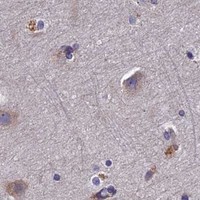


Comprehensive Human Protein Atlas IHC scoring per tissue (reliability: Uncertain — Presumed off target binding observed and disregarded. Pending external verification.). Rows are taken directly from the HPA tissue chart — click any row's HPA link to view the source.
| Tissue | Cell type | Level | Evidence | Source |
|---|---|---|---|---|
| Cerebellum | Purkinje cells | High | Protein (IHC) | HPA → |
| Esophagus | Squamous epithelial cells | High | Protein (IHC) | HPA → |
| Lymph node | Germinal center cells | High | Protein (IHC) | HPA → |
| Nasopharynx | Respiratory epithelial cells | High | Protein (IHC) | HPA → |
| Placenta | Trophoblastic cells | High | Protein (IHC) | HPA → |
| Tissue | Cell type | Level | Evidence | Source |
|---|---|---|---|---|
| Adipose tissue | Adipocytes | Not detected | Protein (IHC) | HPA → |
| Liver | Cholangiocytes | Not detected | Protein (IHC) | HPA → |
| Parathyroid gland | Glandular cells | Not detected | Protein (IHC) | HPA → |
| Seminal vesicle | Glandular cells | Not detected | Protein (IHC) | HPA → |
| Skeletal muscle | Myocytes | Not detected | Protein (IHC) | HPA → |
Troubleshoot NCAPG chromogenic IHC by checking retrieval, staining compartment, and tissue context; interpret tissue patterns cautiously because external verification is pending (HPA: reliability Uncertain).
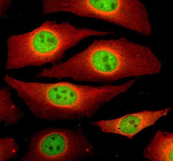
A05501-1 has IHC data from a human tonsil paraffin section and IF/ICC data from HeLa cells (catalog image captions); its listed reactivity is human (catalog).
A05501-1 is listed for human IHC and IF/ICC (catalog applications and reactivity). Its images show NCAPG staining in a human tonsil paraffin section by IHC and in HeLa cells by IF/ICC (catalog image captions).
Which to pick: Choose A05501-1 for paraffin-section IHC: its own tonsil image used EDTA retrieval at pH 8.0, 2 μg/ml primary antibody and DAB detection; the fixative is unreported (A05501-1 IHC image caption). Choose A05501-1 for IF/ICC at the listed 5 μg/ml; its own IF image shows staining in HeLa cells (catalog IF dilution and A05501-1 IF image caption). No cross-species option is documented here: A05501-1 lists human reactivity, and its clonality is unreported (catalog reactivity and clone field).