This website uses cookies to ensure you get the best experience on our website.


- Table of Contents
This guide outlines chromogenic IHC-P for NCBP2 using the catalog antibody at 2–5 μg/ml (datasheet A08031-2). Use its general nuclear tissue pattern and high- and low-staining cell populations to plan controls and interpret staining (HPA tissue IHC).

Expected localisation, validated protocols, controls and antibodies — the at-a-glance facts below, then the full design guide.
| Expected localisation | General nuclear tissue staining (HPA tissue IHC); cytoplasm annotated (UniProt) | |
| Staining pattern | Nuclei of glandular, epithelial and hematopoietic cells (HPA tissue IHC) | |
| Antigen retrieval | EDTA pH 8.0 HIER, heat-mediated (datasheet A08031-2) | |
| Positive control | Adrenal gland+4 more · see all | |
| Negative control | None in HPA (detected in all 45 tissues); use no-primary + isotype controls |
| Fixation | Keep paraffin-section fixation consistent across samples (standard IHC practice; not target-specific) | |
| Caveat | Broad nuclear staining limits negative tissue controls (HPA tissue IHC) | |
| Regulation | No specific expression regulator annotated (UniProt) | |
| Isoform / epitope | 3 isoforms; epitope coverage needs mapping (UniProt) |
The catalog antibody protocol is followed by two published NCBP2 IHC examples (PMC10195147; PMC11930219).
| Sample | Paraffin-embedded human bladder urothelial carcinoma tissue; fixative not specified (datasheet A08031-2) |
| Fixation | Image fixative and duration unreported (datasheet A08031-2); verify before use. |
| Sectioning | 4–5 µm sections on charged slides (standard) |
| Deparaffinisation | Xylene, graded ethanol series to water (standard) |
| Antigen retrieval | Heat retrieval: EDTA pH 8.0 (datasheet A08031-2); 20 min, 95–100 °C (standard) |
| Peroxidase block | 3% H2O2, 10 min, room temperature (standard) |
| Blocking | 10% goat serum (datasheet A08031-2) |
| Primary antibody | Rabbit anti-NCBP2, 2-5 μg/ml (datasheet A08031-2) |
| Primary incubation | Overnight at 4 °C (datasheet A08031-2) |
| Detection | HRP-conjugated secondary, DAB chromogen (datasheet A08031-2) |
| Counterstain | Hematoxylin, blue, dehydrate and mount (standard) |
| Expected result | NCBP2-positive staining in glandular cells of adrenal gland (HPA tissue IHC: High). HPA tissue profile: General nuclear expression. No signal in the no-primary control. |
NCBP2 is expected to show predominantly nuclear staining in paraffin sections, consistent with its nuclear cap-binding role and lack of a transmembrane segment (UniProt P52298: function, subcellular location, topology). HPA reports general nuclear expression, with high staining in several glandular, epithelial and hematopoietic cell populations (HPA: tissue IHC). Treat the pattern as supported with medium consistency between staining and RNA data (HPA: reliability).
| Clear nuclear staining in colon glandular cells or bone marrow hematopoietic cells. | This matches reported high staining in those cell populations and the general nuclear pattern (HPA: tissue IHC). Judge localization within identifiable cells, since tissue-wide color alone cannot establish a nuclear result (standard IHC practice). |
| Predominantly membrane-like or extracellular staining, with little nuclear signal. | This conflicts with the reported general nuclear IHC pattern and with a protein lacking a transmembrane segment (HPA: tissue IHC; UniProt P52298: topology). Review morphology, background and controls before calling it specific staining (standard IHC practice). |
| Strong staining appears mainly in cells expected to have low staining, while a reported high-staining population is weak. | Consider cross-reactivity or endogenous detection activity (standard IHC practice). HPA calls parathyroid glandular cells, cardiomyocytes, smooth muscle cells and adipocytes low, not absent; a single discrepant section cannot prove an off-target reaction (HPA: tissue IHC). |
| Brown signal spreads across cells and tissue spaces without clear nuclear boundaries. | Diffuse background limits interpretation of the expected nuclear pattern (HPA: tissue IHC; standard IHC practice). Check the negative control and detection steps before scoring nuclei; broad color deposition alone does not identify NCBP2-positive cells (standard IHC practice). |
| No nuclear signal appears in a section containing a reported high-staining cell population. | First check tissue identity, preservation and the assay controls (standard IHC practice). HPA reports high staining in bronchial respiratory epithelial cells and esophageal squamous epithelial cells, among others, but its supported rating is not a guarantee for every specimen (HPA: tissue IHC, reliability). |
| Tissue and cell selection | HPA reports high staining in adrenal, cervical, colonic, duodenal and epididymal glandular cells, plus bone marrow hematopoietic, bronchial respiratory epithelial and esophageal squamous epithelial cells (HPA: tissue IHC). Its listed low-staining cells are comparison tissue, not established negative controls (HPA: tissue IHC). |
| Strength of IHC evidence | The tissue pattern is rated Supported, with medium consistency between antibody staining and RNA expression (HPA: tissue IHC). HPA044850 has Supported IHC status; the supplied antibody list gives no IHC status for HPA062483 (HPA: antibodies). Interpret disagreement as a reason to verify the assay, not proof of absence (standard IHC practice). |
| Isoforms and epitope coverage | UniProt lists three NCBP2 isoforms and an RNA recognition motif at residues 40–118 (UniProt P52298: isoforms, domains). No antibody epitope or isoform coverage is supplied, so this record cannot predict isoform-specific staining or an antigen-retrieval requirement. |
| Detection background | For chromogenic IHC, endogenous activity and nonspecific detection can create color unrelated to the primary antibody (standard IHC practice). Use appropriate reagent controls when unexplained staining competes with the reported nuclear pattern (HPA: tissue IHC; standard IHC practice). |
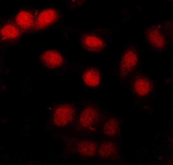
| IF/ICC Q: What localization should I expect? | A: Mainly nucleoplasmic, with additional cytosol and basal body localization (HPA: subcellular ICC-IF). HPA044850 and HPA062483 have Supported ICC status (HPA: antibodies). These ICC-IF observations inform localization; they do not establish an IHC-P protocol or require visible basal bodies in tissue sections. |
| Situation | Likely cause | Next action |
|---|---|---|
| Reported high-staining tissue has no clear nuclear signal. | The section may lack the relevant cell population, or the staining run may have failed (HPA: tissue IHC; standard IHC practice). | Confirm cell identity and inspect a known-positive section and run controls; then review the IHC-validated antibody's supplied IHC-P instructions and the performed steps (standard IHC practice). |
| Signal is mostly cytoplasmic rather than nuclear. | Some cytoplasmic localization is annotated (UniProt P52298: subcellular location; HPA: ICC-IF), but cytoplasm-dominant tissue staining departs from HPA's general nuclear IHC pattern (HPA: tissue IHC). | Compare nuclear and cytoplasmic signal in identifiable cells, assess the negative control, and avoid scoring cytoplasm alone as a definitive IHC positive (standard IHC practice). |
| Membrane-like or extracellular color dominates the section. | The compartment conflicts with the general nuclear IHC profile; NCBP2 has no annotated transmembrane segment (HPA: tissue IHC; UniProt P52298: topology). Background or cross-reactivity remains possible (standard IHC practice). | Check section morphology and primary-antibody negative controls, then reassess detection background before interpreting the color as target staining (standard IHC practice). |
| Unexpectedly strong color occurs in an HPA low-staining cell population. | Low HPA staining does not mean zero expression; cross-reactivity or endogenous detection activity is also possible (HPA: tissue IHC; standard IHC practice). | Compare with a reported high-staining cell population in the same run and use appropriate primary-antibody and detection controls to investigate the discrepancy (HPA: tissue IHC; standard IHC practice). |
| Color is diffuse across the section or obscures nuclei. | High background can prevent compartment-level interpretation (standard IHC practice). The supplied NCBP2 sources do not identify a target-specific fixation effect. | Check blocking, washing, detection and counterstain balance against the assay instructions; score only cells whose nuclear boundaries remain interpretable (standard IHC practice). |
| An IF/ICC image shows cytosolic or basal body signal that differs from the IHC slide. | HPA reports those additional ICC-IF locations while describing general nuclear tissue IHC staining (HPA: subcellular ICC-IF, tissue IHC). | Assess each application against its own reported pattern; for this paraffin IHC result, prioritize identifiable nuclear staining and the tissue IHC controls (HPA: tissue IHC; standard IHC practice). |
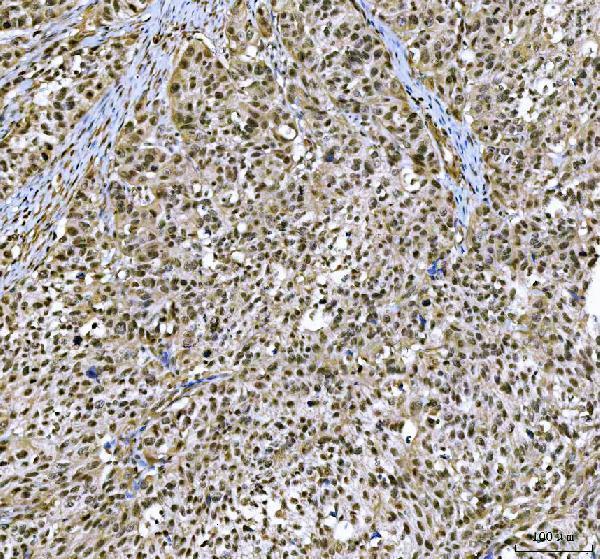
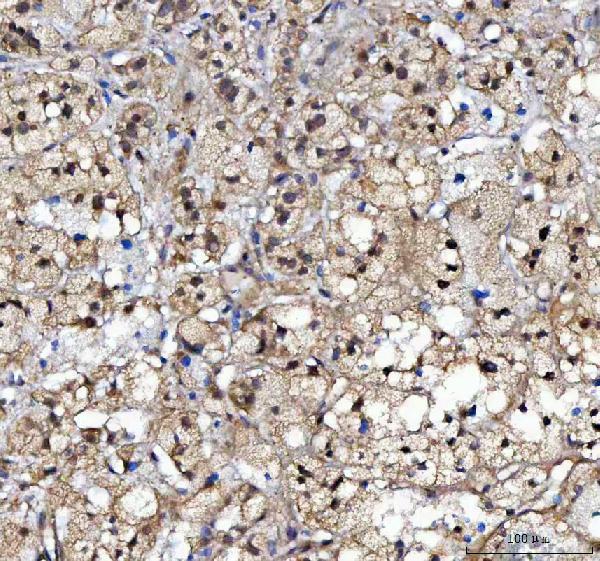


Comprehensive Human Protein Atlas IHC scoring per tissue (reliability: Supported — Medium consistency between antibody staining and RNA expression data.). Rows are taken directly from the HPA tissue chart — click any row's HPA link to view the source.
| Tissue | Cell type | Level | Evidence | Source |
|---|---|---|---|---|
| Adrenal gland | Glandular cells | High | Protein (IHC) | HPA → |
| Bone marrow | Hematopoietic cells | High | Protein (IHC) | HPA → |
| Bronchus | Respiratory epithelial cells | High | Protein (IHC) | HPA → |
| Cervix | Glandular cells | High | Protein (IHC) | HPA → |
| Colon | Glandular cells | High | Protein (IHC) | HPA → |
| Tissue | Cell type | Level | Evidence | Source |
|---|---|---|---|---|
| None in HPA: NCBP2 is detected in all 45 scored tissues. Use a no-primary (secondary-only) and an isotype control instead. | ||||
Troubleshoot NCBP2 staining in paraffin sections by checking nuclear localisation, retrieval conditions, background controls and cell-specific scoring (UniProt P52298; HPA tissue IHC).
A08031-2 has IHC images of human paraffin sections and an IF/ICC image of A549 cells (A08031-2 image captions). Its stated reactivity includes human, mouse, and rat (catalog reactivity).
A08031-2 has IHC images from human bladder urothelial carcinoma and adrenocortical adenoma paraffin sections (A08031-2 IHC captions). Its IF/ICC image uses A549 cells (A08031-2 IF caption).
Which to pick: For tissue IHC, choose A08031-2: its IHC captions document EDTA retrieval at pH 8.0 and 2 μg/ml primary antibody on human paraffin sections; the fixative is unreported (A08031-2 IHC captions). For IF/ICC, A08031-2 has an A549 cell image using 5 μg/ml primary antibody (A08031-2 IF caption). For mouse or rat studies, A08031-2 is listed as reactive, but its supplied IHC and IF images show human material (catalog reactivity; A08031-2 image captions).