This website uses cookies to ensure you get the best experience on our website.


- Table of Contents
Plan paraffin-section NR1D1 IHC with a catalog antibody dilution of 2–5 μg/ml (datasheet A01077-2). Interpret cytoplasmic staining in light of the tissue profile’s uncertain reliability (HPA tissue IHC).

Expected localisation, validated protocols, controls and antibodies — the at-a-glance facts below, then the full design guide.
| Expected localisation | Cytoplasmic tissue staining (HPA tissue IHC) | |
| Staining pattern | Cytoplasmic signal across most tissues (HPA tissue IHC) | |
| Antigen retrieval | EDTA pH 8.0 HIER, heat-mediated (datasheet A01077-2) | |
| Positive control | Adrenal gland+4 more · see all | |
| Negative control | Adipose tissue+4 more · see all |
| Fixation | Keep fixation consistent across sections. (standard IHC practice; not target-specific) | |
| Caveat | Staining has low consistency with RNA data (HPA tissue IHC) | |
| Regulation | Diurnal expression oscillation (UniProt) | |
| Isoform / epitope | No annotated isoforms; one full-length chain (UniProt) |
The catalog antibody’s IHC protocol and these four published NR1D1 IHC protocols provide starting conditions for paraffin sections (datasheet A01077-2; PMC6883674; PMC10531686; PMC12339034; PMC5438901).
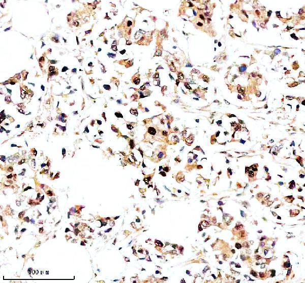
| Sample | Paraffin-embedded human breast cancer tissue; fixative not specified (datasheet A01077-2) |
| Fixation | Image fixative and duration unreported (datasheet A01077-2); verify before use. |
| Sectioning | 4–5 µm sections on charged slides (standard) |
| Deparaffinisation | Xylene, graded ethanol series to water (standard) |
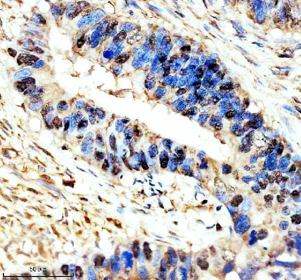
| Antigen retrieval | Heat retrieval: EDTA pH 8.0 (datasheet A01077-2); 20 min, 95–100 °C (standard) |
| Peroxidase block | 3% H2O2, 10 min, room temperature (standard) |
| Blocking | 10% goat serum (datasheet A01077-2) |
| Primary antibody | Rabbit anti-NR1D1, 2-5 μg/ml (datasheet A01077-2) |
| Primary incubation | Overnight at 4 °C (datasheet A01077-2) |
| Detection | HRP-conjugated secondary, DAB chromogen (datasheet A01077-2) |
| Counterstain | Hematoxylin, blue, dehydrate and mount (standard) |
| Expected result | NR1D1-positive staining in glandular cells of adrenal gland (HPA tissue IHC: Medium). HPA tissue profile: Cytoplasmic expression most tissues. No signal in the no-primary control. |
NR1D1 can localize to the nucleus and cytoplasm, with context-dependent distribution (UniProt P20393: subcellular location). In paraffin tissue IHC, HPA reports predominantly cytoplasmic staining across tissues, including medium staining in several glandular and endothelial cell populations (HPA: tissue IHC). Interpret that pattern cautiously: HPA rates its tissue staining Uncertain because antibody staining and RNA expression have low consistency (HPA: tissue IHC reliability). NR1D1 has no transmembrane segment (UniProt P20393: topology).
| Cytoplasmic staining in adrenal, appendix, duodenal or endometrial glandular cells, or colon endothelial cells. | This resembles HPA’s reported medium staining in those cell populations and its predominantly cytoplasmic tissue pattern (HPA: tissue IHC). Record the stained cell type and compartment. The resemblance supports interpretation but does not confirm specificity: the tissue IHC assessment remains Uncertain (HPA: tissue IHC reliability). |
| Nuclear staining, or a prominent membranous or extracellular outline. | Nuclear staining can fit NR1D1 biology; UniProt lists the nucleus, and HPA supports nuclear bodies in ICC-IF (UniProt P20393: subcellular location; HPA: subcellular ICC-IF). A membrane or extracellular outline is less consistent with a protein lacking a transmembrane segment (UniProt P20393: topology). Check controls before calling it artefact (general IHC practice). |
| Strong staining in cells listed by HPA as not detected, such as adipocytes or ovarian stromal cells. | This conflicts with those HPA tissue IHC observations and raises concern for cross-reactivity or endogenous detection activity (HPA: tissue IHC; general IHC practice). It is not decisive by itself: HPA’s tissue assessment is Uncertain, and UniProt reports high expression in adipose tissue without resolving staining in individual adipocytes (HPA: tissue IHC reliability; UniProt P20393: tissue specificity). |
| Diffuse color across tissue structures, with little distinction between cells. | A uniform deposit is difficult to assign to the reported glandular or endothelial populations and may reflect background from the staining workflow (HPA: tissue IHC; general IHC practice). Compare the no-primary control, review blocking and detection steps, and judge any remaining signal by its cellular distribution (general IHC practice). |
| No staining in an adrenal gland or appendix section expected to provide a positive comparison. | HPA reports medium staining in glandular cells in both tissues, so a blank result calls for a workflow and specimen check (HPA: tissue IHC; general IHC practice). A negative section alone does not establish absence of NR1D1: tissue staining is rated Uncertain, and UniProt reports diurnal variation in expression (HPA: tissue IHC reliability; UniProt P20393: tissue specificity). |
| IHC compartment versus IF/ICC: what should IF/ICC show? | HPA describes cytoplasmic expression in most tissue IHC samples but supports localization to nuclear bodies in ICC-IF; UniProt lists both nuclear and cytoplasmic locations (HPA: tissue IHC and subcellular ICC-IF; UniProt P20393: subcellular location). Assess the two applications against their respective observations rather than requiring identical patterns. |
| Time and cellular context | UniProt reports predominantly nuclear localization at ZT8 and cytoplasmic localization at ZT20, alongside diurnal expression in the suprachiasmatic nucleus and peripheral tissues (UniProt P20393: subcellular location and tissue specificity). Where collection time is known, keep it with the staining record; these statements do not predict an IHC intensity at a particular time. |
| Confidence in tissue comparisons | HPA rates tissue IHC Uncertain because staining and RNA expression have low consistency; the listed antibody’s IHC status is also Uncertain (HPA: tissue IHC reliability and antibody validation). Treat medium and not-detected entries as observed comparisons, not definitive positive or negative standards. |
| Topology and processing | UniProt lists no transmembrane segment, signal peptide or propeptide, and one chain spanning residues 1–614 (UniProt P20393: topology and processing). This supports scrutiny of an isolated membrane pattern; the supplied record does not establish epitope accessibility or a target-specific retrieval requirement. |
| Situation | Likely cause | Next action |
|---|---|---|
| Expected positive tissue is blank or very faint. | The staining run may have failed, or the section may differ from HPA’s observed medium-staining example (HPA: tissue IHC; general IHC practice). | Check section integrity, retrieval and detection run controls, then compare another HPA-listed medium-staining cell population if available (HPA: tissue IHC; general IHC practice). Do not infer a target-specific retrieval condition from these sources. |
| Signal appears mainly on membranes or outside cells. | An isolated surface pattern is hard to reconcile with NR1D1’s lack of a transmembrane segment (UniProt P20393: topology). | Inspect tissue morphology and no-primary controls; review detection background before scoring the outline as NR1D1 (general IHC practice). Nuclear signal alone should be assessed separately because UniProt also lists nuclear localization (UniProt P20393: subcellular location). |
| The whole section has diffuse chromogenic color. | Nonspecific background or endogenous detection activity can obscure cellular localization (general IHC practice). | Compare a no-primary control and review blocking, washing and detection conditions (general IHC practice). Score only signal that can be assigned to cells and compartments; HPA’s tissue pattern is predominantly cytoplasmic (HPA: tissue IHC). |
| A purported negative cell population stains strongly. | The result differs from HPA’s not-detected observations, but those observations carry an Uncertain reliability rating (HPA: tissue IHC). | Check the exact cell type, no-primary control and nearby tissue morphology before attributing the signal to NR1D1 (general IHC practice). For adipocytes, note the distinction between HPA’s not-detected IHC result and UniProt’s adipose tissue expression statement (HPA: tissue IHC; UniProt P20393: tissue specificity). |
| Nuclear and cytoplasmic proportions differ between specimens. | NR1D1 localization can vary with context and circadian time; UniProt reports nuclear predominance at ZT8 and cytoplasmic localization at ZT20 (UniProt P20393: subcellular location). | Record compartment, cell type and collection time when known, and compare matched specimens and staining runs (general IHC practice). Do not diagnose a technical failure from this difference alone (UniProt P20393: subcellular location). |
| An IHC result differs from an IF/ICC image. | HPA reports a predominantly cytoplasmic tissue IHC profile and supported nuclear-body localization in ICC-IF (HPA: tissue IHC and subcellular ICC-IF). | Interpret each result in its application and cellular context, while retaining HPA’s Uncertain tissue IHC rating (HPA: tissue IHC reliability and subcellular ICC-IF). |


Comprehensive Human Protein Atlas IHC scoring per tissue (reliability: Uncertain — Low consistency between antibody staining and RNA expression data. Pending external verification.). Rows are taken directly from the HPA tissue chart — click any row's HPA link to view the source.
| Tissue | Cell type | Level | Evidence | Source |
|---|---|---|---|---|
| Adrenal gland | Glandular cells | Medium | Protein (IHC) | HPA → |
| Appendix | Glandular cells | Medium | Protein (IHC) | HPA → |
| Bone marrow | Hematopoietic cells | Medium | Protein (IHC) | HPA → |
| Bronchus | Respiratory epithelial cells | Medium | Protein (IHC) | HPA → |
| Cerebellum | Cells in granular layer | Medium | Protein (IHC) | HPA → |
| Tissue | Cell type | Level | Evidence | Source |
|---|---|---|---|---|
| Adipose tissue | Adipocytes | Not detected | Protein (IHC) | HPA → |
| Breast | Adipocytes | Not detected | Protein (IHC) | HPA → |
| Ovary | Ovarian stroma cells | Not detected | Protein (IHC) | HPA → |
| Salivary gland | Glandular cells | Not detected | Protein (IHC) | HPA → |
| Smooth muscle | Smooth muscle cells | Not detected | Protein (IHC) | HPA → |
Troubleshoot NR1D1 staining in paraffin sections by checking retrieval, compartment, controls and scoring before interpreting differences between specimens.
The catalog lists human, mouse and rat reactive NR1D1 antibodies for IHC and IF/ICC; the supplied image evidence covers human paraffin section IHC only (catalog applications/reactivity; A01077-2 IHC captions).
A01077-2 has IHC images from human breast cancer, colon cancer and colon paraffin sections; its listed IHC dilution is 2–5 μg/ml for human samples (A01077-2 IHC captions; catalog dilution). M01077-1 lists IHC and IF/ICC applications and human, mouse and rat reactivity, but has no supplied IHC or IF image (M01077-1 catalog applications/reactivity; image fields).
Which to pick: For tissue IHC, choose A01077-2 when a documented human paraffin section example matters; its captions describe EDTA retrieval at pH 8.0, 2 μg/ml primary antibody and DAB detection, with the fixative unreported (A01077-2 IHC captions). For IF/ICC, M01077-1 is the listed option because it is a rabbit monoclonal with IF/ICC among its applications, although no IF image is supplied (M01077-1 catalog clone/applications; image fields). Both list human, mouse and rat reactivity, but the supplied tissue images document only A01077-2 in human paraffin sections (catalog reactivity; A01077-2 IHC captions).