NSDHL / Sterol-4-alpha-carboxylate 3-dehydrogenase, decarboxylating · IHC design guide

Design Immunohistochemistry for NSDHL

Plan NSDHL chromogenic IHC in paraffin sections using cytoplasmic staining and strongly staining hepatocytes as references (HPA tissue IHC). This guide covers fixation, detection and scoring for a protein annotated at the ER membrane and lipid droplets (UniProt).

Evidence assembled Oct 2026 · For research use; verify linked source records and product datasheet before use
Immunohistochemistry protocol sheet for NSDHL (IHC for NSDHL): expected localisation Cytoplasmic in tissue (HPA tissue IHC); ER membrane and lipid droplet at protein level (UniProt), antibody A04902-1, validated IHC image, and IHC protocol steps
Printable NSDHL IHC protocol sheet — expected localisation Cytoplasmic in tissue (HPA tissue IHC); ER membrane and lipid droplet at protein level (UniProt), antibody A04902-1, controls and protocol steps. Open the full NSDHL IHC guide →

NSDHL Immunohistochemistry Experimental Design Guide

Expected localisation, validated protocols, controls and antibodies — the at-a-glance facts below, then the full design guide.

Must know before staining
Expected localisation Cytoplasmic in tissue (HPA tissue IHC); ER membrane and lipid droplet at protein level (UniProt)
Staining pattern Cytoplasmic staining across tissues (HPA tissue IHC)
Antigen retrieval EDTA pH 8.0 HIER, heat-mediated (datasheet A04902-1)
Positive control ⓘ Epididymis+4 more · see all
Negative control ⓘ None in HPA (detected in all 45 tissues); use no-primary + isotype controls
Important caveats
Reasons your staining may differ from the expected pattern.
Fixation Keep fixation consistent across sections. (standard IHC practice; not target-specific)
Caveat High in hepatocytes (HPA tissue IHC); endogenous peroxidase may raise DAB background (standard IHC practice)
Regulation No staining regulator specified (UniProt)
Isoform / epitope No annotated isoforms; one 1–373 chain (UniProt)
Section 1

Recommended NSDHL IHC & IF Protocols

The catalog antibody uses EDTA pH 8.0 heat retrieval (datasheet A04902-1). Published IHC protocols below cover gastric cancer, breast cancer, and mouse tissues (PMC7891516; PMC7197182; PMC2783206).

Recommended immunohistochemistry (IHC-P) protocol parameters
SampleParaffin-embedded human liver cancer tissue; fixative not specified (datasheet A04902-1)
FixationImage fixative and duration unreported (datasheet A04902-1); verify before use.
Sectioning4–5 µm sections on charged slides (standard)
DeparaffinisationXylene, graded ethanol series to water (standard)
Antigen retrievalHeat retrieval: EDTA pH 8.0 (datasheet A04902-1); 20 min, 95–100 °C (standard)
Peroxidase block3% H2O2, 10 min, room temperature (standard)
Blocking10% goat serum (datasheet A04902-1)
Primary antibodyRabbit anti-NSDHL, 2-5 μg/ml (datasheet A04902-1)
Primary incubationOvernight at 4 °C (datasheet A04902-1)
DetectionHRP-conjugated secondary, DAB chromogen (datasheet A04902-1)
CounterstainHematoxylin, blue, dehydrate and mount (standard)
Expected resultNSDHL-positive staining in glandular cells of epididymis (HPA tissue IHC: High). HPA tissue profile: Cytoplasmic expression in all tissues. No signal in the no-primary control.
💡Decision noteStart with EDTA pH 8.0 heat retrieval for the catalog antibody (datasheet A04902-1). Use each published protocol’s stated retrieval conditions when reproducing that study (PMC7197182; PMC2783206).
Section 2

What Is the Expected NSDHL Staining Pattern?

NSDHL is an ER membrane protein also found at lipid droplets (UniProt Q15738; HPA subcellular). In paraffin IHC, expect cytoplasmic staining, including strong signal in hepatocytes, Leydig cells, esophageal squamous epithelium, and epididymal glandular cells (HPA tissue IHC). Its single transmembrane segment spans residues 298–318 (UniProt Q15738 topology). HPA rates the tissue pattern Enhanced, with medium consistency between antibody staining and RNA expression (HPA tissue IHC).

What am I looking at on my slide?
Cytoplasmic staining is strong in liver hepatocytes or testis Leydig cells.This matches the cell type and High staining level reported for each tissue (HPA tissue IHC). Judge signal against a reagent negative control and surrounding tissue; HPA intensity categories are reference observations, not fixed scoring thresholds (general IHC practice; HPA tissue IHC).
Signal appears mainly nuclear, extracellular, or confined to cell borders.That distribution does not fit the cytoplasmic tissue pattern (HPA tissue IHC) or ER membrane and lipid droplet locations (UniProt Q15738; HPA subcellular). Check morphology and controls before assigning it to NSDHL; chromogenic IHC alone does not resolve ER membranes or lipid droplets (general IHC practice).
An unexpected cell population stains much more strongly than nearby expected cells.Compare the exact cell type with HPA observations: cerebral cortex endothelial cells and parathyroid glandular cells, for example, are Low (HPA tissue IHC). Unexpected intensity can prompt a cross-reactivity or detection-background check, but HPA reports cytoplasmic expression across tissues and supplies no negative tissue here (HPA tissue IHC).
Brown color spreads across tissue, stroma, or spaces without a clear cellular pattern.Treat this as possible detection background rather than a positive NSDHL call (general IHC practice). HPA describes cytoplasmic expression in cells (HPA tissue IHC); compare a reagent negative control and inspect whether deposits follow tissue structure before scoring (general IHC practice).
A known high-staining cell type shows no detectable signal.Hepatocytes and Leydig cells are High HPA reference populations (HPA tissue IHC). First check tissue identity, staining run controls, antibody application, retrieval, and detection steps (general IHC practice). An absent result alone does not establish loss of NSDHL, and no target-specific fixation sensitivity is reported in the supplied sources.
💡Expected NSDHL appearanceCall a positive result when identifiable hepatocytes or Leydig cells show clear, predominantly cytoplasmic chromogenic staining near the High HPA reference pattern; isolated nuclear, extracellular, or structureless brown signal is suspect (HPA tissue IHC; UniProt Q15738; general IHC practice).
How each factor affects the staining
Cell and tissue referenceHPA reports cytoplasmic expression across tissues and low RNA tissue specificity; High staining is documented in hepatocytes, Leydig cells, esophageal squamous epithelial cells, and epididymal glandular cells, while several other listed cell populations are Medium or Low (HPA tissue IHC). Use the named cell population when comparing slides, rather than treating an entire organ as uniformly positive.
Membrane topology and epitope uncertaintyNSDHL has one transmembrane segment at residues 298–318 and is assigned to the ER membrane and lipid droplets (UniProt Q15738). The supplied evidence does not locate the antibody epitope or show how retrieval changes its accessibility; topology alone cannot predict an optimal retrieval condition (UniProt Q15738 topology; general IHC practice).
Antibody evidenceBoth listed rabbit polyclonal antibodies, HPA000248 and HPA000571, have Enhanced IHC and ICC validation status (HPA antibodies). HPA also rates the tissue IHC profile Enhanced but describes only medium staining–RNA consistency (HPA tissue IHC). These ratings support comparison with HPA patterns; they do not validate every unexpected cell type or staining condition.
Chromogenic detection backgroundEndogenous enzyme activity or nonspecific reagent binding can contribute color in chromogenic IHC (general IHC practice). A matched reagent negative control and the detection system's recommended blocking steps help distinguish background from cell-associated staining (general IHC practice); neither UniProt nor HPA identifies an NSDHL-specific blocking requirement.
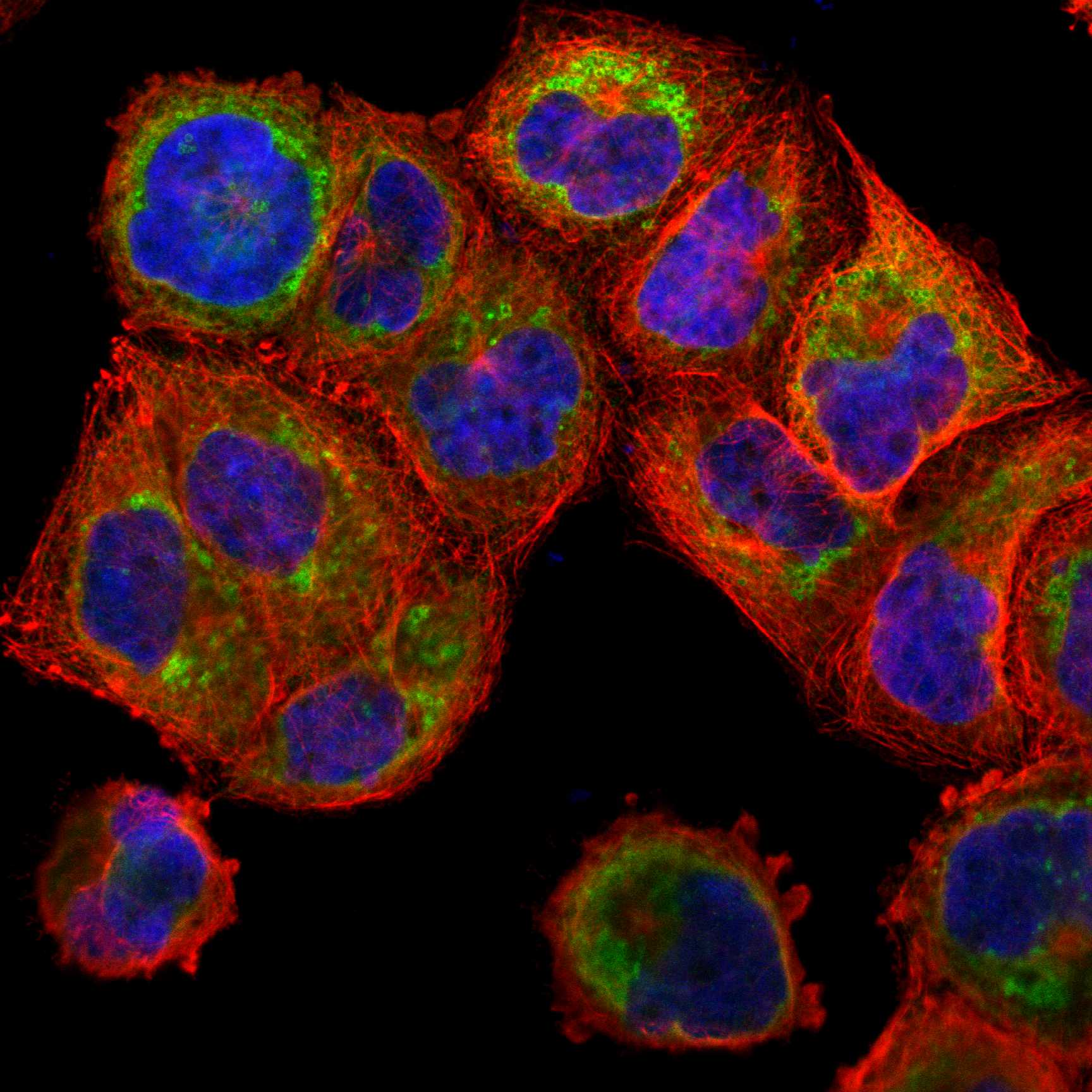
IF/ICC: where should NSDHL appear?HPA places the main IF/ICC signal in the ER, with additional lipid droplet localization; its subcellular assessment is Enhanced (HPA subcellular). UniProt likewise assigns ER membrane and lipid droplet locations (UniProt Q15738). This is the expected IF/ICC localization; the organelle-level pattern should not be demanded of routine chromogenic tissue IHC (general IHC practice).
Why is my staining missing, weak or wrong?
SituationLikely causeNext action
No signal in liver hepatocytes or testis Leydig cellsA high-staining HPA reference population is absent, but the observation alone cannot identify a failed step or biological loss (HPA tissue IHC).Confirm the cell type and run control, then check antibody application, retrieval, detection reagents, and counterstain visibility (general IHC practice). Record any changes before repeating; the supplied sources specify no NSDHL retrieval setting.
Predominantly nuclear or extracellular stainingThe compartment conflicts with HPA cytoplasmic IHC and the ER membrane or lipid droplet assignments (HPA tissue IHC; UniProt Q15738). Background or cross-reactivity is possible (general IHC practice).Inspect section morphology and the reagent negative control; compare a known high-staining cell population before interpreting the unusual compartment as NSDHL (HPA tissue IHC; general IHC practice).
Uniform brown haze obscures cellular boundariesDiffuse chromogenic deposit can arise from detection background, including endogenous activity or nonspecific reagent binding (general IHC practice).Check reagent negative controls and the detection system's blocking and wash steps, then score only clearly cell-associated staining (general IHC practice; HPA tissue IHC: cytoplasmic expression).
Strong signal in a population listed as Low by HPAA mismatch may reflect cross-reactivity, background, or a context difference; HPA Low is not a formal negative designation (HPA tissue IHC).Verify the exact cell type and compare staining with an HPA High reference and reagent negative control (HPA tissue IHC; general IHC practice). Report the discrepancy instead of treating it as established NSDHL expression.
Uneven staining across a sectionAn inconsistent chromogenic run or uneven tissue condition can complicate interpretation (general IHC practice); the supplied UniProt and HPA records do not establish an NSDHL-specific fixation effect.Compare affected and unaffected areas, inspect section integrity and run controls, and repeat the stain if the pattern prevents cell-level scoring (general IHC practice).
A reagent negative control also develops brown colorThe control implicates detection background rather than a reliable NSDHL-specific positive call (general IHC practice).Review endogenous-activity blocking, detection reagents, and washes under the detection system's instructions; resolve control staining before comparing intensities with HPA tissue references (general IHC practice; HPA tissue IHC).

Sample controls for NSDHL IHC & IF

🧪Run liver first and expect hepatocyte staining (HPA: High in hepatocytes). HPA detects NSDHL in all 45 scored tissues, so there is no supported negative tissue; use no-primary and isotype controls, and regard cells without specific chromogen on the positive slide as internal background references rather than proven NSDHL-negative cells (HPA: detected in all 45 scored tissues; standard IHC practice).
Positive control tissue: Epididymis (Glandular cells, HPA High)
Negative control tissue: None in HPA: NSDHL is detected in all 45 scored tissues. Use a no-primary (secondary-only) and an isotype control instead.
ICC-IF cell lines (HPA subcellular resource): HPA ICC-IF images show NSDHL in A-431, U-251MG, U2OS, with annotated localisation: Endoplasmic reticulum (enhanced) (HPA subcellular).
Technical controls: Include a no-primary, secondary-only slide and a concentration-matched rabbit IgG isotype control for the rabbit primary; a validated NSDHL knockout provides a biological specificity control (caption: rabbit anti-NSDHL; standard IHC practice). For liver sections, quench endogenous peroxidase before HRP/DAB detection and assess residual background on the no-primary slide (caption: liver tissue and HRP/DAB detection; standard IHC practice).
⚠️Feasibility: The selected A04902-1 paraffin-section caption reports heat-mediated EDTA retrieval at pH 8.0 and 2 μg/ml primary antibody overnight at 4°C, but its fixative is unreported; no target-specific fixation window or fixation effect is reported in the supplied evidence (A04902-1 tissue-IHC caption). Retrieval is therefore documented for this example, while the evidence does not establish whether frozen sections or IF are easier; ICC-IF images support evaluating ER and lipid-droplet localization if IF is pursued separately (A04902-1 tissue-IHC caption; HPA subcellular: ER and lipid droplets). Liver HRP/DAB interpretation needs the endogenous-peroxidase control described above (caption: liver tissue and HRP/DAB detection; standard IHC practice).

HPA tissue IHC evidence for NSDHL

Comprehensive Human Protein Atlas IHC scoring per tissue (reliability: Enhanced — Medium consistency between antibody staining and RNA expression data.). Rows are taken directly from the HPA tissue chart — click any row's HPA link to view the source.

Positive expression · recommended positive controls

TissueCell typeLevelEvidenceSource
Epididymis Glandular cells High Protein (IHC) HPA →
Esophagus Squamous epithelial cells High Protein (IHC) HPA →
Liver Hepatocytes High Protein (IHC) HPA →
Testis Leydig cells High Protein (IHC) HPA →
Adrenal gland Glandular cells Medium Protein (IHC) HPA →

Undetected expression · recommended negative controls

TissueCell typeLevelEvidenceSource
None in HPA: NSDHL is detected in all 45 scored tissues. Use a no-primary (secondary-only) and an isotype control instead.
Section 3

Advanced NSDHL IHC Tips

Troubleshoot NSDHL staining in paraffin sections using the documented IHC conditions, expected cytoplasmic pattern, and compartment-specific controls.

Where should I start if NSDHL staining is weak after antigen retrieval?
Start with heat-mediated retrieval in EDTA at pH 8.0 for paraffin-section IHC (datasheet A04902-1). The documented liver cancer section then received 2 μg/mL primary antibody overnight at 4°C (caption A04902-1). Keep section thickness, heating, cooling, and detection consistent while comparing retrieval runs (general IHC practice). If staining remains weak, first confirm that a parallel liver section shows staining in hepatocytes, a high-expression cell population (HPA: High in hepatocytes). Evaluate any alternative retrieval buffer only as a controlled fallback, because the selected image documents EDTA at pH 8.0 (caption A04902-1).
How can I troubleshoot variable staining when the tissue fixative is unknown?
The selected NSDHL image documents a paraffin-embedded human liver cancer section, but its fixative is unreported (caption A04902-1). Target-specific sensitivity to fixation is therefore unknown; neither tissue expression nor membrane topology establishes a fixation effect (HPA: tissue IHC; UniProt Q15738 topology). Record the actual fixative and processing history for each specimen, then compare sections processed under matched conditions (general IHC practice). Keep the documented EDTA retrieval at pH 8.0 and 2 μg/mL primary concentration constant during that comparison (caption A04902-1). Include a similarly processed liver control to help distinguish specimen processing variation from an assay-wide failure (HPA: High in hepatocytes; general IHC practice).
What staining pattern should count as plausible NSDHL localisation in tissue?
Expect predominantly cytoplasmic chromogenic staining across tissues, with stronger hepatocyte staining among the reported examples (HPA: cytoplasmic expression in all tissues; High in hepatocytes). At finer scale, NSDHL is associated mainly with the endoplasmic reticulum and additionally with lipid droplets (HPA: subcellular localisation, enhanced). Its 298–318 transmembrane segment supports interpreting a cytoplasmic compartment pattern rather than a nuclear-only pattern (UniProt Q15738 topology; HPA: tissue IHC). Review staining against cell boundaries and a matched counterstain before assigning the positive compartment (general IHC practice). Uniform nuclear colour or staining restricted to tissue edges warrants investigation before reporting NSDHL localisation (HPA: expected localisation; general IHC practice).
How should epitope uncertainty affect interpretation of this antibody's tissue staining?
The supplied NSDHL record lists a 373-residue chain and no annotated isoforms, but does not locate the catalog antibody’s epitope (UniProt Q15738 record; caption A04902-1). NSDHL has a transmembrane segment at residues 298–318, plus annotated N-acetylmethionine at residue 1 and phosphothreonine at residue 22 (UniProt Q15738 topology and modified residues). Those annotations alone do not show whether retrieval exposes or alters the antibody’s binding site (UniProt Q15738 record; general IHC practice). Use the documented EDTA pH 8.0 retrieval as the reference condition (caption A04902-1). If staining changes across processing conditions, compare matched controls before attributing that change to an isoform or modification (general IHC practice).
How should I check NSDHL localisation in a multiplex IF experiment?
Use IF to compare NSDHL with an endoplasmic reticulum marker and a marker identifying the expected cell type, such as hepatocytes in liver (HPA: enhanced endoplasmic reticulum localisation; High in hepatocytes). Lipid droplet association is also reported, so assess overlap at the object level rather than requiring every NSDHL signal to coincide with one marker (HPA: enhanced lipid droplet localisation). Select spectrally separated fluorophores and favour a far-red channel when tissue autofluorescence obscures shorter wavelengths (general IF practice). Because the antibody epitope and its membrane-facing side are unspecified, establish permeabilisation empirically with a positive control instead of assuming a detergent condition (UniProt Q15738 topology; general IF practice). Judge IF localisation independently of the paraffin-section chromogenic result (caption A04902-1; general IF practice).
What should I change when DAB produces diffuse NSDHL background?
First compare the stained section with a no-primary control and inspect whether colour follows tissue structures, section edges, or damaged areas (general IHC practice). The documented assay used 10% goat serum blocking, a peroxidase-conjugated secondary for 30 minutes at 37°C, and DAB development (caption A04902-1). Check blocking, wash stringency, primary concentration, and DAB development separately while keeping the EDTA pH 8.0 retrieval fixed (caption A04902-1; general IHC practice). Include a peroxidase-blocking step when using HRP/DAB, since endogenous enzyme activity can create colour independent of antibody binding (general IHC practice). Accept a pattern only if it exceeds control background and remains compatible with NSDHL’s cytoplasmic localisation (HPA: tissue IHC; general IHC practice).
How should I score NSDHL staining across paraffin-section samples? ⚠ ANSWER MARKED FOR VERIFICATION
Define the cell population and cytoplasmic compartment before scoring, since HPA reports cytoplasmic NSDHL expression across tissues (HPA: tissue IHC profile). For a consistent chromogenic assay, record the percentage of positive cells and staining intensity, then calculate an H-score if graded intensity is reproducible (general IHC practice). Alternatively, report positive-cell density per mm² of viable tissue when cell counts and sampled area are reliable (general IHC practice). Normalise comparisons to the same eligible cell type, viable area, batch controls, and detection settings (general IHC practice). Keep the documented 2 μg/mL primary concentration and EDTA pH 8.0 retrieval consistent across scored sections (caption A04902-1).
How can I distinguish genuine NSDHL positivity from staining artefacts?
Give greatest weight to reproducible cytoplasmic staining in intact cells, because HPA reports cytoplasmic expression and enhanced endoplasmic reticulum localisation (HPA: tissue IHC and subcellular localisation). Hepatocytes are a useful positive reference in liver, where their staining is reported as high (HPA: High in hepatocytes). A nuclear-only pattern, colour confined to section edges or necrotic regions, or signal in the no-primary control should prompt an artefact check (HPA: expected localisation; general IHC practice). With HRP/DAB detection, verify that endogenous peroxidase has been blocked before assigning brown colour to NSDHL (caption A04902-1; general IHC practice). Interpret differences cautiously: HPA rates tissue staining Enhanced but reports medium consistency with RNA expression (HPA: reliability description).
Boster reagents

Best NSDHL / Sterol-4-alpha-carboxylate 3-dehydrogenase, decarboxylating IHC Antibodies

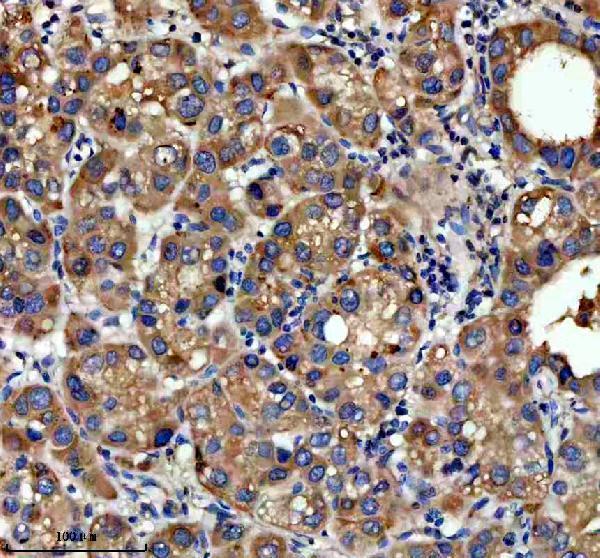
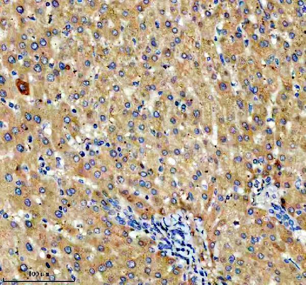
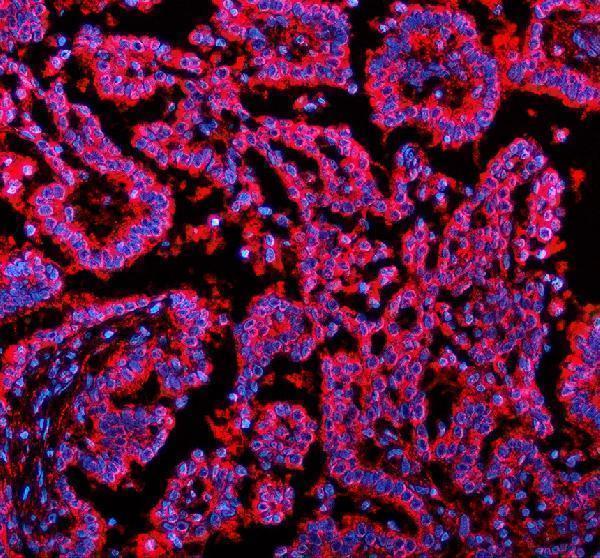
The catalog antibody has IHC images from human and rat paraffin sections and IF images from human paraffin sections (A04902-1 image captions).

Real IHC data IHC analysis of NSDHL using anti-NSDHL antibody (A04902-1). NSDHL was detected in a paraffin-embedded section of human liver cancer tissue. Heat mediated antigen retrieval was performed in EDTA buffer (pH 8.0, epitope retrieval solution). The tissue section was blocked with 10% goat serum. The tissue section was then incubated with 2 μg/ml rabbit anti-NSDHL Antibody (A04902-1) overnight at 4°C. Peroxidase Conjugated Goat Anti-rabbit IgG was used as secondary antibody and incubated for 30 minutes at 37°C. The tissue section was developed using HRP Conjugated Rabbit IgG Super Vision Assay Kit (Catalog # SV0002) with DAB as the chromogen.
Anti-NSDHL Antibody ®
Cat # A04902-1

A04902-1 has IHC images from human liver, human liver cancer, human lung cancer and rat liver paraffin sections, plus IF images from human liver and lung cancer paraffin sections (catalog image captions). M04902 is listed for human IHC and ICC/IF, but has no IHC or IF image in the payload (catalog applications, reactivity and image lists).

Which to pick: For tissue IHC, choose A04902-1: its paraffin-section captions document EDTA retrieval at pH 8.0 and primary antibody at 2 μg/ml; the fixative is unreported (A04902-1 IHC captions). For human ICC, M04902 is a rabbit monoclonal listed for ICC/IF, while A04902-1 has documented tissue IF images (catalog clone, applications and IF image captions). For cross-species IHC, A04902-1 has human and rat paraffin-section images; mouse reactivity is listed, but the supplied IHC captions do not show mouse tissue (catalog reactivity and A04902-1 IHC captions).

Each figure is that product's own IHC / IF validation image from its datasheet.

References

  1. UniProt Consortium. UniProt entry Q15738 (NSDHL_HUMAN, Sterol-4-alpha-carboxylate 3-dehydrogenase, decarboxylating).
  2. Human Protein Atlas. NSDHL tissue IHC expression (reliability: Enhanced).
  3. Human Protein Atlas. NSDHL subcellular location (ICC-IF): Mainly localized to the endoplasmic reticulum. In addition localized to the lipid droplets..
  4. Human Protein Atlas. NSDHL antibody validation summary (2 antibodies).
  5. NAD(P)-dependent steroid dehydrogenase-like protein and neutral cholesterol ester hydrolase 1 serve as novel markers for early detection of gastric cancer identified using quantitative proteomics. Journal of clinical laboratory analysis 2021 — PMC7891516.
  6. NAD(P)-dependent steroid dehydrogenase-like is involved in breast cancer cell growth and metastasis. BMC cancer 2020 — PMC7197182.
  7. Developmental expression pattern of the cholesterogenic enzyme NSDHL and negative selection of NSDHL-deficient cells in the heterozygous Bpa(1H)/+ mouse. Molecular genetics and metabolism 2009 — PMC2783206.
  8. NSDHL as a regulator of lipid peroxidation and ER stress-mediated apoptosis in ovarian cancer. Biology direct 2026 — PMC13173746.
  9. PubMed PMID:10854409 — UniProt-cited evidence.
  10. PubMed PMID:15164054 — UniProt-cited evidence.
  11. PubMed PMID:15772651 — UniProt-cited evidence.