This website uses cookies to ensure you get the best experience on our website.


- Table of Contents
Plan NSUN6 chromogenic IHC around granular cytoplasmic staining, with strong signal reported in ciliated cell bodies (HPA tissue IHC). Compare these cells with HPA tissue-IHC cell populations where staining was not detected, and start the catalog antibody at 2–5 μg/ml (HPA tissue IHC; datasheet A13404-3).

Expected localisation, validated protocols, controls and antibodies — the at-a-glance facts below, then the full design guide.
| Expected localisation | Granular cytoplasm in most tissues (HPA tissue IHC) | |
| Staining pattern | Strong granular cytoplasmic staining in ciliated cell bodies (HPA tissue IHC) | |
| Antigen retrieval | EDTA pH 8.0 HIER, heat-mediated (datasheet A13404-3) | |
| Positive control | Bronchus+4 more · see all | |
| Negative control | Adipose tissue+3 more · see all |
| Fixation | Keep fixation consistent across paraffin sections (standard IHC practice; not target-specific) | |
| Caveat | Staining has medium consistency with RNA data (HPA tissue IHC) | |
| Regulation | No expression regulator annotated (UniProt) | |
| Isoform / epitope | No isoforms annotated; one 1–469 chain (UniProt) |
The catalog antibody’s IHC-P protocol (datasheet: A13404-3) is accompanied by published NSUN6 IHC methods for ovarian, renal, and cervical cancer samples (PMC13454582; PMC13270792; PMC11225205).
| Sample | Paraffin-embedded human liver cancer tissue; fixative not specified (datasheet A13404-3) |
| Fixation | Image fixative and duration unreported (datasheet A13404-3); verify before use. |
| Sectioning | 4–5 µm sections on charged slides (standard) |
| Deparaffinisation | Xylene, graded ethanol series to water (standard) |
| Antigen retrieval | Heat retrieval: EDTA pH 8.0 (datasheet A13404-3); 20 min, 95–100 °C (standard) |
| Peroxidase block | 3% H2O2, 10 min, room temperature (standard) |
| Blocking | 10% goat serum (datasheet A13404-3) |
| Primary antibody | Rabbit anti-NSUN6, 2-5 μg/ml (datasheet A13404-3) |
| Primary incubation | Overnight at 4 °C (datasheet A13404-3) |
| Detection | HRP-conjugated secondary, DAB chromogen (datasheet A13404-3) |
| Counterstain | Hematoxylin, blue, dehydrate and mount (standard) |
| Expected result | NSUN6-positive staining in ciliated cells (cell body) of bronchus (HPA tissue IHC: High). HPA tissue profile: Granular cytoplasmic expression in most tissues. No signal in the no-primary control. |
In paraffin-section IHC, expect granular cytoplasmic NSUN6 staining in most tissues, with high staining in the cell bodies of ciliated cells in bronchus, fallopian tube, and nasopharynx (HPA: tissue IHC). NSUN6 is cytoplasmic and has no transmembrane segment (UniProt Q8TEA1). HPA rates its tissue IHC profile Approved, with medium consistency between staining and RNA data; external verification remains pending (HPA: tissue IHC reliability).
| Granular cytoplasmic staining is high in bronchial, fallopian tube, or nasopharyngeal ciliated cell bodies. | This matches the reported location and strongest listed cell-level examples (HPA: granular cytoplasmic profile; High in ciliated cells). Judge staining in the named cells, not from a whole-section average: neighboring cell types need not show the same intensity. |
| Staining is predominantly nuclear or confined to cell borders, without the expected cytoplasmic granules. | That distribution conflicts with the paraffin-section profile and cytoplasmic annotation (HPA: tissue IHC profile; UniProt Q8TEA1 subcellular location). Treat an isolated compartment mismatch as suspect; review morphology, the negative control, and a known-positive section before assigning it to NSUN6. |
| Strong staining appears in adipocytes, ovarian stroma, smooth muscle cells, or splenic red-pulp cells. | Those specific cells were reported as not detected in the listed tissues (HPA: tissue IHC negatives). Consider cross-reactivity or endogenous chromogenic detection activity, both general IHC possibilities; compare a reagent control and inspect whether pigment or staining follows cell boundaries. |
| Diffuse color covers cells and extracellular spaces without a clear granular cytoplasmic pattern. | This is difficult to score as NSUN6 because HPA describes granular cytoplasmic expression (HPA: tissue IHC profile). General IHC causes include nonspecific reagent binding, residual detection activity, or precipitated chromogen; assess a negative control and examine whether color tracks tissue structures. |
| No staining is visible in the ciliated cell bodies of a bronchus positive-control section. | This disagrees with the reported High staining for those cells (HPA: High in bronchial ciliated cells). First confirm that the section contains intact ciliated cells, then check the run controls and reagent steps. HPA's Approved rating still carries pending external verification (HPA: tissue IHC reliability). |
| Cell-level positive controls | Bronchus, fallopian tube, and nasopharynx have High staining specifically in ciliated cell bodies (HPA: tissue IHC). Use the relevant cells as the reference; a tissue name alone does not identify which cells should stain. |
| Cell-level negative and lower-signal comparisons | Adipocytes and smooth muscle cells are listed as not detected, while cerebral-cortex neurons and lung alveolar cells are Low (HPA: tissue IHC). Low is a weaker expected observation, not a negative-control designation. |
| Evidence strength | The tissue pattern is Approved but has medium staining-versus-RNA consistency and awaits external verification (HPA: tissue IHC reliability). One antibody, HPA045902, is listed as IHC Approved (HPA: antibody validation); do not treat the pattern as independently confirmed. |
| Topology and processing | NSUN6 has no transmembrane segment, signal peptide, propeptide, or annotated isoforms; its recorded chain spans residues 1–469 (UniProt Q8TEA1). These facts support intracellular interpretation but do not establish an epitope location or a target-specific retrieval requirement. |
| IF/ICC question: should its pattern match paraffin-section IHC? | HPA reports an approved Golgi apparatus location by ICC-IF, while tissue IHC describes granular cytoplasm (HPA: subcellular ICC-IF; HPA: tissue IHC). Interpret each assay against its own reported pattern; this section supplies no IF/ICC protocol. |
| Situation | Likely cause | Next action |
|---|---|---|
| The bronchus positive control is blank. | The expected ciliated cells may be absent from the plane of section, or the staining run may have failed (HPA: High in bronchial ciliated cells; general IHC practice). | Confirm ciliated-cell morphology, inspect a run control, then verify antibody application, detection reagents, and chromogen development (general IHC practice). Avoid calling the test tissue NSUN6-negative until the control works. |
| Color is mainly nuclear, membranous, or extracellular. | This disagrees with the granular cytoplasmic tissue pattern (HPA: tissue IHC) and cytoplasmic annotation (UniProt Q8TEA1). | Recheck compartment assignment at higher magnification; compare the positive tissue and a negative reagent control. If the mismatch persists, report it as discordant staining rather than a confirmed NSUN6 distribution (general IHC practice). |
| Listed negative cell types stain strongly. | Strong color in adipocytes, ovarian stroma, smooth muscle cells, or splenic red-pulp cells conflicts with their not-detected entries (HPA: tissue IHC negatives). Cross-reactivity or endogenous activity is possible (general IHC practice). | Compare a negative reagent control and inspect the distribution of color. Where the detection system permits, check its endogenous-activity control before interpreting strong staining as target expression (general IHC practice). |
| Background is diffuse across the section. | A diffuse film does not resemble the reported granules (HPA: tissue IHC profile); nonspecific binding or chromogen deposit can obscure cells (general IHC practice). | Check negative controls, wash and blocking steps, and chromogen deposition. Score only distinguishable cellular staining against the background (general IHC practice). |
| A low-signal tissue appears nearly blank. | HPA lists cerebral-cortex neurons, lung alveolar cells, and several other cell types as Low, so weak visibility can be consistent with the reference profile (HPA: tissue IHC low entries). | Use a High ciliated-cell reference to assess run performance, then document the low-signal tissue at cell level. Do not promote a weak reference into a definitive negative control. |
| IHC appears granular, but an ICC-IF image emphasizes Golgi staining. | The observations come from different assay contexts: granular cytoplasm in tissue IHC and approved Golgi localization in ICC-IF (HPA: tissue IHC; HPA: subcellular ICC-IF). | Compare each image with the HPA pattern for its own assay. For this paraffin-section IHC decision, prioritize cell-level tissue staining and its controls (HPA: tissue IHC; general IHC practice). |


Comprehensive Human Protein Atlas IHC scoring per tissue (reliability: Approved — Medium consistency between antibody staining and RNA expression data. Pending external verification.). Rows are taken directly from the HPA tissue chart — click any row's HPA link to view the source.
| Tissue | Cell type | Level | Evidence | Source |
|---|---|---|---|---|
| Bronchus | Ciliated cells (cell body) | High | Protein (IHC) | HPA → |
| Fallopian tube | Ciliated cells (cell body) | High | Protein (IHC) | HPA → |
| Nasopharynx | Ciliated cells (cell body) | High | Protein (IHC) | HPA → |
| Adrenal gland | Glandular cells | Medium | Protein (IHC) | HPA → |
| Appendix | Glandular cells | Medium | Protein (IHC) | HPA → |
| Tissue | Cell type | Level | Evidence | Source |
|---|---|---|---|---|
| Adipose tissue | Adipocytes | Not detected | Protein (IHC) | HPA → |
| Ovary | Ovarian stroma cells | Not detected | Protein (IHC) | HPA → |
| Smooth muscle | Smooth muscle cells | Not detected | Protein (IHC) | HPA → |
| Spleen | Cells in red pulp | Not detected | Protein (IHC) | HPA → |
Use the catalog antibody’s paraffin-section result as a starting point, then assess staining by cell type, compartment and control performance.
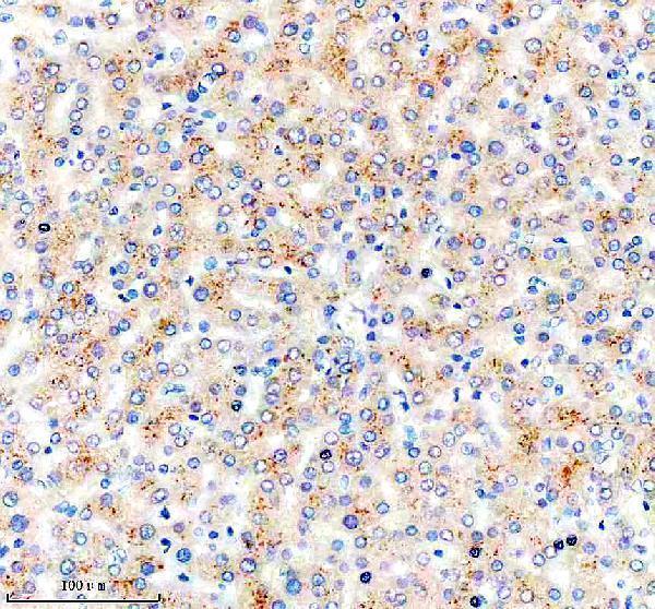
A13404-3 has an IHC image from a paraffin-embedded human liver cancer section (catalog image caption). Human, Mouse, and Rat reactivity is listed (catalog reactivity); no IF image is supplied (catalog IF images).
A13404-3 is listed for IHC and for Human, Mouse, and Rat reactivity (catalog applications/reactivity). Its IHC image documents a paraffin-embedded human liver cancer section, EDTA retrieval at pH 8.0, and 2 μg/ml primary antibody (A13404-3 image caption).
Which to pick: Choose A13404-3 for chromogenic IHC on paraffin sections: its own caption documents DAB detection in human liver cancer tissue; the fixative is unreported (A13404-3 image caption). No IF/ICC-validated SKU is supplied, and clonality is unreported (catalog applications, IF images, and clone field). For cross-species planning, A13404-3 lists Human, Mouse, and Rat reactivity, but its IHC image documents human tissue only (catalog reactivity; A13404-3 image caption).