This website uses cookies to ensure you get the best experience on our website.


- Table of Contents
Plan paraffin-section NVL IHC using the nucleolar and cytoplasmic tissue pattern (HPA tissue IHC). Kidney distal tubules show high staining (HPA tissue IHC); check epitope coverage across the 5 annotated isoforms (UniProt).

Expected localisation, validated protocols, controls and antibodies — the at-a-glance facts below, then the full design guide.
| Expected localisation | IHC: nucleolar/cytoplasmic (HPA tissue IHC); protein: predominantly nucleolar (UniProt) | |
| Staining pattern | Nucleolar/cytoplasmic in several tissues; high in kidney distal tubules (HPA tissue IHC) | |
| Antigen retrieval | EDTA pH 8.0 HIER, heat-mediated (datasheet A05446-3) | |
| Positive control | Kidney+4 more · see all | |
| Negative control | Adipose tissue+4 more · see all |
| Fixation | Keep fixation consistent across compared paraffin sections. (selected-SKU IHC image A05446-3) | |
| Caveat | Antibody staining and RNA show medium consistency (HPA tissue IHC) | |
| Regulation | Specific expression regulation not reported (UniProt) | |
| Isoform / epitope | 5 isoforms; isoform-specific epitope coverage unknown (UniProt) |
The catalog antibody’s IHC-P protocol (datasheet: A05446-3) is accompanied by one published NVL IHC protocol in human prostate adenocarcinoma sections (PMC12220030).
| Sample | Paraffin-embedded human liver cancer tissue; fixative not specified (datasheet A05446-3) |
| Fixation | Image fixative and duration unreported (datasheet A05446-3); verify before use. |
| Sectioning | 4–5 µm sections on charged slides (standard) |
| Deparaffinisation | Xylene, graded ethanol series to water (standard) |
| Antigen retrieval | Heat retrieval: EDTA pH 8.0 (datasheet A05446-3); 20 min, 95–100 °C (standard) |
| Peroxidase block | 3% H2O2, 10 min, room temperature (standard) |
| Blocking | 10% goat serum (datasheet A05446-3) |
| Primary antibody | Rabbit anti-NVL, 2-5 μg/ml (datasheet A05446-3) |
| Primary incubation | Overnight at 4 °C (datasheet A05446-3) |
| Detection | HRP-conjugated secondary, DAB chromogen (datasheet A05446-3) |
| Counterstain | Hematoxylin, blue, dehydrate and mount (standard) |
| Expected result | NVL-positive staining in distal tubules of kidney (HPA tissue IHC: High). HPA tissue profile: Nucleolar and cytoplasmic expression in several tissues. No signal in the no-primary control. |
NVL is predominantly nucleolar, with nucleoplasmic localization also reported (UniProt O15381); it has no transmembrane segment (UniProt O15381 topology). In paraffin-section IHC, expect nucleolar staining in some cells, with cytoplasmic staining also reported across several tissues (HPA tissue IHC). Distal tubular cells in kidney show the strongest listed staining; several epithelial cell types show medium staining (HPA tissue IHC). The tissue IHC assessment is Supported, with medium consistency between staining and RNA data (HPA tissue IHC).
| Distinct nucleolar staining in kidney distal tubules, with or without cytoplasmic staining. | This fits the strongest listed tissue result, High staining in distal tubules (HPA tissue IHC), and the predominant nucleolar localization (UniProt O15381; HPA subcellular ICC-IF). Assess the nuclear compartment and cell identity together; staining intensity alone does not establish specificity (general IHC interpretation). |
| Strong, uniform staining confined to membranes or extracellular material, without recognizable nucleolar signal. | Treat this as a suspect compartment pattern: NVL is predominantly nucleolar and has no transmembrane segment (UniProt O15381 topology; HPA subcellular ICC-IF). Check the control slide and detection system before interpreting it as NVL; the supplied sources do not establish a membrane or extracellular NVL pattern. |
| Strong staining in adipocytes or cardiomyocytes while kidney distal tubules stain as expected. | Consider cross-reactivity or endogenous detection activity, then review whether staining truly belongs to those cells (general IHC interpretation). HPA lists NVL as Not detected in adipocytes and cardiomyocytes (HPA tissue IHC). A single unexpected positive cell population cannot by itself identify the cause. |
| Diffuse color across many compartments, including areas without a discrete nucleolar pattern. | This is difficult to score as specific NVL staining. HPA describes nucleolar and cytoplasmic expression in several tissues (HPA tissue IHC), so cytoplasmic color alone is insufficient evidence of artefact. Compare the distribution with a detection control and a kidney distal-tubule section before changing the interpretation (general IHC practice). |
| No detectable staining in kidney distal tubules. | A technical false negative is possible because those cells are listed as High (HPA tissue IHC). Check section integrity, primary-antibody conditions, retrieval and detection controls as general IHC practice. Absence of signal does not establish that NVL is biologically absent from the sampled kidney. |
| Compartment and cell identity | Predominant nucleolar localization provides the main compartment check (UniProt O15381; HPA subcellular ICC-IF). HPA tissue IHC also reports cytoplasmic expression in several tissues, so cytoplasmic staining should be judged alongside nucleolar signal, cell identity and controls, not rejected automatically. |
| Tissue choice and source disagreement | Kidney distal tubules are a useful positive reference because HPA scores them High; breast and gallbladder glandular cells are among the listed Medium examples (HPA tissue IHC). UniProt reports high expression in heart, yet HPA scores cardiomyocytes Not detected (UniProt O15381 tissue specificity; HPA tissue IHC). Do not turn either observation into a universal tissue prediction. |
| Antibody validation | HPA reports IHC Supported for HPA028207, HPA028224 and HPA028654 (HPA antibodies). That status supports use of their recorded IHC patterns, but the tissue profile has only medium consistency with RNA data (HPA tissue IHC). Antibody-specific results should be interpreted within their own validation and staining context. |
| Isoforms and processing | UniProt lists 5 NVL isoforms and a full-length chain of residues 1–856, without a signal peptide or propeptide (UniProt O15381). These facts do not identify the epitope recognized by the chosen IHC antibody or predict which isoforms it detects; make no isoform-specific staining claim without antibody-specific evidence. |
| Situation | Likely cause | Next action |
|---|---|---|
| Kidney distal tubules are blank. | Possible section, retrieval, antibody or detection failure; the cause is not established by the blank result (general IHC practice). | Verify tissue morphology and detection controls, then optimize retrieval and primary-antibody conditions using the selected antibody’s IHC instructions (general IHC practice). Kidney distal tubules are the listed High reference (HPA tissue IHC). |
| Color is widespread and obscures nuclei. | Background from the detection workflow or nonspecific binding is possible (general IHC practice). HPA also reports cytoplasmic NVL staining in several tissues (HPA tissue IHC). | Run an appropriate primary-omission or detection control, review blocking and washes, and reassess whether any discrete nucleolar pattern remains (general IHC practice). Do not equate all cytoplasmic staining with background. |
| The dominant signal appears membranous. | The compartment conflicts with predominant nucleolar localization and the absence of a transmembrane segment (UniProt O15381; HPA subcellular ICC-IF). | Confirm the stained structure at higher magnification and compare with a positive tissue and detection control (general IHC practice). Avoid scoring membrane-only signal as NVL on this evidence. |
| Adipocytes or cardiomyocytes stain strongly. | Cross-reactivity, endogenous detection activity or cell misidentification is possible (general IHC interpretation); HPA scores those cells Not detected (HPA tissue IHC). | Check cell identity and control staining, then compare the result with kidney distal tubules on the same run (general IHC practice; HPA tissue IHC). Treat the unexpected result as unresolved until verified. |
| A tissue expected from expression summaries gives weak or no staining. | Tissue-wide expression and cell-specific IHC can disagree: UniProt lists high heart expression, while HPA reports cardiomyocytes Not detected (UniProt O15381 tissue specificity; HPA tissue IHC). | Record the exact cell population and assay result. Use a listed HPA-positive cell population as the IHC reference, and avoid assigning a target-specific fixation or retrieval explanation without evidence. |
| Can the IHC pattern be applied directly to IF/ICC? | The assays show related localization evidence, but HPA tissue IHC reports nucleolar and cytoplasmic expression, while HPA ICC-IF reports mainly nucleolar localization (HPA tissue IHC; HPA subcellular ICC-IF). | Use the IF/ICC guide for that assay. Here, regard a nucleolar signal as the shared localization check; do not treat an IHC chromogenic intensity or tissue score as an IF/ICC acceptance threshold (general assay interpretation). |
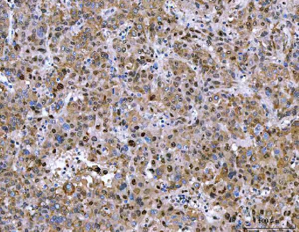
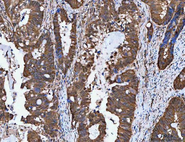


Comprehensive Human Protein Atlas IHC scoring per tissue (reliability: Supported — Medium consistency between antibody staining and RNA expression data.). Rows are taken directly from the HPA tissue chart — click any row's HPA link to view the source.
| Tissue | Cell type | Level | Evidence | Source |
|---|---|---|---|---|
| Kidney | Distal tubules | High | Protein (IHC) | HPA → |
| Breast | Glandular cells | Medium | Protein (IHC) | HPA → |
| Gallbladder | Glandular cells | Medium | Protein (IHC) | HPA → |
| Liver | Cholangiocytes | Medium | Protein (IHC) | HPA → |
| Oral mucosa | Squamous epithelial cells | Medium | Protein (IHC) | HPA → |
| Tissue | Cell type | Level | Evidence | Source |
|---|---|---|---|---|
| Adipose tissue | Adipocytes | Not detected | Protein (IHC) | HPA → |
| Adrenal gland | Glandular cells | Not detected | Protein (IHC) | HPA → |
| Appendix | Glandular cells | Not detected | Protein (IHC) | HPA → |
| Bone marrow | Hematopoietic cells | Not detected | Protein (IHC) | HPA → |
| Caudate | Glial cells | Not detected | Protein (IHC) | HPA → |
Troubleshoot chromogenic NVL IHC in paraffin sections by checking retrieval, compartment-specific staining, controls and scoring before interpreting differences between samples.
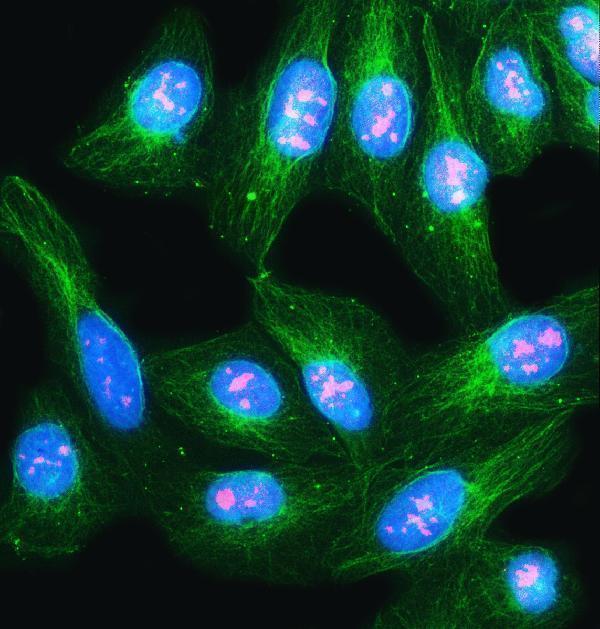
The IHC-validated anti-NVL antibody has paraffin-section data from human liver cancer tissue and IF/ICC data from U2OS cells (catalog image captions); its listed reactivity is human and rat (catalog).
A05446-3 is listed for IHC and was shown on a paraffin-embedded human liver cancer section with EDTA retrieval and DAB detection (catalog IHC caption). A05446-3 is also listed for IF/ICC, with an IF image from U2OS cells (catalog IF caption).
Which to pick: Choose A05446-3 for human tissue IHC: its paraffin-section image used EDTA retrieval at pH 8.0 and 2 μg/ml primary antibody (catalog IHC caption); the fixative was unreported (catalog IHC caption). For IF/ICC, the same rabbit antibody has a U2OS cell image at 5 μg/ml (catalog IF caption). For rat samples, A05446-3 lists rat reactivity for ICC/IF, while its listed paraffin-section IHC application and supplied IHC images are human (catalog dilution listing and IHC captions).