This website uses cookies to ensure you get the best experience on our website.


- Table of Contents
Plan OPN3 paraffin-section IHC around observed cytoplasmic tissue staining (HPA tissue IHC) and annotated membrane localization (UniProt). Use the catalog antibody at 2–5 μg/ml (datasheet), include tissue controls, and interpret staining cautiously because HPA rates its reliability as uncertain (HPA tissue IHC).

Expected localisation, validated protocols, controls and antibodies — the at-a-glance facts below, then the full design guide.
| Expected localisation | Cytoplasmic staining (HPA tissue IHC); membrane location annotated (UniProt) | |
| Staining pattern | Cytoplasmic in glandular, airway epithelial and neuronal cells (HPA tissue IHC) | |
| Antigen retrieval | EDTA pH 8.0 HIER, heat-mediated (datasheet A09353-3) | |
| Positive control | Adrenal gland+4 more · see all | |
| Negative control | Lymph node+3 more · see all |
| Fixation | Keep fixation consistent across sections (standard IHC practice; not target-specific) | |
| Caveat | Staining has low consistency with RNA expression (HPA tissue IHC) | |
| Regulation | Basal epidermal enrichment (UniProt) | |
| Isoform / epitope | 2 isoforms; check epitope side and isoform coverage (UniProt) |
The catalog antibody protocol (datasheet A09353-3) is accompanied by two published OPN3 IHC protocols (PMC8342238; PMC8857800).
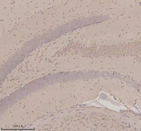
| Sample | Paraffin-embedded mouse brain tissue; fixative not specified (datasheet A09353-3) |
| Fixation | Image fixative and duration unreported (datasheet A09353-3); verify before use. |
| Sectioning | 4–5 µm sections on charged slides (standard) |
| Deparaffinisation | Xylene, graded ethanol series to water (standard) |
| Antigen retrieval | Heat retrieval: EDTA pH 8.0 (datasheet A09353-3); 20 min, 95–100 °C (standard) |
| Peroxidase block | 3% H2O2, 10 min, room temperature (standard) |
| Blocking | 10% goat serum (datasheet A09353-3) |
| Primary antibody | Rabbit anti-OPN3, 2-5 μg/ml (datasheet A09353-3) |
| Primary incubation | Overnight at 4 °C (datasheet A09353-3) |
| Detection | HRP-conjugated secondary, DAB chromogen (datasheet A09353-3) |
| Counterstain | Hematoxylin, blue, dehydrate and mount (standard) |
| Expected result | OPN3-positive staining in glandular cells of adrenal gland (HPA tissue IHC: Medium). HPA tissue profile: General cytoplasmic expression. No signal in the no-primary control. |
OPN3 is a seven-transmembrane protein annotated at the cell membrane and in the cytoplasm (UniProt Q9H1Y3 topology and subcellular location). In paraffin-section IHC, expect chiefly cytoplasmic staining in selected epithelial, glandular and neuronal cells (HPA: general cytoplasmic expression; listed medium-staining cells). Treat this as a provisional pattern: HPA rates tissue IHC reliability uncertain because antibody staining and RNA expression have low consistency (HPA: tissue IHC reliability).
| Cytoplasmic chromogen in bronchial respiratory epithelium, colon glands or cerebellar Purkinje cells, with identifiable cell boundaries. | This fits the reported cellular distribution; HPA scores each example medium (HPA: bronchus, colon and cerebellum tissue IHC). Membrane accentuation is plausible from OPN3 topology, but a sharp membrane rim is not required by the reported tissue pattern (UniProt Q9H1Y3 topology; HPA: general cytoplasmic expression). |
| A strong, exclusively nuclear IHC signal replaces the expected cytoplasmic pattern. | Investigate an artifactual or off-target signal before scoring it as OPN3 (HPA: general cytoplasmic tissue IHC expression). HPA reports nucleoplasm in ICC/IF with uncertain confidence, so that separate assay does not establish an exclusively nuclear paraffin-section IHC pattern (HPA: subcellular ICC/IF). |
| The strongest chromogen appears in lymph-node germinal center cells or ovarian stromal cells. | These cells are reported as not detected by HPA tissue IHC (HPA: lymph node and ovary). Consider antibody cross-reactivity or endogenous chromogenic activity, especially if the expected cells are weak. Because overall tissue IHC reliability is uncertain, this mismatch prompts controls rather than proving the signal false (HPA: tissue IHC reliability). |
| Broad, hazy color covers cells and surrounding section tissue without a discernible cellular pattern. | This is background rather than an interpretable OPN3 distribution; compare it with the cell-restricted, generally cytoplasmic HPA pattern (HPA: tissue IHC profile). In chromogenic IHC, residual detection activity, insufficient blocking or overly strong primary-antibody signal can produce background; a no-primary control helps separate detection background from antibody-dependent staining. |
| No staining appears in a section containing an HPA-listed medium-staining cell population. | First check that the relevant cells are present and identifiable: for example, bronchial respiratory epithelium, not merely bronchial tissue (HPA: bronchus tissue IHC). A blank result may reflect assay failure or a weak sample; HPA's uncertain tissue-IHC reliability means its medium score is a reference pattern, not a guaranteed positive control (HPA: tissue IHC reliability). |
| Membrane topology and compartment | OPN3 has seven transmembrane segments and is annotated at the cell membrane and in the cytoplasm (UniProt Q9H1Y3 topology and subcellular location). The tissue-IHC reference pattern is generally cytoplasmic (HPA: tissue IHC profile). Topology alone cannot predict how prominently a particular antibody will outline membranes in paraffin sections. |
| Epitope position | UniProt assigns extracellular and cytoplasmic stretches across the OPN3 sequence (UniProt Q9H1Y3 topology). The supplied evidence does not map an IHC antibody epitope to either side. Interpret a weak or absent signal using assay controls; do not infer an epitope-specific antigen-retrieval requirement from topology. |
| Isoforms and modification | UniProt lists two isoforms and glycosylation sites at residues 5 and 198 (UniProt Q9H1Y3 isoforms and glycosylation). No supplied antibody epitope or isoform-specific staining evidence establishes whether these features alter the observed IHC pattern; a stained section cannot identify an isoform from appearance alone. |
| Tissue-IHC evidence | HPA calls the overall tissue pattern uncertain because staining and RNA expression show low consistency; its IHC listing for CAB013682 is also uncertain (HPA: tissue IHC reliability; HPA: antibody validation). Use named positive and negative cell populations as comparison points, not definitive specificity standards. |
| IF/ICC: should its localisation guide this IHC call? | HPA ICC/IF supports cytosol and plasma membrane localisation, while nucleoplasm, primary cilium and basal body are uncertain (HPA: subcellular ICC/IF). HPA lists ICC support for HPA049243, whereas its tissue-IHC listing concerns CAB013682 (HPA: antibody validation); IF/ICC localisation is context, not paraffin-IHC validation. |
| Situation | Likely cause | Next action |
|---|---|---|
| The expected epithelial or neuronal cells show no chromogen. | The selected field may lack the specific cells scored by HPA, or the chromogenic assay may have failed; HPA's medium observations are not guaranteed in every section (HPA: listed positive cells; HPA: uncertain tissue IHC reliability). | Confirm cell identity on the counterstained section and compare a section containing an HPA-listed medium-staining population processed in the same run; then review the assay's retrieval and detection controls as general IHC checks. |
| Color appears in the no-primary control. | Antibody-independent detection activity or nonspecific chromogen deposition can create apparent staining in chromogenic IHC. | Address endogenous detection activity and reagent background using the detection system's standard controls, then repeat the comparison. Do not score antibody-treated sections as OPN3 positive while the no-primary control has the same pattern. |
| Lymph-node germinal center cells stain more strongly than the candidate positive cells. | HPA reports germinal center cells as not detected; cross-reactivity or detection background is possible, although the tissue-IHC reference itself is uncertain (HPA: lymph node; HPA: tissue IHC reliability). | Compare cell morphology, the no-primary control and an HPA-listed medium-staining population in the same run (HPA: listed positives). Treat the germinal-center signal as unresolved until those comparisons support a cell-specific pattern. |
| Only nuclei stain in a paraffin section. | This departs from HPA's general cytoplasmic tissue-IHC profile; the uncertain nucleoplasm call comes from a separate ICC/IF assay (HPA: tissue IHC profile; HPA: subcellular ICC/IF). | Check the no-primary control and whether cytoplasmic signal is visible in an HPA-listed positive cell population (HPA: listed positives). Record nuclear-only staining separately from the provisional tissue-IHC positive pattern. |
| The whole section has uniform haze, obscuring cell boundaries. | General IHC possibilities include excessive primary-antibody signal, inadequate blocking or incomplete removal of detection reagents; haze does not match HPA's generally cytoplasmic, cell-associated profile (HPA: tissue IHC profile). | Compare no-primary and antibody-treated sections, review blocking and wash steps, and adjust antibody concentration only within the assay's validated range. Reassess whether the remaining signal follows the specified cell populations (HPA: listed positives). |
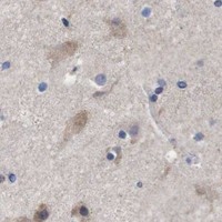


Comprehensive Human Protein Atlas IHC scoring per tissue (reliability: Uncertain — Low consistency between antibody staining and RNA expression data.). Rows are taken directly from the HPA tissue chart — click any row's HPA link to view the source.
| Tissue | Cell type | Level | Evidence | Source |
|---|---|---|---|---|
| Adrenal gland | Glandular cells | Medium | Protein (IHC) | HPA → |
| Appendix | Glandular cells | Medium | Protein (IHC) | HPA → |
| Bronchus | Respiratory epithelial cells | Medium | Protein (IHC) | HPA → |
| Cerebellum | Purkinje cells | Medium | Protein (IHC) | HPA → |
| Cerebral cortex | Neuronal cells | Medium | Protein (IHC) | HPA → |
| Tissue | Cell type | Level | Evidence | Source |
|---|---|---|---|---|
| Lymph node | Germinal center cells | Not detected | Protein (IHC) | HPA → |
| Ovary | Ovarian stroma cells | Not detected | Protein (IHC) | HPA → |
| Soft tissue | Chondrocytes | Not detected | Protein (IHC) | HPA → |
| Vagina | Squamous epithelial cells | Not detected | Protein (IHC) | HPA → |
Troubleshoot OPN3 staining in paraffin sections by checking retrieval, antibody specificity, cell identity and compartment before comparing signal across samples.
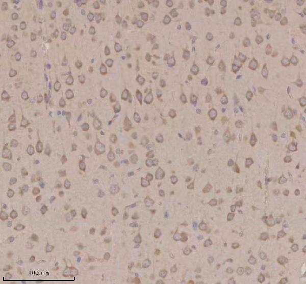
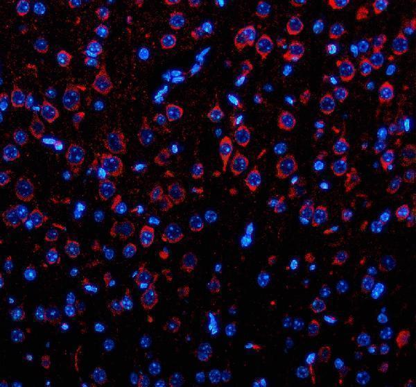
A09353-3 has IHC and IF images from mouse and rat paraffin-embedded brain sections (catalog image captions); human, mouse and rat are listed as reactive (catalog reactivity).
A09353-3 will render with its IHC image from a mouse paraffin-embedded brain section (catalog IHC image caption). Additional IHC images show rat brain, and IF images show mouse and rat brain sections (catalog IHC and IF image captions).
Which to pick: For tissue IHC, choose the polyclonal A09353-3 (catalog antibody details): its IHC caption documents a paraffin-embedded mouse brain section, EDTA retrieval at pH 8.0 and 2 μg/ml primary antibody; the fixative is unreported (catalog IHC image caption). For IF on sections, the same SKU has mouse and rat brain images at 5 μg/ml; ICC is not listed among its tested applications (catalog IF image captions; catalog applications). For work across species, A09353-3 lists human, mouse and rat reactivity, with direct tissue images for mouse and rat (catalog reactivity; catalog image captions).