This website uses cookies to ensure you get the best experience on our website.


- Table of Contents
Plan chromogenic OPTN IHC in paraffin sections with the catalog antibody at 2–5 μg/ml (datasheet: PB9343). Assess cytoplasmic staining in glandular and brain cells (HPA tissue IHC), and account for the reported low consistency between antibody staining and RNA expression (HPA tissue IHC).

Expected localisation, validated protocols, controls and antibodies — the at-a-glance facts below, then the full design guide.
| Expected localisation | Cytoplasmic tissue staining (HPA tissue IHC); perinuclear and Golgi localisation (UniProt) | |
| Staining pattern | Cytoplasmic signal in glandular, neuronal and glial cells (HPA tissue IHC) | |
| Antigen retrieval | EDTA pH 8.0 HIER, heat-mediated (datasheet PB9343) | |
| Positive control | Adrenal gland+4 more · see all | |
| Negative control | None in HPA (detected in all 45 tissues); use no-primary + isotype controls |
| Fixation | Keep fixation consistent across sections. (standard IHC practice; not target-specific) | |
| Caveat | Staining has low consistency with RNA expression (HPA tissue IHC) | |
| Regulation | RNA enriched in skeletal muscle, tongue (HPA tissue RNA) | |
| Isoform / epitope | 3 isoforms; no extracellular domain; epitope map unknown (UniProt) |
Compare the catalog antibody’s IHC-P protocol with 4 published OPTN IHC protocols (PMC7940236; PMC4470809; PMC5833650; PMC9167278).
| Sample | Paraffin-embedded human gall bladder adenosquamous carcinoma tissue; fixative not specified (datasheet PB9343) |
| Fixation | Image fixative and duration unreported (datasheet PB9343); verify before use. |
| Sectioning | 4–5 µm sections on charged slides (standard) |
| Deparaffinisation | Xylene, graded ethanol series to water (standard) |
| Antigen retrieval | Heat retrieval: EDTA pH 8.0 (datasheet PB9343); 20 min, 95–100 °C (standard) |
| Peroxidase block | 3% H2O2, 10 min, room temperature (standard) |
| Blocking | 10% goat serum (datasheet PB9343) |
| Primary antibody | Rabbit anti-OPTN, 2-5 μg/ml (datasheet PB9343) |
| Primary incubation | Overnight at 4 °C (datasheet PB9343) |
| Detection | Streptavidin-biotin complex (SABC), DAB chromogen (datasheet PB9343) |
| Counterstain | Hematoxylin, blue, dehydrate and mount (standard) |
| Expected result | OPTN-positive staining in glandular cells of adrenal gland (HPA tissue IHC: High). HPA tissue profile: General cytoplasmic expression. No signal in the no-primary control. |
OPTN should show predominantly cytoplasmic staining, with possible perinuclear or Golgi-associated accentuation; it has no transmembrane segment (UniProt Q96CV9 localization and topology). HPA reports general cytoplasmic IHC expression and high staining in several glandular and epithelial populations, as well as selected neural cells (HPA: tissue IHC). Its tissue-IHC reliability is Approved, with low consistency between antibody staining and RNA expression (HPA: tissue IHC).
| Cytoplasmic staining in adrenal, appendix, breast or colon glandular cells; or bronchial respiratory epithelium. | This fits the reported high-staining cell populations and general cytoplasmic profile (HPA: tissue IHC). Judge the signal in the named cells, rather than treating staining anywhere in the section as confirmation. |
| Cytoplasmic staining in caudate glial cells, cerebellar granular-layer cells or cerebral-cortex neurons. | These are additional reported high-staining populations (HPA: tissue IHC). Compare the observed cell type with the relevant HPA entry; a positive region alone does not establish the expected cellular pattern. |
| Predominantly nuclear, sharply membrane-bound or exclusively extracellular staining. | That distribution conflicts with OPTN's reported cytoplasmic, perinuclear, Golgi and vesicular locations and absence of a transmembrane segment (UniProt Q96CV9). Treat it as a possible artefact and check controls before assigning it to OPTN. |
| Strong signal in cells outside the reported positive population, or widespread diffuse staining over tissue and empty areas. | The cell mismatch raises concern for cross-reactivity or endogenous detection activity; diffuse signal also suggests nonspecific background (general IHC practice). HPA supplies no negative-cell list here, so an unlisted cell cannot be declared OPTN-negative from this record. |
| No discernible cytoplasmic signal in an HPA high-staining cell population. | First consider a failed or insensitive IHC run, while allowing for specimen differences (general IHC practice; HPA: high staining in listed cells). The Approved HPA rating has low agreement with RNA data, so one blank section does not settle OPTN absence (HPA: tissue IHC). |
| Compartment and topology | OPTN is reported in cytoplasm, perinuclear region, Golgi, vesicles and recycling endosomes, with no transmembrane segment (UniProt Q96CV9). At light-microscope IHC resolution, cytoplasmic staining is the primary call; do not require a distinct organelle pattern. |
| Tissue reference and evidence limit | HPA lists high staining in specific glandular, respiratory and neural cell populations, but supplies no negative tissues in this payload (HPA: tissue IHC). Its Approved rating includes low consistency with RNA expression, so use the listed patterns as comparators rather than absolute cutoffs (HPA: tissue IHC). |
| Antibody validation | All 3 listed antibodies have Approved IHC status; none is listed as IHC Enhanced (HPA: antibody validation). An Enhanced ICC rating for HPA003279 or HPA003360 applies to ICC, not automatically to paraffin-section IHC (HPA: antibody validation). |
| Isoforms and epitope coverage | UniProt lists 3 OPTN isoforms (UniProt Q96CV9). No antibody epitope or isoform coverage is supplied, so the IHC signal cannot be assigned to a particular isoform from this record. |
| IF/ICC Q: What localisation should be expected? | A: HPA describes OPTN as mainly cytosolic in ICC-IF, consistent with UniProt's broader cytoplasmic localisation (HPA: subcellular ICC-IF; UniProt Q96CV9). That observation informs compartment interpretation; it is not a paraffin-IHC protocol or proof of identical staining in tissue sections. |
| Situation | Likely cause | Next action |
|---|---|---|
| The expected positive cells are blank. | The staining run may have failed or the antibody may be too insensitive under the chosen conditions (general IHC practice). | Check a listed HPA high-staining tissue in the same run and verify that detection and counterstain worked; then review the catalog antibody's IHC-P instructions (HPA: tissue IHC; general IHC practice). |
| Signal appears only in nuclei or along cell borders. | This conflicts with the reported OPTN distribution and topology (UniProt Q96CV9). | Compare with a negative reagent control and inspect whether the signal tracks tissue edges or other artefacts before scoring cells (general IHC practice). |
| Colour covers the section or persists outside cells. | Nonspecific binding, incomplete washing or endogenous detection activity can produce background (general IHC practice). | Inspect the negative reagent control; review blocking, washes and detection controls, then score only distinct cellular staining (general IHC practice). |
| A strongly stained cell type does not match the HPA entry for that tissue. | Cross-reactivity or endogenous detection activity is possible; HPA reports high staining by cell type, not merely by organ (HPA: tissue IHC; general IHC practice). | Recheck cell identity on the counterstain and compare a negative reagent control. Treat unlisted cells as unresolved, since this payload has no HPA negative list (HPA: tissue IHC). |
| A faint cytoplasmic wash is difficult to score. | Low contrast can obscure a cellular pattern (general IHC practice); the HPA record does not define a numeric OPTN intensity cutoff (HPA: tissue IHC). | Compare the named positive cells with background and the negative reagent control in the same run; record equivocal staining instead of forcing a positive call (general IHC practice). |
| Only one specimen shows a different pattern from the reference. | HPA tissue-IHC staining has low consistency with RNA expression, and its listed positives are observations rather than universal thresholds (HPA: tissue IHC). | Review the cell type and compartment, repeat with a listed positive comparator, and report the discordance without inferring OPTN absence or a new localization from one section (HPA: tissue IHC; UniProt Q96CV9 localization). |


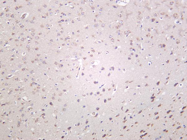
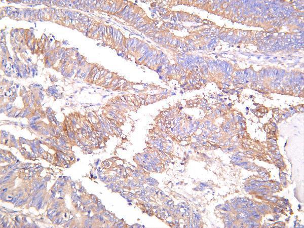


Comprehensive Human Protein Atlas IHC scoring per tissue (reliability: Approved — Low consistency between antibody staining and RNA expression data.). Rows are taken directly from the HPA tissue chart — click any row's HPA link to view the source.
| Tissue | Cell type | Level | Evidence | Source |
|---|---|---|---|---|
| Adrenal gland | Glandular cells | High | Protein (IHC) | HPA → |
| Appendix | Glandular cells | High | Protein (IHC) | HPA → |
| Breast | Glandular cells | High | Protein (IHC) | HPA → |
| Bronchus | Respiratory epithelial cells | High | Protein (IHC) | HPA → |
| Caudate | Glial cells | High | Protein (IHC) | HPA → |
| Tissue | Cell type | Level | Evidence | Source |
|---|---|---|---|---|
| None in HPA: OPTN is detected in all 45 scored tissues. Use a no-primary (secondary-only) and an isotype control instead. | ||||
Troubleshoot OPTN staining in paraffin section IHC by checking retrieval, staining controls, cellular location, and how positive cells are scored.
Both anti-OPTN antibodies list IHC for human, mouse and rat samples; tissue images support those species for PB9343 and human and mouse for M00952-2 (catalog: applications, reactivity, image alts).
PB9343 has IHC images from human gall bladder adenosquamous carcinoma and mouse and rat brain (catalog: PB9343 IHC image alts). M00952-2 has IHC images from human colon carcinoma and human and mouse brain, and lists IF as an application without an IF image (catalog: M00952-2 image alts, applications).
Which to pick: Choose PB9343 for paraffin-section IHC when its documented conditions are useful: the caption reports EDTA retrieval at pH 8.0 and 2 μg/ml antibody; the fixative is unreported (catalog: PB9343 IHC image alt). For IF, choose M00952-2 because it lists IF at 1:200–1:1000 and is a rabbit monoclonal antibody; ICC-specific validation is unreported (catalog: M00952-2 applications, dilution, title). For human, mouse or rat tissue IHC, both list reactivity, while PB9343 alone supplies IHC images for all three species (catalog: reactivity, IHC image alts).