This website uses cookies to ensure you get the best experience on our website.


- Table of Contents
Use the cytoplasmic tissue pattern and high-staining cell types to plan OXR1 paraffin-section IHC (HPA tissue IHC). Start antibody titration within 1:100–1:300 and compare staining across cell types (datasheet: 1:100–1:300; HPA tissue IHC).

Expected localisation, validated protocols, controls and antibodies — the at-a-glance facts below, then the full design guide.
| Expected localisation | Cytoplasmic staining across tissues (HPA tissue IHC) | |
| Staining pattern | Variable cytoplasmic staining across tissue cell types (HPA tissue IHC) | |
| Antigen retrieval | Tris-EDTA pH 9.0 HIER, 95–98 °C, 20 min (rule: nuclear antigen) | |
| Positive control | Adrenal gland+4 more · see all | |
| Negative control | Ovary+2 more · see all |
| Fixation | Keep fixation consistent across samples. (standard IHC practice; not target-specific) | |
| Caveat | Antibody staining and RNA show medium consistency (HPA tissue IHC) | |
| Regulation | Staining levels vary across tissues (HPA tissue IHC) | |
| Isoform / epitope | 8 isoforms; epitope coverage depends on sequence (UniProt) |
The catalog antibody’s IHC-P protocol is followed by one published OXR1 kidney IHC protocol (PMC4435960).
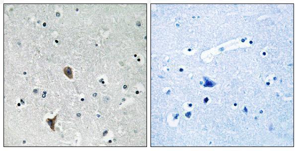
| Sample | Paraffin-embedded human brain tissue; fixative not specified (datasheet A04583-1) |
| Fixation | Image fixative and duration unreported (datasheet A04583-1); verify before use. |
| Sectioning | 4–5 µm sections on charged slides (standard) |
| Deparaffinisation | Xylene, graded ethanol series to water (standard) |
| Antigen retrieval | Heat-induced epitope retrieval in Tris-EDTA buffer, pH 9.0, 20 min at 95–98 °C (standard rule: nuclear antigen) |
| Peroxidase block | 3% H2O2, 10 min, room temperature (standard) |
| Blocking | 10% normal serum of the secondary host, 30 min, room temperature (standard) |
| Primary antibody | Rabbit anti-OXR1, 1:100-1:300 (datasheet A04583-1) |
| Primary incubation | Overnight at 4 °C (standard) |
| Detection | HRP-polymer secondary, DAB chromogen 5–10 min (standard) |
| Counterstain | Hematoxylin, blue, dehydrate and mount (standard) |
| Expected result | OXR1-positive staining in glandular cells of adrenal gland (HPA tissue IHC: High). HPA tissue profile: Cytoplasmic expression in all tissues at variable levels. No signal in the no-primary control. |
OXR1 should appear mainly as variable cytoplasmic staining in tissue IHC, with strong signal in selected glandular, neuronal, and germ-cell populations (HPA: tissue IHC, Enhanced reliability). Its reported locations include mitochondria, the nucleolus, and Golgi-associated membranes (UniProt Q8N573: subcellular location). OXR1 has no transmembrane segment, so a continuous cell-surface rim is unexpected (UniProt Q8N573: topology).
| Cytoplasmic signal is strong in adrenal or endometrial glandular cells, cortical neurons, or testicular pachytene spermatocytes (HPA: High in each). | This fits the observed tissue pattern. Compare the named cells with adjacent structures on the same section; OXR1 intensity varies by tissue, so equal staining across all cells is unnecessary (HPA: variable cytoplasmic expression). |
| Signal is predominantly a continuous cell-surface rim, with little cytoplasmic staining. | Check for an artefact or off-target staining: OXR1 lacks a transmembrane segment (UniProt Q8N573: topology), while tissue IHC is chiefly cytoplasmic (HPA: tissue IHC). Discrete organelles may be difficult to resolve by chromogenic IHC (general IHC practice). |
| Strong staining appears in ovarian stromal cells, skin keratinocytes, or vaginal squamous epithelium (HPA: Not detected in each). | Treat this as unexpected for the named cell population, rather than proof of cross-reactivity. Review morphology and controls for off-target binding or endogenous detection activity (HPA: tissue IHC; general IHC practice). |
| Color is spread across the section or tissue-free areas, obscuring cell boundaries. | This is background rather than a readable OXR1 cell pattern (general IHC practice). Check reagent-only controls, blocking, washes, and detection conditions before assigning intensity scores (general IHC practice). |
| No signal appears in a well-preserved, expected-positive cell population. | A negative result is inconclusive if the run fails in a High group, such as cerebellar granular cells or salivary glandular cells (HPA: High). Check the positive control and assay conditions before scoring the test section negative (general IHC practice). |
| Cell population and tissue (HPA: tissue IHC) | HPA reports High staining in adrenal, endometrial, and salivary glandular cells, cerebral cortical neurons, cerebellar granular cells, and pachytene spermatocytes; adipocytes are Medium. Use the specific cell population, rather than the organ alone, when comparing slides (HPA: tissue IHC). |
| Compartment and resolution (HPA: tissue IHC; UniProt Q8N573: subcellular location) | Tissue IHC is described as cytoplasmic. UniProt reports mitochondria in neuronal cells, nucleoli in fibroblasts and myoblasts, and Rab-dependent recruitment to Golgi membranes; these finer assignments should not be required from routine chromogenic sections (HPA: tissue IHC; UniProt Q8N573: subcellular location; general IHC practice). |
| Strength of tissue evidence (HPA: Enhanced reliability) | HPA labels tissue staining Enhanced, yet describes only medium consistency between staining and RNA data. Its three listed antibodies have Enhanced IHC status; that supports the reported pattern without making any single positive or negative cell an absolute diagnostic rule (HPA: tissue IHC; HPA: HPA027375, HPA027380, HPA027395). |
| Isoforms and epitope coverage (UniProt Q8N573: isoforms) | UniProt lists 8 isoforms. Whether a particular IHC antibody detects each one depends on its epitope, which is not supplied here; do not infer an isoform-specific tissue pattern or use one antibody's negative result to exclude every isoform (UniProt Q8N573: isoforms; general immunostaining practice). |
| IF/ICC expectation? (HPA: subcellular ICC-IF) | HPA's approved main ICC-IF location is vesicles, with images in U2OS and A-431. That is useful context for a separate IF/ICC design, while tissue IHC should be read against its cytoplasmic cell pattern; two listed antibodies have Uncertain ICC status (HPA: subcellular ICC-IF; HPA: HPA027380, HPA027395). |
| Situation | Likely cause | Next action |
|---|---|---|
| Positive-control tissue is blank. | The run may lack detectable signal; absence in a High population is unexpected (HPA: tissue IHC). | Verify tissue identity and the IHC-validated antibody's documented conditions; inspect detection and counterstain controls before interpreting test negatives (general IHC practice). |
| Test section is blank but the positive control stains. | The sampled cells may express less OXR1: HPA reports variable tissue levels and some cell populations as Not detected (HPA: tissue IHC). | Identify the actual cell population and compare it with HPA's cell-specific entries; record a negative only for the sampled cells under the validated run conditions (HPA: tissue IHC; general IHC practice). |
| Strong color appears in tissue-free areas or across many unrelated structures. | Diffuse deposition can arise from nonspecific detection or inadequate washing (general IHC practice). | Review the reagent-only control, blocking, washes, and detection time; score cells only once background permits reliable boundaries (general IHC practice). |
| Unexpected cell-surface rims dominate the section. | That distribution conflicts with predominantly cytoplasmic tissue staining and a protein without a transmembrane segment (HPA: tissue IHC; UniProt Q8N573: topology). | Review morphology and controls, then compare with staining from another IHC-validated antibody if available; do not call the rim specific from color alone (HPA: antibody IHC status; general IHC practice). |
| HPA-listed Not detected cells stain strongly. | Off-target binding or endogenous detection activity is possible; HPA's absence call is cell-specific rather than an organ-wide rule (HPA: tissue IHC; general IHC practice). | Confirm cell identity, inspect a reagent-only control, and assess endogenous detection activity appropriate to the chromogenic system before interpreting OXR1 positivity (general IHC practice). |
| Fine puncta or nucleolar color are inconsistent between sections. | UniProt reports several context-dependent locations, while chromogenic section resolution and tissue morphology can limit compartment assignment (UniProt Q8N573: subcellular location; general IHC practice). | Prioritize reproducible, cell-associated cytoplasmic staining in expected populations; use the separate IF/ICC guide for finer localisation questions (HPA: tissue IHC; HPA: subcellular ICC-IF). |

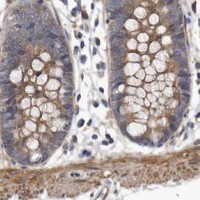
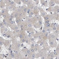
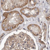


Comprehensive Human Protein Atlas IHC scoring per tissue (reliability: Enhanced — Medium consistency between antibody staining and RNA expression data.). Rows are taken directly from the HPA tissue chart — click any row's HPA link to view the source.
| Tissue | Cell type | Level | Evidence | Source |
|---|---|---|---|---|
| Adrenal gland | Glandular cells | High | Protein (IHC) | HPA → |
| Cerebellum | Granular cells - cytoplasm/membrane | High | Protein (IHC) | HPA → |
| Cerebral cortex | Neuronal cells | High | Protein (IHC) | HPA → |
| Endometrium | Glandular cells | High | Protein (IHC) | HPA → |
| Salivary gland | Glandular cells | High | Protein (IHC) | HPA → |
Troubleshoot OXR1 staining in paraffin sections by checking retrieval, cell type, compartment, and controls before interpreting chromogenic signal.
A04583-1 has a human brain paraffin-section IHC image (IHC caption), listed IF use, and Human, Mouse, and Rat reactivity (catalog applications and reactivity).
A04583-1 has an IHC image of paraffin-embedded human brain tissue with a peptide-blocked comparison (IHC caption). IF is listed as an application, and Human, Mouse, and Rat are listed as reactive species; no IF image is supplied (catalog applications, reactivity, and image records).
Which to pick: For tissue IHC, choose A04583-1: its own image shows staining in paraffin-embedded human brain tissue with a peptide-blocked comparison; the fixative is unreported (IHC caption). For IF, A04583-1 is listed at 1:50, but ICC is not separately listed and no IF image is supplied (catalog applications, dilution, and image records). For Human, Mouse, or Rat samples, A04583-1 lists reactivity across those species; the supplied IHC image documents Human tissue only, and clonality is unreported (catalog reactivity, IHC caption, and clone field).