This website uses cookies to ensure you get the best experience on our website.
- Table of Contents
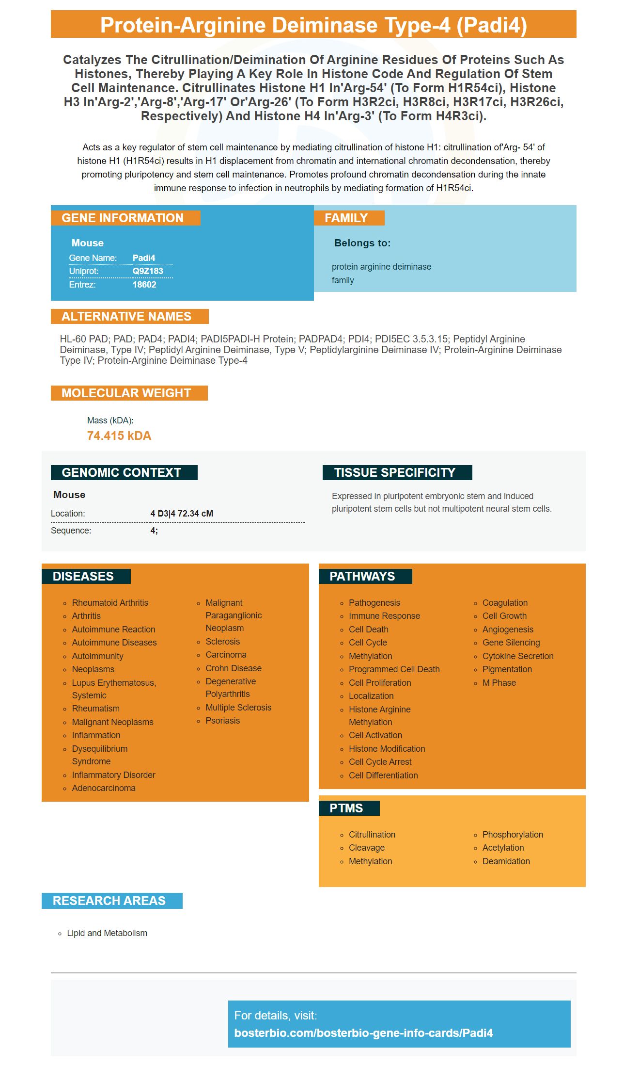
Facts about Protein-arginine deiminase type-4.
Acts as a key regulator of stem cell maintenance by mediating citrullination of histone H1: citrullination of'Arg- 54' of histone H1 (H1R54ci) results in H1 displacement from chromatin and international chromatin decondensation, thereby promoting pluripotency and stem cell maintenance. Promotes profound chromatin decondensation during the innate immune response to infection in neutrophils by mediating formation of H1R54ci.
Mouse | |
---|---|
Gene Name: | Padi4 |
Uniprot: | Q9Z183 |
Entrez: | 18602 |
Belongs to: |
---|
protein arginine deiminase family |
HL-60 PAD; PAD; PAD4; PADI4; PADI5PADI-H protein; PADPAD4; PDI4; PDI5EC 3.5.3.15; peptidyl arginine deiminase, type IV; peptidyl arginine deiminase, type V; Peptidylarginine deiminase IV; Protein-arginine deiminase type IV; protein-arginine deiminase type-4
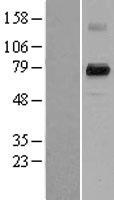
Mass (kDA):
74.415 kDA
Mouse | |
---|---|
Location: | 4 D3|4 72.34 cM |
Sequence: | 4; |
Expressed in pluripotent embryonic stem and induced pluripotent stem cells but not multipotent neural stem cells.
Boster Bio customers have the option to use PADI4 markers antibodies in their research. Boster's high-affinity prima antibodies are validated on multiple platforms with known positive or negative samples to ensure maximum specificity and affinity. Boster offers product credits to scientists who are amongst the first to review its product. Here are some of the most common uses of this marker antibody.
Researchers are turning towards boster Bio for high affinity primary antibodies against PADA4 in their quest to understand PADI4 function within the human body. The company produces highly specific antibodies that are validated for WB, IHC, and Flow. Boster antibodies are tested against a variety of cell lines and tissues. Additionally, these antibodies can be used for ELISA, Western Blotting, and immunohistochemistry applications.
Mutations in the lightchain of an antibody can increase its affinity to the antigen. These mutations usually involve the hot spot residue Phe101. L-N31R, which has a 4.6-fold higher affinity than the wild type antibody, is the single mutation with the highest affinity. All single-mutations which exhibit enhanced affinity contain a charged residue. The thermodynamic parameters are also analyzed to determine the energetic mechanism. The formation of new electrostatic interactions can also improve the durability and stability of the antibody-antigen combination.
A calibration curve was produced using a variety monoclonal anti-PADI4 antibodies. These preparations have measurable antigen binding activity, and are compatible with ELISA. Boster Bio offers high-affinity primaries against the PADI4 mark. They are available for purchase in many formats. The one that suits your research needs is the best option. Alternatively, you can also opt for a monoclonal antibody that has been specifically designed to target PADI4 as a biomarker for the autoimmune disease.
The PAD4 gene encodes an essential protein that plays a part in intranuclear histone citrullination. Citrullinated H4R3 is an important part of the response to DNA damages, activating c-Fos. This protein also regulates the transcription process of the PADI4 gene.
The PAD4 protein encodes PADI4 genes. The synovium of RA mice also contains Padi2 mRNA, but it is not present in wild-type mice. The Padi4 gene was expressed in both macrophages and granulocytes, and immunization induced its expression in both cells. Padi4 mRNA can be expressed in a mouse model of the disease using CII.
A variety of studies have found that the presence or absence of the PADI4 genetic gene is linked to RA vulnerability. A Japanese study recently found that a PADI4 susceptible haplotype produced more transcripts in vitro and in vivo. Furthermore, PADI4 produces citrullinated antigens in RA-specific autoantibodies, though its pathophysiological role is still unknown.
These mice are at higher risk for developing peripheral neuropathies, as they have lower levels IL-10 than wild-type mice. They also have lower levels IL-10, a cytokine, which acts as an antiinflammatory cytokine. Hence, the Boster Bio offers high-affinity primary antibodies against the PADI4 marker to help researchers in their research.
In mouse spleens, the expression patterns of Padi2 and PADI4 are similar. The Padi4-/ mice have higher levels in spleens, pancreatic cancer cells, and more. Wild-type mice have lower PAD2 levels. However, the expression of both markers was not significantly different in wild-type mice. Although the results are encouraging, further research is needed in order to determine whether they influence immune response.
Primary antibodies are crucial in many applications. Primary antibodies are specifically specific to the antigen they are targeting. The quality of an antibody's non-covalent bonds is what determines its strength. Specificity of an antibody is determined by its ability recognize the antigen and not to bind to unintended substances. If your primary antibodies can recognize the antigen in question with high specificity you can use them for detection, purification, measurement, and analysis.
Boster Bio's anti ACE2 antibody is catalog no. A00756. It reacts well with Mouse and can be stored at 20°C. A blocking peptide can be purchased for a nominal price if you have to use it.
Boster validates all antibodies on WB, IHC and EC to maximize productivity. If you can prove the accuracy and reliability of your results, you will be able to share your findings with colleagues. Publication of your results can earn you product credits. These are just some of many benefits to working with Boster antibodies. It allows scientists to share their discoveries and receive compensation.
All Boster antibody have been tested and validated in a specialized Histology laboratory. In addition to detecting the protein of interest, these antibodies also detect denatured forms of the protein of interest. To validate this, the company uses a 10ug/ml anti-body. The company is committed at providing high quality products for its customers. Their reputation has been built upon the reliability and efficiency of its antibodies.
Boster also offers a wide array of secondary antibodies and isotype controls. These detection systems are optimized for WB/ICC/IHC, ELISA, ELISA and other applications. Boster offers complimentary secondary antibodies and reagents in addition to high-quality products. Boster antibodies offer the highest quality primary antibodies.
The IHC protocol developed by Boster Bio contains step-by-step illustrations, recommended reagents, and an immunohistochemistry workflow guide. IHC sample preparation is a crucial step in the process and is an integral part of any test. The guide will also contain tips for preparing tissues for optimal staining. Boster Bio's guide contains specific procedures for different types and types of preserved tissue, including whole blood.
Immunohistochemistry is a technique that detects antigens within tissue sections. Antibodies can bind to specific cells antigens, which is why they are so specific. Improper preparation of the sample, or an antibody problem may result in non-specific staining. There are many ways to detect antigens. It is important to know what type of specimens you have.
The most common isotype control is the isotype. This provides a clear idea of the background level, but can be misleading. There are two types of isotype controls, polyclonal and monoclonal. Polyclonal control is derived from unimmunized animal and tends to have a higher background than monoclonal antibody.
PMID: 10092850 by Rusd A.A., et al. Molecular cloning of cDNAs of mouse peptidylarginine deiminase type I, type III and type IV, and the expression pattern of type I in mouse.
PMID: 15087120 by Chavanas S., et al. Comparative analysis of the mouse and human peptidylarginine deiminase gene clusters reveals highly conserved non-coding segments and a new human gene, PADI6.