This website uses cookies to ensure you get the best experience on our website.


- Table of Contents
Plan PER3 IHC in paraffin sections around the general cytoplasmic tissue pattern (HPA tissue IHC). Start with the catalog antibody’s 1:50–1:200 IHC dilution (datasheet), and record sampling time when comparing staining because PER3 participates in circadian regulation (UniProt).

Expected localisation, validated protocols, controls and antibodies — the at-a-glance facts below, then the full design guide.
| Expected localisation | Cytoplasmic tissue staining (HPA tissue IHC); nuclear transit (UniProt) | |
| Staining pattern | General cytoplasmic staining (HPA tissue IHC) | |
| Antigen retrieval | Tris-EDTA pH 9.0 HIER, 95–98 °C, 20 min (rule: nuclear antigen) | |
| Positive control | Adrenal gland+4 more · see all | |
| Negative control | None in HPA (detected in all 44 tissues); use no-primary + isotype controls |
| Fixation | Keep fixation consistent across sections. (standard IHC practice; not target-specific) | |
| Caveat | Low staining–RNA consistency; verify the pattern (HPA tissue IHC) | |
| Regulation | Circadian timing can affect expression (UniProt) | |
| Isoform / epitope | 2 isoforms; epitope impact is unknown (UniProt) |
The catalog antibody’s IHC-P protocol (datasheet) is accompanied by four published PER3 IHC protocols (PMC4206725; PMC12644904; PMC11439802; PMC11743850).
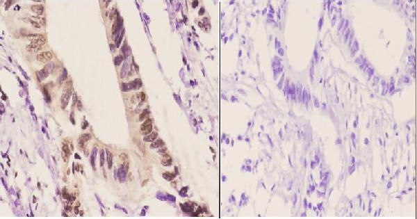
| Sample | Paraffin-embedded human rectum carcinoma tissue; fixative not specified (datasheet A01835) |
| Fixation | Image fixative and duration unreported (datasheet A01835); verify before use. |
| Sectioning | 4–5 µm sections on charged slides (standard) |
| Deparaffinisation | Xylene, graded ethanol series to water (standard) |
| Antigen retrieval | Heat-induced epitope retrieval in Tris-EDTA buffer, pH 9.0, 20 min at 95–98 °C (standard rule: nuclear antigen) |
| Peroxidase block | 3% H2O2, 10 min, room temperature (standard) |
| Blocking | 10% normal serum of the secondary host, 30 min, room temperature (standard) |
| Primary antibody | Rabbit anti-PER3, 1:50-1:200 (datasheet A01835) |
| Primary incubation | Overnight at 4 °C (standard) |
| Detection | HRP-polymer secondary, DAB chromogen 5–10 min (standard) |
| Counterstain | Hematoxylin, blue, dehydrate and mount (standard) |
| Expected result | PER3-positive staining in glandular cells of adrenal gland (HPA tissue IHC: High). HPA tissue profile: General cytoplasmic expression. No signal in the no-primary control. |
PER3 should appear mainly in the cytoplasm, with possible nuclear staining because it can enter the nucleus through binding clock partners (UniProt P56645). HPA reports general cytoplasmic tissue staining, including high staining in several glandular, hematopoietic and neural cell populations (HPA tissue IHC). PER3 has no transmembrane segment (UniProt P56645 topology). Interpret this pattern cautiously: HPA rates its tissue IHC evidence Approved, with low staining–RNA consistency and external verification pending (HPA tissue IHC).
| Cytoplasmic staining in adrenal glandular cells, bronchial respiratory epithelial cells or bone marrow hematopoietic cells. | This matches HPA's reported High staining in those populations and its general cytoplasmic profile (HPA tissue IHC). Compare staining with cell morphology and a matched control; HPA's Approved rating carries a low staining–RNA consistency caveat (HPA tissue IHC). |
| Cytoplasmic staining with a nuclear component in otherwise plausible positive cells. | Nuclear PER3 is biologically plausible: UniProt places PER3 in both compartments and describes nuclear entry through binding PER1, PER2, CRY1 or CRY2 (UniProt P56645). Judge the nuclear signal alongside cytoplasmic staining and controls; HPA describes the overall tissue pattern as cytoplasmic (HPA tissue IHC). |
| Predominantly cell-surface, extracellular or structure-shaped staining without convincing intracellular signal. | That distribution conflicts with the reported cytoplasmic pattern (HPA tissue IHC) and PER3's lack of a transmembrane segment (UniProt P56645 topology). Treat it as suspect; check an adjacent section and a no-primary control before assigning PER3 localisation. |
| Strong staining in adipocytes, especially when expected cell populations lack signal. | HPA lists adipocytes as Low, while reporting High staining in several other cell populations (HPA tissue IHC). This mismatch raises concern for nonspecific staining or endogenous detection activity; it does not establish that every stained adipocyte is a false positive. |
| No detectable staining in a tissue and cell population listed as High by HPA. | A negative section is inconclusive: HPA's tissue observations have low staining–RNA consistency and await external verification (HPA tissue IHC). First assess section quality, control performance and the IHC workflow; avoid calling the specimen PER3-negative from one failed stain. |
| Cell population and tissue | HPA reports High staining in adrenal and breast glandular cells, bronchial respiratory epithelial cells, bone marrow hematopoietic cells and selected brain cells, but Low staining in adipocytes (HPA tissue IHC). Score the intended cell population rather than treating the entire section as uniformly positive. |
| Compartment and clock partners | PER3 is mainly cytoplasmic and can translocate to the nucleus through binding PER1, PER2, CRY1 or CRY2 (UniProt P56645). Record cytoplasmic and nuclear staining separately; a nuclear component alone does not establish altered PER3 expression. |
| Circadian timing | PER3 participates in the circadian clock, whose components show approximately 24-hour rhythms (UniProt P56645). If comparing specimens collected at different times, record collection time and avoid attributing an intensity difference to disease or treatment without considering timing. |
| Strength of tissue evidence | HPA calls the tissue IHC profile Approved, reports low consistency between staining and RNA expression, and says external verification is pending; its listed antibody HPA019530 is IHC Approved (HPA tissue IHC; HPA antibodies). Use controls and confirm unexpected patterns before interpreting them biologically. |
| IF/ICC: should the pattern match? | HPA reports supported cytosolic localisation by ICC-IF, consistent with the main cytoplasmic expectation in tissue IHC (HPA subcellular; HPA tissue IHC). UniProt also allows nuclear PER3 through partner binding (UniProt P56645); compare compartment patterns without assuming identical intensities across applications. |
| Situation | Likely cause | Next action |
|---|---|---|
| No signal in an HPA High cell population. | The run may lack detectable signal, or the selected tissue pattern may not reproduce; HPA notes low staining–RNA consistency (HPA tissue IHC). | Check a positive control on the same run, then review the catalog antibody's IHC-P dilution, antigen retrieval and detection steps. These are general IHC checks, not evidence that PER3 has a known retrieval requirement. |
| Strong nuclear staining with little or no cytoplasmic staining. | Nuclear entry is possible (UniProt P56645), but a predominantly nuclear slide differs from HPA's general cytoplasmic tissue profile (HPA tissue IHC). | Score compartments separately, compare with a plausible positive tissue and examine the no-primary control. If the pattern persists, describe it as unexpected rather than assigning a mechanism from staining alone. |
| Colour extends across stroma, empty spaces or section edges. | The distribution does not resemble intracellular PER3 (HPA tissue IHC; UniProt P56645); general IHC causes include nonspecific reagent deposition or uneven detection. | Inspect morphology and no-primary control staining, then review blocking, washes and chromogen development as general IHC workflow checks. Exclude noncellular colour from PER3 scoring. |
| Many unrelated cell types stain similarly, including strong adipocyte staining. | HPA reports Low staining in adipocytes and High staining in selected populations (HPA tissue IHC). Broad colour could reflect nonspecific binding or endogenous detection activity. | Compare the no-primary and appropriate detection controls, assess endogenous enzyme blocking if using enzyme detection, and score each cell population separately. Controls distinguish detection background from antibody-associated staining. |
| Comparable sections show different staining intensity. | Collection timing could contribute because PER3 belongs to the circadian clock (UniProt P56645); differences in cell composition or staining runs can also affect an IHC comparison. | Record collection time, compare the same cell population, and include a common control across runs. Do not infer a biological change from intensity alone, especially given HPA's low staining–RNA consistency (HPA tissue IHC). |
| A crisp membrane-like outline dominates the stain. | A membrane-dominant pattern conflicts with HPA's cytoplasmic profile and PER3's lack of a transmembrane segment (HPA tissue IHC; UniProt P56645 topology). | Check whether the outline remains in the no-primary control, review morphology under the counterstain, and repeat with an appropriate control if needed. Do not score the outline as confirmed PER3. |


Comprehensive Human Protein Atlas IHC scoring per tissue (reliability: Approved — Low consistency between antibody staining and RNA expression data. Pending external verification.). Rows are taken directly from the HPA tissue chart — click any row's HPA link to view the source.
| Tissue | Cell type | Level | Evidence | Source |
|---|---|---|---|---|
| Adrenal gland | Glandular cells | High | Protein (IHC) | HPA → |
| Appendix | Glandular cells | High | Protein (IHC) | HPA → |
| Bone marrow | Hematopoietic cells | High | Protein (IHC) | HPA → |
| Breast | Glandular cells | High | Protein (IHC) | HPA → |
| Bronchus | Respiratory epithelial cells | High | Protein (IHC) | HPA → |
| Tissue | Cell type | Level | Evidence | Source |
|---|---|---|---|---|
| None in HPA: PER3 is detected in all 44 scored tissues. Use a no-primary (secondary-only) and an isotype control instead. | ||||
Troubleshoot PER3 chromogenic IHC in paraffin sections by checking retrieval, compartment patterns, controls, and circadian sampling context.
A01835 has IHC data from paraffin-embedded human rectum carcinoma (catalog IHC image caption); Human, Mouse, and Rat reactivity is listed (catalog reactivity). No IF data is supplied (catalog).
A01835 is listed for IHC and WB in Human, Mouse, and Rat samples (catalog applications and reactivity). Its IHC image shows nuclear and cytoplasmic staining in paraffin-embedded human rectum carcinoma at 1:50, with a PBS primary-antibody omission control (catalog IHC image caption).
Which to pick: For tissue IHC, choose A01835: it is a rabbit antibody listed for IHC at 1:50–1:200, with its own paraffin-section image at 1:50 (catalog host, applications, dilution, and IHC image caption). The caption does not report a fixative (catalog IHC image caption). No SKU in this payload lists IF/ICC, so there is no IF/ICC pick; A01835 lists Human, Mouse, and Rat reactivity, but its supplied IHC image documents only human tissue (catalog applications, reactivity, and IHC image caption).