This website uses cookies to ensure you get the best experience on our website.
- Table of Contents
1 Q&As
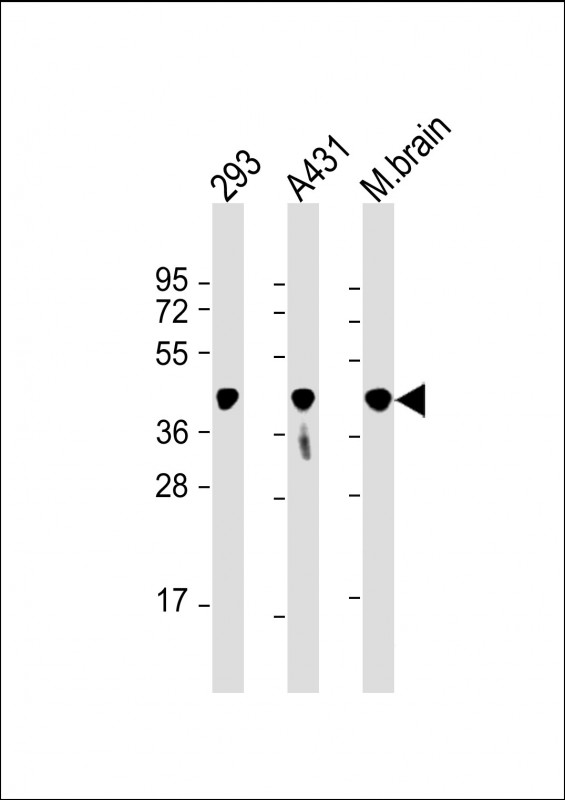
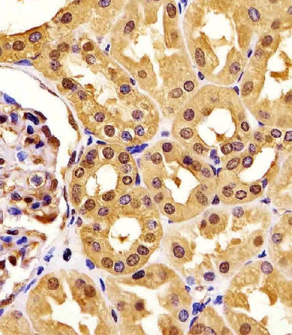
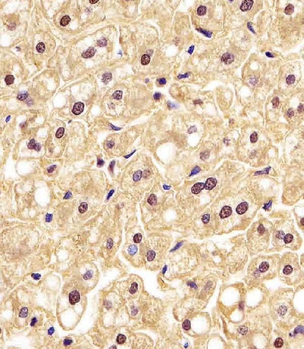
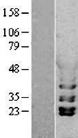
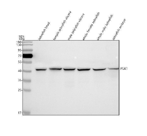
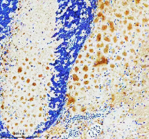
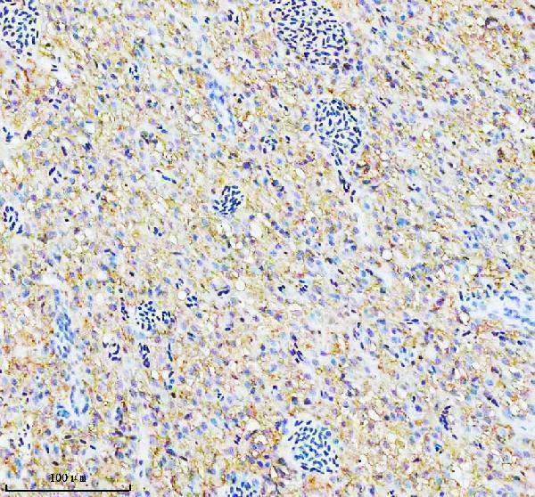
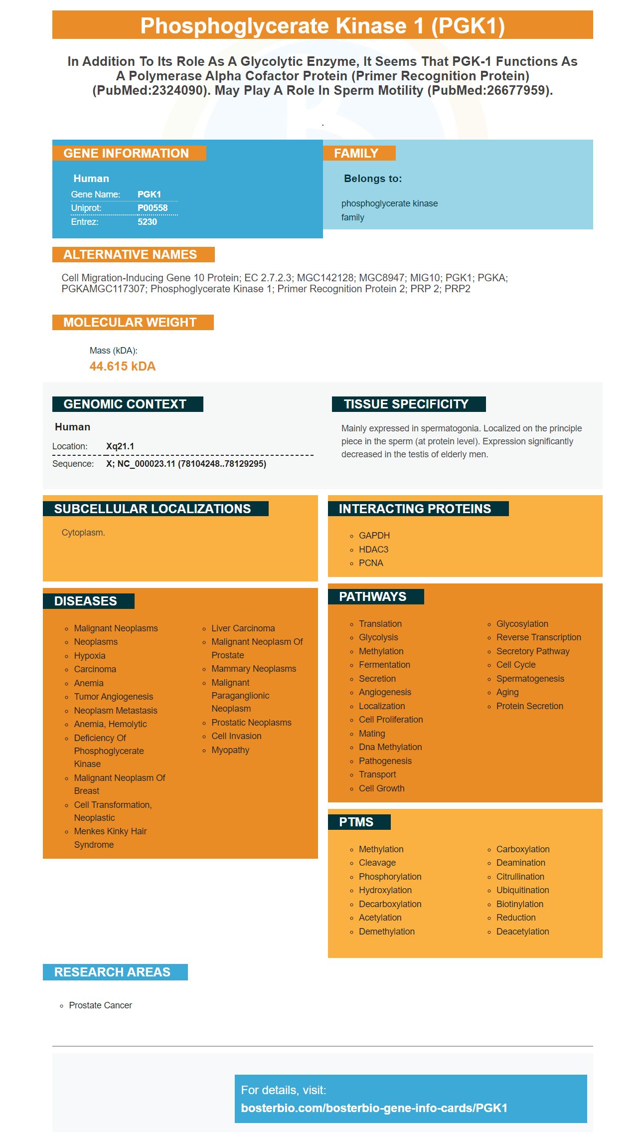
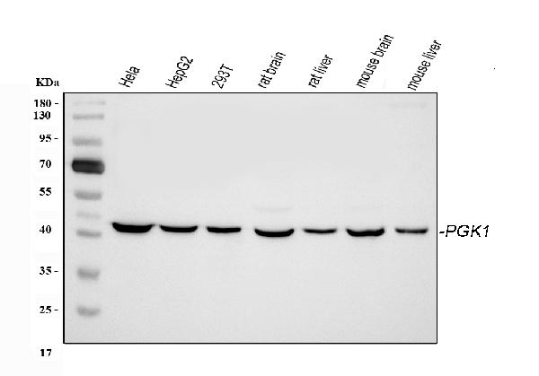
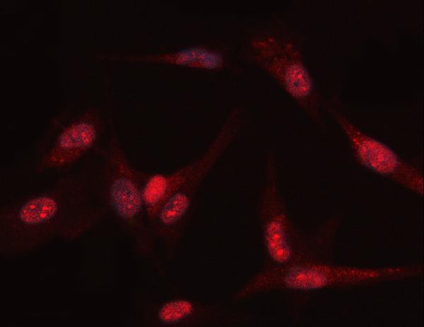
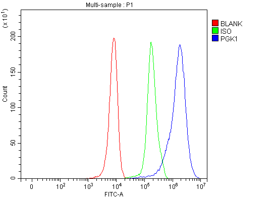
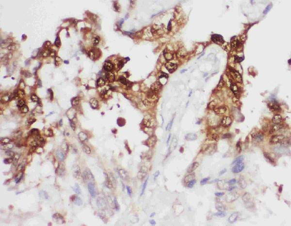
Facts about Phosphoglycerate kinase 1.
.
Human | |
---|---|
Gene Name: | PGK1 |
Uniprot: | P00558 |
Entrez: | 5230 |
Belongs to: |
---|
phosphoglycerate kinase family |
Cell migration-inducing gene 10 protein; EC 2.7.2.3; MGC142128; MGC8947; MIG10; PGK1; PGKA; PGKAMGC117307; phosphoglycerate kinase 1; Primer recognition protein 2; PRP 2; PRP2
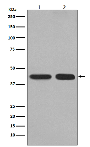
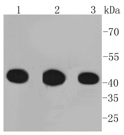
Mass (kDA):
44.615 kDA
Human | |
---|---|
Location: | Xq21.1 |
Sequence: | X; NC_000023.11 (78104248..78129295) |
Mainly expressed in spermatogonia. Localized on the principle piece in the sperm (at protein level). Expression significantly decreased in the testis of elderly men.
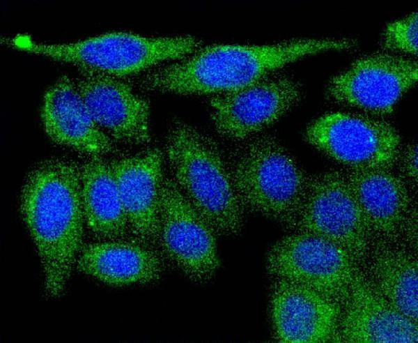
Cytoplasm.
PGK1 is an enzyme involved in glycolysis. This enzyme is also recognized for its ability to inhibit apoptosis. The inhibitors TZ, Ta, and PGK1 are all effective anti-apoptotic agents. Boster has tested its antibodies on various platforms by using known positive and negative samples to ensure best quality. Boster also provides product credits to scientists who analyze Boster antibodies first.
Cellular metabolic stress regulates the function of PGK1. O-GlcNAcylation increases PGK1 interaction with TOM20 and increases its to substrates. The function of PGK1 in cells is unknown, however, it is believed to play an important role in the metabolism sugars. Here are a few possible roles for PGK1.
Proliferating cancer cells exhibit an abnormal glucose metabolism. They exhibit an increased rate of glycolysis and glucose uptake. They also maintain the highest rate of this metabolism, converting most of the glucose into lactic acid. Interestingly, O-GlcNAcylation increased the enzyme activity of PGK1 and caused its transfer to mitochondria. O-GlcNAcylation also slowed down the PDH enzyme and caused it to lose its capability to function.
The overexpression of PGK1 in human tumors increased cellular levels of 3-phosphoserine, a essential metabolite in the de novo serine synthesis pathway. This could be due to a decrease in the metabolism of 3-PG an enzyme that is absorbed into the SSP. The decrease in T255 O-GlcNAcylation in PGK1 did not affect serine levels in the cells.
Several human tumors, including breast cancer, have been found to have an increase in PGK1. Although studies of Acetylation by this enzyme have been limited in their applications to mitochondrial function, new research has revealed that metabolic changes in cancer cells alters the epigenetic map, leading both to malignant transformation and the growth of tumors. By preventing the activity of this enzyme could be a good treatment for cancer. It is important to understand how this enzyme is expressed.
TZ has been proven to block apoptosis in cultivated mammalian cell lines. This compound blocks apoptosis via blocking the caspase-mediated pathway. It is able to inhibit apoptosis at low levels of 0.1 millimol. However, a comprehensive mechanism of TZ's antiapoptotic effect remains elusive.
This compound also blocks the function of Pgk1 which is a glycolysis enzyme that suppresses apoptosis in yeast. It also has positive effects on human cell viability. To determine its effectiveness, purified Pgk1 was used in a forward reaction assay. Furthermore, higher levels of TZ inhibited the activity of Pgk1, while lower levels of TZ activated it.
The compound was intraperitoneally administered to mice. The mice were given the dose of 240 mg/kg Avertin i.p. for seven days. The mice were monitored every 12 hours over the course of seven days. In addition to the effect of TZ on apoptosis, it also inhibited the activity of Hsp90. This study highlights the need for further studies on the function of Hsp90 in the process of apoptosis.
In a separate study the present inventors investigated whether TZ inhibited apoptosis in cells that are in an ischemic state. A condition known as ischemic is when cells are not receiving enough blood flow to supply oxygen and glucose. In the hypoxic condition, glucose is converted to lactic acid, which supplies little energy. However, the process of energy generation resumes when adequate oxygen is delivered to the cells.
Tubocapsenolide (TA) blocks apoptosis in human cancer cells through inducing death and stopping the cell cycle at G1 stage. This was in turn correlated with a decrease in glutathione content and a brief increase in reactive oxygen species. TA was found to interfere with chaperone machinery Hsp90 and Hsp70.
Several previous studies have shown that TA has anticancer properties. It triggers cell death by suppressing the production of and reducing levels of mediators of oxidative damage. It has also been proven to trigger apoptosis in IPECJ2 cells and block the proliferation of various cancerous cells. These findings suggest that TA reduces apoptosis through induction of apoptosis.
In addition, TA inhibits the mitochondrial pathway for apoptosis within IPEC-J2 cells. ZLEHD-FMK, a caspase-9 inhibitor, significantly inhibited TA-induced apoptosis. Additionally, TA induced the ratio of Bcl-2 to Bax and increased the cleavage of caspase-3 as well as caspase-9.
TA hinders Hsp70 activity chaperone compound that is crucial for the apoptotic process. It binds to Hsp90, which is a client protein, and works to block chaperone activity. The result is that cells become apoptotic and are unable complete the cycle of cells. Thus, TA could be a novel cancer treatment strategy. However, further research is required to establish the mechanism behind TA's anti-apoptosis effects.
The Libdock docking method used in Bostro Bio is a fast and effective method to detect ligand-protein interactions. This method generates conformations based upon distance geometry using clique-finding algorithms. Essential points allows users to define atoms that need to connect to a ligand. If the interaction is well-known essential points are useful. Different scoring algorithms are available to determine if the conformation is acceptable.
In this study, five polyunsaturated fatty acid were chosen to further investigate. Linolenic acid and Arachidonic acid were chosen for further research. The compounds were screened using Discover studio 4.0, Libdock docking, and PMF docking. These ligands scored better than cocrystallized drugs that were redocked.
The process of molecular docking has become a popular method to discover new drugs. This technique is utilized to simulate small molecule interactions with proteins, as well as to understand their behavior in binding sites, and reveal fundamental biochemical processes. The docking process is comprised of two steps. Prediction of the ligand’s shape and location, and evaluation of the ability to bind between the protein and ligand. The resulting docked structure can be visualized using scoring strategies.
Bostro Bio's Libdock docking technique is able to accurately detect ligand/protein interactions in a variety biological processes. The Libdock docking method is more precise and powerful than other methods and has a higher score that CDOCKER. The Lidock docking system also includes AMBER-derived Force-field scoring, as well as the GB/SA score for solvation.
Boster Bio PGK1 ELSA kit is thoroughly tested to ensure that the results are reliable. To ensure consistency the company measures CV percent variance (or variation from lot to lot) of the tests. Its assay kits are highly precise, with a coefficient of variation (CV percent) less than 10%. These assays are able to assess intra-assay precision and accuracy between assays.
This is an ELISA that is competitive. ELISA, with one antibody that focuses on a specific part of the antigen. In a sandwich ELISA the first antibody, referred to as the capture antibody, covers the wells. The results are graphed in terms of optical density or fluorescence vs. concentration. The test results are then evaluated against the standard curve. This is a sequence of dilutes of an antigen. This is a great way to determine the amount of antigen present.
Boster Bio PGK1 ELSA validation kit concentration was determined using a technique called the tyrosinekinase inhibition. After amplification of the mPGK1 CDNA, mPGK1 was cloned into an E2 vector and expressed in the BL21 (DE3) chemically competent cells that are chemically competent. Following this cell lysis, the cells were then lysed using an extraction buffer that contained five milligrams imidazole and a protease inhibition cocktail. The final fractions of pure mPGK1 were pooled in a buffer containing 100 mM NaCl and 2 mM DTT, and then flash-cooled using liquid nitrogen.
The Boster Bio PGK1 ELSA has been validated to verify its reproducibility. Its sensitivity was evaluated using three independent tests. The results were comparable to those obtained by Boster Bio's ELISA. Boster Bio's ELISA Kit validation was also performed using two methods - direct and competitive. The direct method was used when commercial ELISA kits were not available for the targeted protein.
The PGK1 marker is used for a variety of purposes. The protein is released into extracellular space, making it an ideal liquid biopsy. Rapid diagnostic tools are offered by abnormal PGK1 levels that are found in tumor tissues peripheral blood, saliva and saliva. It is also possible to use alongside other tumor markers to improve the sensitivity of diagnosis. For instance, serum PGK1 levels could be used together with the levels of AFP and cKit to enhance the accuracy of diagnosis.
The PGK1 gene is extremely expressed in cells that have high levels of lactic acid. The levels of expression are high when the medium is rich in glucose, but are low in carbon sources, such as yeast. The PGK mRNAs include zeocin sensitive probes which indicate that the vector of the virus is not intact. The pB3 sensitive to zeocin PGK marker is also found in the pCRE3 vector indicating that a plasmid carrying the PGK gene has been destroyed. Surose gradient fractionation and Northern Blotting can be utilized to identify intermediates in PGK1 degradation of mRNA.
PGK1 is involved in angiogenesis and glucose metabolism. This causes increased tumorigenesis and the Warburg Effect. Extracellular PGK1 activates the production of angiostatin (a protein that inhibits blood vessel formation) by acting as a disulfide reducer. Its high levels in cells encourage tumor angiogenesis.
PMID: 6188151 by Michelson A.M., et al. Isolation and DNA sequence of a full-length cDNA clone for human X chromosome-encoded phosphoglycerate kinase.
PMID: 2995995 by Michelson A.M., et al. Structure of the human phosphoglycerate kinase gene and the intron- mediated evolution and dispersal of the nucleotide-binding domain.