This website uses cookies to ensure you get the best experience on our website.


- Table of Contents
Plan paraffin-section PHF8 IHC with the catalog antibody at 1:50 (datasheet M03288). Compare nuclear staining with colon glandular cells or kidney tubular cells, where staining is high, while allowing for occasional cytoplasmic signal (HPA tissue IHC).

Expected localisation, validated protocols, controls and antibodies — the at-a-glance facts below, then the full design guide.
| Expected localisation | Predominantly nuclear, with occasional cytoplasmic staining (HPA tissue IHC) | |
| Staining pattern | Variable nuclear staining across cell types; occasional cytoplasm (HPA tissue IHC) | |
| Antigen retrieval | EDTA pH 8.0 HIER, heat-mediated (datasheet M03288) | |
| Positive control | Cerebellum+4 more · see all | |
| Negative control | Liver+1 more · see all |
| Fixation | Matched tissue-IHC evidence does not establish the fixation claim. Validate the specimen-specific method before use. (selected-SKU IHC image M03288) | |
| Caveat | Tissue staining has medium consistency with RNA expression (HPA tissue IHC) | |
| Regulation | Tissue-specific expression regulation is not annotated (UniProt) | |
| Isoform / epitope | 5 isoforms; check epitope coverage across variants (UniProt) |
The catalog antibody’s IHC-P protocol (datasheet M03288) is accompanied by published PHF8 IHC methods for gallbladder carcinoma and melanoma (PMC10047028; PMC8856617).
| Sample | Paraffin-embedded human prostate cancer tissue; fixative not specified (datasheet M03288) |
| Fixation | Image fixative and duration unreported (datasheet M03288); verify before use. |
| Sectioning | 4–5 µm sections on charged slides (standard) |
| Deparaffinisation | Xylene, graded ethanol series to water (standard) |
| Antigen retrieval | Heat retrieval: EDTA pH 8.0 (datasheet M03288); 20 min, 95–100 °C (standard) |
| Peroxidase block | 3% H2O2, 10 min, room temperature (standard) |
| Blocking | 10% goat serum (datasheet M03288) |
| Primary antibody | Rabbit monoclonal (clone ADAI-16) anti-PHF8, 1:50 (datasheet M03288) |
| Primary incubation | Overnight at 4 °C (datasheet M03288) |
| Detection | HRP-conjugated secondary, DAB chromogen (datasheet M03288) |
| Counterstain | Hematoxylin, blue, dehydrate and mount (standard) |
| Expected result | PHF8-positive staining in cells in molecular layer of cerebellum (HPA tissue IHC: High). HPA tissue profile: General nuclear with occasional cytoplasmic expression at variable levels. No signal in the no-primary control. |
PHF8 should stain mainly nuclei in paraffin sections, with high staining reported in colon glandular cells, kidney tubule cells and several other cell types (HPA tissue IHC). HPA describes a general nuclear pattern with occasional cytoplasmic expression, but rates tissue IHC only Approved: antibody staining and RNA show medium consistency, and external verification is pending (HPA tissue IHC). PHF8 is nuclear, can occur in nucleoli, and has no transmembrane segment (UniProt Q9UPP1).
| Clear nuclear chromogen in colon glandular cells or kidney tubule cells. | This matches cell types scored High by HPA (HPA tissue IHC: colon glandular cells; kidney tubule cells). Judge the cell population and nuclear compartment together; occasional cytoplasmic staining can occur, but the reported overall pattern is nuclear (HPA tissue IHC). |
| Predominantly membranous or broadly cytoplasmic staining, with little nuclear signal. | This does not fit PHF8’s nuclear localization or lack of a transmembrane segment (UniProt Q9UPP1). HPA reports occasional cytoplasmic expression, so a small cytoplasmic component alone is inconclusive (HPA tissue IHC). Reassess specificity when the nonnuclear pattern dominates. |
| Strong signal in liver cholangiocytes or oral mucosal squamous cells. | Both populations were scored Not detected by HPA, although esophageal squamous cells were scored High (HPA tissue IHC). Check cell identity and controls; unexpected chromogen may reflect cross-reactivity or endogenous detection activity (general IHC practice). |
| Diffuse chromogen across nuclei, cytoplasm, stroma or empty spaces. | A widespread haze obscures the cell and compartment pattern expected for PHF8 (HPA tissue IHC; UniProt Q9UPP1). Uneven blocking, excess primary antibody or detection background can produce nonspecific staining (general IHC practice); intensity alone cannot confirm a positive result. |
| No nuclear signal in colon glands or kidney tubules. | These are reported High cell populations, so a blank result warrants a technical check (HPA tissue IHC). Examine tissue preservation, retrieval, antibody dilution and detection controls as general IHC checks; HPA does not establish PHF8-specific fixation or retrieval sensitivity. |
| Cell population | PHF8 staining varies by cell type: colon glandular cells and kidney tubule cells are High, while liver cholangiocytes are Not detected (HPA tissue IHC). Score named populations rather than treating an entire organ as uniformly positive or negative. |
| Cell-cycle state | PHF8 is recruited to H3K4me3 chromatin in interphase and dissociates from chromatin on entry into mitosis (UniProt Q9UPP1). Nuclear distribution may therefore differ in mitotic cells; this annotation does not predict a specific chromogenic intensity. |
| Subnuclear distribution | UniProt lists nucleus and nucleolus (UniProt Q9UPP1). HPA’s ICC-IF summary instead identifies the nucleoplasm as the supported main location (HPA subcellular ICC-IF). Do not require a prominent nucleolar focus to score tissue IHC. |
| Antibody evidence | The listed IHC antibody HPA038779 is Approved; the HPA tissue profile reports medium staining–RNA consistency and pending external verification (HPA antibodies; HPA tissue IHC). Compare compartment and cell type with controls before treating an isolated signal as specific. |
| Isoforms and processing | UniProt lists five PHF8 isoforms, no signal peptide or propeptide, and a full-length chain (UniProt Q9UPP1). These annotations do not establish which isoforms the IHC antibody detects; interpret unexpected staining through controls, not assumed isoform coverage. |
| IF/ICC Q: Where should PHF8 appear? | A: Mainly in the nucleoplasm (HPA subcellular ICC-IF: supported); UniProt also lists nucleolar localization (UniProt Q9UPP1). This location check is for IF/ICC interpretation, not an alternative tissue IHC protocol. |
| Situation | Likely cause | Next action |
|---|---|---|
| The expected nuclear signal is absent in a reported High cell population. | Retrieval, primary-antibody dilution or detection may have failed (general IHC practice); the HPA High designation describes an observed population, not a guarantee for every section (HPA tissue IHC). | Check the catalog antibody’s IHC-P instructions and run a reported High population, such as colon glands, beside the specimen (HPA tissue IHC). Verify each detection step with appropriate controls (general IHC practice). |
| Chromogen fills cytoplasm or outlines cell membranes more strongly than nuclei. | The dominant distribution conflicts with nuclear PHF8 and its lack of a transmembrane segment (UniProt Q9UPP1), though occasional cytoplasmic expression is reported (HPA tissue IHC). | Confirm morphology and nuclear counterstain, then compare a no-primary control and a reported High tissue (general IHC practice; HPA tissue IHC). Reassess antibody dilution and detection background before scoring. |
| A reported Not detected population appears strongly positive. | Liver cholangiocytes and oral mucosal squamous cells were Not detected in the HPA profile (HPA tissue IHC). Misidentified cells, nonspecific binding or endogenous detection activity can explain an unexpected IHC signal (general IHC practice). | Recheck the cell population and inspect no-primary and detection controls (general IHC practice). Compare with a reported High population in the same staining run (HPA tissue IHC). |
| The whole section has a brown haze or deposits outside cells. | Diffuse background prevents a compartment call and can arise from excessive antibody or inadequate blocking or washing (general IHC practice). It does not resemble HPA’s general nuclear profile (HPA tissue IHC). | Inspect a no-primary control, review blocking and washes, and adjust antibody dilution within the validated IHC workflow (general IHC practice). Score PHF8 only where individual stained nuclei remain distinguishable. |
| A nucleolar focus is absent despite convincing nuclear staining. | HPA identifies the nucleoplasm as the supported main IF/ICC location, while UniProt also lists nucleoli (HPA subcellular ICC-IF; UniProt Q9UPP1). Neither source requires a visible nucleolar focus in tissue IHC. | Score the nuclear pattern in the correct cell population against tissue controls (HPA tissue IHC; general IHC practice). Avoid rejecting a clear nuclear result solely because nucleoli are not distinct. |
| Replicate sections show different staining intensity. | HPA reports variable tissue levels and only medium agreement between staining and RNA; external verification remains pending (HPA tissue IHC). Section quality or run conditions can also affect chromogenic intensity (general IHC practice). | Compare the same named cell population across runs and include a reported High tissue control (HPA tissue IHC). Review section quality, retrieval, dilution and detection consistency without assuming a PHF8-specific fixation effect (general IHC practice). |
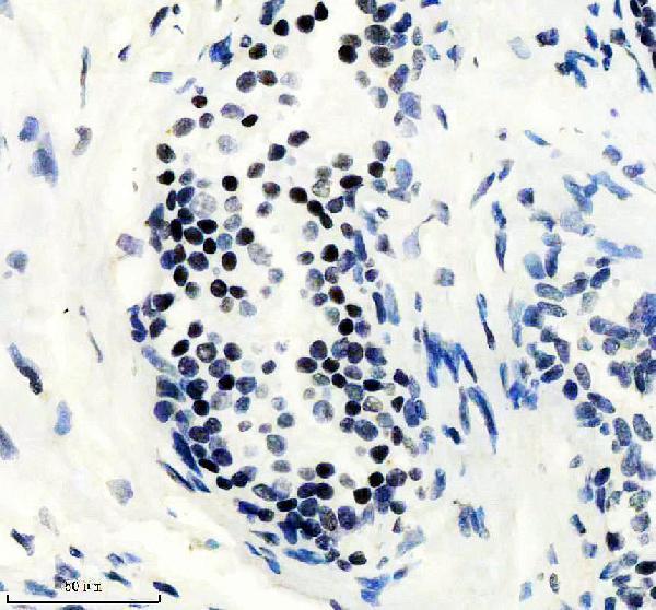
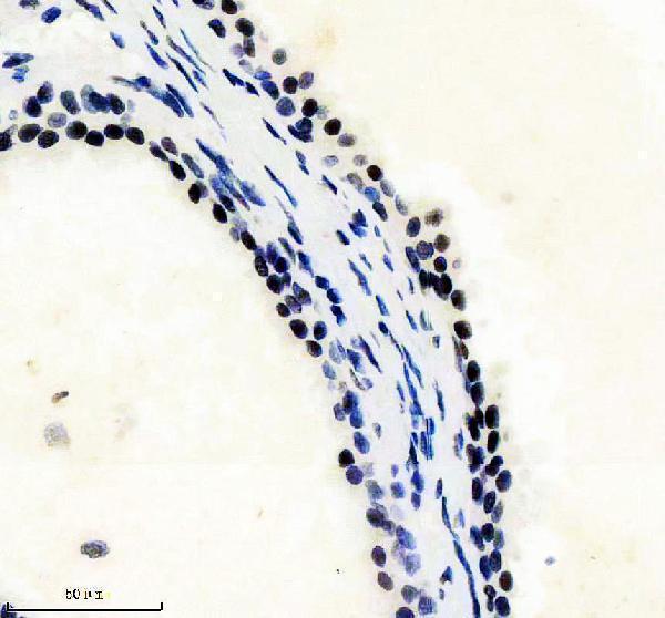
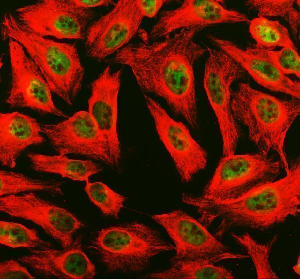


Comprehensive Human Protein Atlas IHC scoring per tissue (reliability: Approved — Medium consistency between antibody staining and RNA expression data. Pending external verification.). Rows are taken directly from the HPA tissue chart — click any row's HPA link to view the source.
| Tissue | Cell type | Level | Evidence | Source |
|---|---|---|---|---|
| Cerebellum | Cells in molecular layer | High | Protein (IHC) | HPA → |
| Cerebral cortex | Glial cells | High | Protein (IHC) | HPA → |
| Colon | Glandular cells | High | Protein (IHC) | HPA → |
| Esophagus | Squamous epithelial cells | High | Protein (IHC) | HPA → |
| Kidney | Cells in tubules | High | Protein (IHC) | HPA → |
Troubleshoot PHF8 staining in paraffin sections using the catalog antibody’s tissue IHC conditions, nuclear localisation evidence, and matched controls.
PHF8 image data cover human paraffin-embedded prostate cancer tissue and HELA cells; M03288 lists Human, Mouse and Rat reactivity, while A03288-2 lists Human (catalog captions; catalog reactivity).
M03288 will render with an IHC image from a paraffin-embedded human prostate cancer section (M03288 IHC caption). A03288-2 will render with an IF/ICC image from HELA cells (A03288-2 IF caption).
Which to pick: For tissue IHC, choose rabbit monoclonal M03288: its image documents a 1:50 dilution and EDTA pH 8.0 retrieval on a paraffin section; the fixative is unreported (M03288 catalog; M03288 IHC caption). For human IF/ICC, choose A03288-2, shown in HELA cells at 5 μg/mL (A03288-2 IF caption). For mouse or rat samples, M03288 is the option with listed reactivity, although its depicted IHC sample is human (M03288 catalog reactivity; M03288 IHC caption).