This website uses cookies to ensure you get the best experience on our website.


- Table of Contents
Plan chromogenic PICK1 IHC in paraffin sections using the catalog antibody’s documented workflow (datasheet A02457-2). Expect predominantly cytoplasmic staining, with high signal in endocrine glandular cells and CNS neurons (HPA tissue IHC).

Expected localisation, validated protocols, controls and antibodies — the at-a-glance facts below, then the full design guide.
| Expected localisation | Predominantly cytoplasmic tissue staining (HPA tissue IHC) | |
| Staining pattern | Cytoplasmic staining in endocrine glandular cells and CNS neurons (HPA tissue IHC) | |
| Antigen retrieval | EDTA pH 8.0 HIER, heat-mediated (datasheet A02457-2) | |
| Positive control | Adrenal gland+4 more · see all | |
| Negative control | Spleen+1 more · see all |
| Fixation | Keep paraffin-section fixation consistent across samples. (standard IHC practice; not target-specific) | |
| Caveat | Staining and RNA expression show medium consistency (HPA tissue IHC) | |
| Regulation | No specific expression regulator reported (UniProt) | |
| Isoform / epitope | 2 isoforms; epitope differences unspecified; no transmembrane segment (UniProt) |
The catalog antibody’s IHC-P protocol is paired with published chromogenic PICK1 staining methods for dewaxed sections (PMC8285178) and mouse frozen sections (PMC3038962).
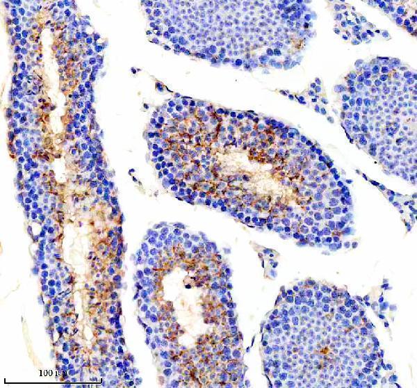
| Sample | Paraffin-embedded mouse testis tissue; fixative not specified (datasheet A02457-2) |
| Fixation | Image fixative and duration unreported (datasheet A02457-2); verify before use. |
| Sectioning | 4–5 µm sections on charged slides (standard) |
| Deparaffinisation | Xylene, graded ethanol series to water (standard) |
| Antigen retrieval | Heat retrieval: EDTA pH 8.0 (datasheet A02457-2); 20 min, 95–100 °C (standard) |
| Peroxidase block | 3% H2O2, 10 min, room temperature (standard) |
| Blocking | 10% goat serum (datasheet A02457-2) |
| Primary antibody | Rabbit anti-PICK1, 2-5 μg/ml (datasheet A02457-2) |
| Primary incubation | Overnight at 4 °C (datasheet A02457-2) |
| Detection | HRP-conjugated secondary, DAB chromogen (datasheet A02457-2) |
| Counterstain | Hematoxylin, blue, dehydrate and mount (standard) |
| Expected result | PICK1-positive staining in glandular cells of adrenal gland (HPA tissue IHC: High). HPA tissue profile: General cytoplasmic expression, most abundant in endocrine tissues and CNS. No signal in the no-primary control. |
PICK1 staining should be predominantly cytoplasmic, with possible perinuclear or membrane-associated signal (UniProt Q9NRD5 localisation; UniProt Q9NRD5 topology: no transmembrane segment). Strong staining is documented in selected neuronal, endocrine glandular, muscle and testicular cells (HPA tissue IHC: High). HPA rates the tissue staining Approved, with medium consistency against RNA expression; use these patterns as guides rather than absolute tissue-wide rules (HPA tissue IHC reliability).
| Cytoplasmic staining is strong in hippocampal or cortical neurons and adrenal or parathyroid glandular cells (HPA tissue IHC: High). | This fits the reported cell and compartment pattern (HPA tissue IHC: neuronal and glandular cells High; UniProt Q9NRD5 localisation). Compare the relevant cells with background on the same section before scoring. |
| Perinuclear accentuation or fine cell-associated puncta accompany cytoplasmic staining (UniProt Q9NRD5 localisation). | These can fit PICK1's perinuclear, membrane-associated and synaptic locations (UniProt Q9NRD5 localisation). Chromogenic puncta alone do not identify a synapse; assess cellular context before assigning that structure. |
| Signal is exclusively nuclear, with no convincing cytoplasm (HPA tissue IHC: general cytoplasmic expression). | This conflicts with the reported pattern (HPA tissue IHC; UniProt Q9NRD5 localisation). Consider nonspecific staining or a reading artefact, and compare a known-positive section and detection controls before calling it PICK1. |
| Strong signal appears in spleen red-pulp cells or vaginal squamous epithelium (HPA tissue IHC: Not detected). | These cell populations were not detected in the HPA survey, so strong staining is discordant rather than definitive proof of cross-reactivity (HPA tissue IHC). Check tissue identity, nonspecific binding and chromogenic endogenous activity using appropriate controls (general IHC practice). |
| The whole section looks hazy, or a known-positive cell population has no signal (HPA tissue IHC: High examples). | Uniform haze limits compartment scoring; an absent signal in documented high-staining cells makes this run inconclusive (general IHC practice; HPA tissue IHC). Review negative and positive controls before inferring biological absence. |
| Tissue and cell selection | HPA reports High staining in neurons of caudate, cortex and hippocampus; adrenal and parathyroid glandular cells; skeletal myocytes; and spermatogonia (HPA tissue IHC). Appendix glandular cells are Medium, while several other listed populations are Low (HPA tissue IHC). Select controls by cell population, not tissue name alone. |
| Subcellular interpretation | PICK1 is cytoplasmic, perinuclear and membrane-associated, including at excitatory synapses; it has no transmembrane segment (UniProt Q9NRD5 localisation and topology). A cell-edge accent does not require a continuous membrane outline, and chromogenic IHC cannot establish synaptic identity from puncta alone (general IHC practice). |
| Evidence strength | The tissue IHC profile is Approved with medium staining–RNA consistency (HPA tissue IHC reliability). The listed IHC antibody HPA072674 is Approved; no Enhanced IHC status is supplied (HPA antibodies). Treat unexpected patterns as findings to verify, especially where staining is Low or Not detected (HPA tissue IHC). |
| Isoforms and epitope coverage | Two isoforms are listed, but the payload gives no antibody epitope or isoform-specific staining data (UniProt Q9NRD5 isoforms; HPA antibodies). The pattern cannot distinguish isoforms; use epitope documentation if an isoform-specific conclusion is needed. |
| IF/ICC Q&A: what localisation should IF show? | Supported HPA ICC-IF localisation is cytosol, with images listed for HAP1, HEK293 and U2OS (HPA subcellular ICC-IF). That observation supports a cytosolic IF expectation, but it does not validate an IHC-P condition or a synaptic-puncta claim (HPA subcellular ICC-IF; HPA antibodies). |
| Situation | Likely cause | Next action |
|---|---|---|
| Known-positive cells lack chromogenic signal (HPA tissue IHC: High examples). | A weak run, unsuitable detection conditions or tissue handling can make a positive control fail (general IHC practice). PICK1-specific fixation sensitivity is unreported in the supplied evidence. | Repeat with a documented High cell population and check section integrity, retrieval and detection controls using the antibody's IHC-P instructions (HPA tissue IHC; general IHC practice). |
| Staining appears only in nuclei (HPA tissue IHC: general cytoplasmic expression). | The compartment disagrees with reported cytoplasmic and perinuclear localisation (HPA tissue IHC; UniProt Q9NRD5 localisation); nonspecific signal or interpretation error is possible. | Recheck cell boundaries against the counterstain and compare positive and negative controls (general IHC practice). Do not score nuclear-only staining as the expected PICK1 pattern. |
| Strong signal appears in an HPA Not detected cell population (HPA tissue IHC). | Cross-reactivity, nonspecific binding or endogenous chromogenic activity is possible (general IHC practice); the HPA observation is not a universal biological negative. | Confirm the cell type; compare omission and detection controls, then repeat with an independently validated reagent if the discrepancy persists (general IHC practice). |
| Diffuse haze obscures cell boundaries (general IHC practice). | Excess background from staining or detection conditions can prevent compartment assignment (general IHC practice). | Inspect the negative control, blocking and washes; optimize staining conditions within the antibody's IHC-P guidance before comparing cell populations (general IHC practice). |
| A Low-staining tissue seems negative (HPA tissue IHC: Low populations). | Low reported staining may be hard to separate from background in that section (HPA tissue IHC; general IHC practice). | Check a High-staining control in the same run before reporting absence; score the specified cell population rather than the entire tissue (HPA tissue IHC; general IHC practice). |
| Puncta are interpreted as confirmed synapses (UniProt Q9NRD5 localisation). | PICK1 can occur at synapses, but chromogenic morphology alone lacks the resolution and markers to assign puncta to that compartment (UniProt Q9NRD5 localisation; general IHC practice). | Report the observed cytoplasmic or punctate pattern; reserve a synaptic assignment for an appropriately controlled colocalisation study (general IF practice). |


Comprehensive Human Protein Atlas IHC scoring per tissue (reliability: Approved — Medium consistency between antibody staining and RNA expression data.). Rows are taken directly from the HPA tissue chart — click any row's HPA link to view the source.
| Tissue | Cell type | Level | Evidence | Source |
|---|---|---|---|---|
| Adrenal gland | Glandular cells | High | Protein (IHC) | HPA → |
| Caudate | Neuronal cells | High | Protein (IHC) | HPA → |
| Cerebral cortex | Neuronal cells | High | Protein (IHC) | HPA → |
| Hippocampus | Neuronal cells | High | Protein (IHC) | HPA → |
| Parathyroid gland | Glandular cells | High | Protein (IHC) | HPA → |
Troubleshooting chromogenic PICK1 IHC in paraffin sections, with one Q&A on IF/ICC.
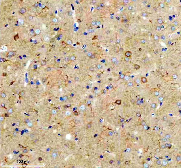
A02457-2 has real paraffin-section IHC images from mouse testis and rat brain (catalog IHC image captions). No IF/ICC image is supplied (catalog images).
A02457-2 is the sole card and lists Human, Mouse, and Rat reactivity (catalog reactivity). Its IHC captions document mouse testis and rat brain paraffin sections; the rendered figure shows mouse testis (catalog IHC image captions; card figure).
Which to pick: Choose A02457-2 for mouse or rat paraffin-section tissue IHC: its captions use 2 μg/ml, within the listed 2–5 μg/ml IHC range (catalog IHC image captions; catalog dilution). The fixative is unreported (catalog IHC image captions). For IF/ICC or human tissue IHC, seek separate validation; IF/ICC is absent from the application list, and the IHC captions show only mouse and rat tissue despite listed Human reactivity (catalog applications; catalog IHC image captions; catalog reactivity).