This website uses cookies to ensure you get the best experience on our website.


- Table of Contents
Plan PLXDC1 chromogenic IHC on paraffin sections using its observed cytoplasmic and membranous tissue pattern (HPA tissue IHC). Start the catalog antibody at 2.5 μg/mL (datasheet), and assess glandular and endothelial staining by cell type (HPA tissue IHC; UniProt).

Expected localisation, validated protocols, controls and antibodies — the at-a-glance facts below, then the full design guide.
| Expected localisation | Cytoplasmic with membranous tissue staining (HPA tissue IHC) | |
| Staining pattern | Cytoplasmic, sometimes membranous, in several cell types (HPA tissue IHC) | |
| Antigen retrieval | Citrate pH 6.0 HIER, 95–98 °C, 20 min (rule: cytoplasmic / membrane antigen) | |
| Positive control | Appendix+4 more · see all | |
| Negative control | Adipose tissue+4 more · see all |
| Fixation | Keep paraffin-section fixation consistent (standard IHC practice; not target-specific) | |
| Caveat | Glandular staining can complicate endothelial scoring (HPA tissue IHC) | |
| Regulation | Tumour endothelium enriched versus normal (UniProt) | |
| Isoform / epitope | 4 isoforms; verify the epitope’s isoform and membrane side (UniProt) |
The catalog antibody’s IHC-P protocol is accompanied by one published gastric cancer protocol (PMC9488617).
| Sample | Tissue sections; selected-image fixative not specified (standard IHC workflow) |
| Fixation | Image fixative and duration unreported (datasheet A08584-1); verify before use. |
| Sectioning | 4–5 µm sections on charged slides (standard) |
| Deparaffinisation | Xylene, graded ethanol series to water (standard) |
| Antigen retrieval | Heat-induced epitope retrieval in citrate buffer, pH 6.0, 20 min at 95–98 °C (standard rule: cytoplasmic / membrane antigen) |
| Peroxidase block | 3% H2O2, 10 min, room temperature (standard) |
| Blocking | 10% normal serum of the secondary host, 30 min, room temperature (standard) |
| Primary antibody | Rabbit anti-PLXDC1, 2.5 μg/mL (datasheet A08584-1) |
| Primary incubation | Overnight at 4 °C (standard) |
| Detection | HRP-polymer secondary, DAB chromogen 5–10 min (standard) |
| Counterstain | Hematoxylin, blue, dehydrate and mount (standard) |
| Expected result | PLXDC1-positive staining in glandular cells of appendix (HPA tissue IHC: High). HPA tissue profile: General cytoplasmic expression with a membranous pattern in several tissues. No signal in the no-primary control. |
PLXDC1 is a transmembrane protein with extracellular and cytoplasmic regions; UniProt places it predominantly at vascular endothelial tight junctions and, to a lesser extent, the luminal surface (UniProt Q8IUK5 topology and subcellular location). On tissue sections, HPA also reports general cytoplasmic staining and membranous staining in several tissues, including high staining in specified glandular and hematopoietic cells (HPA tissue IHC). HPA rates its tissue IHC profile Approved, with medium consistency against RNA data (HPA tissue IHC).
| Vascular cell borders stain, with weaker luminal staining. | This matches UniProt's predominant tight-junction and lesser luminal localization in vascular endothelial cells (UniProt Q8IUK5 subcellular location). Interpret borders in their tissue context: HPA also describes general cytoplasmic and some membranous staining outside vessels (HPA tissue IHC). |
| Glandular cells show cytoplasmic staining, sometimes with a membrane pattern. | That can fit the HPA profile: glandular cells are High in appendix, epididymis, gallbladder and stomach (HPA tissue IHC). Do not require every positive cell to show a crisp endothelial-junction pattern; compare its compartment with the HPA profile (HPA tissue IHC). |
| A strong, predominantly nuclear pattern appears in the cells being scored. | A nuclear pattern does not match the listed membrane, junction, cytoplasm or secreted locations (UniProt Q8IUK5 subcellular location; HPA tissue IHC). Check the no-primary control and staining distribution before calling it PLXDC1 (general IHC practice). |
| Unexpected cells stain while the expected cells in the same section do not. | Check the exact cell population against HPA, which reports both positive and undetected populations within named tissues (HPA tissue IHC). If the pattern remains discordant, investigate antibody cross-reactivity or endogenous detection activity with controls (general IHC practice). |
| A known HPA-positive cell population has no discernible signal. | High HPA staining identifies a useful comparator, such as appendix glandular cells or bone-marrow hematopoietic cells (HPA tissue IHC). Absence in one run calls for checking controls and assay performance before interpreting the specimen as negative (general IHC practice). |
| Cell population and tissue | Use cell-level comparators: HPA calls appendix glandular cells and bone-marrow hematopoietic cells High, but adipocytes and cardiomyocytes Not detected (HPA tissue IHC). Whole-section positivity cannot substitute for identifying which cells stain (general IHC practice). |
| Endothelial context | UniProt reports PLXDC1 in endothelial cells from several primary cancers and not detectable in normal-tissue endothelial cells (UniProt Q8IUK5 tissue specificity). A negative normal vessel therefore cannot by itself invalidate a run; assess a suitable positive comparator (general IHC practice). |
| Topology and compartment | Residues 19–426 are extracellular, 427–447 span the membrane, and 448–500 are cytoplasmic (UniProt Q8IUK5 topology). The record gives no antibody epitope, so it cannot predict which region the catalog antibody recognizes (UniProt Q8IUK5 topology; HPA antibody record). |
| Processing and protein forms | UniProt lists a signal peptide at 1–18, a 19–500 chain, four isoforms and glycosylation sites at 33, 80 and 197 (UniProt Q8IUK5). These annotations do not establish an IHC-specific staining difference or a shedding mechanism (UniProt Q8IUK5). |
| Strength of HPA evidence | The tissue IHC profile is Approved with medium antibody–RNA consistency; the listed antibody HPA012805 is IHC Approved (HPA tissue IHC; HPA antibody record). Treat an unusual pattern as a finding to check, rather than assuming antibody-independent confirmation (HPA tissue IHC; HPA antibody record). |
| IF/ICC: is a membrane signal established? | HPA summarizes PLXDC1 as membrane-localized but provides no ICC-IF images; HPA012805 has no listed ICC status (HPA subcellular; HPA antibody record). Use that as a localization clue, not a validated IF/ICC result or protocol (HPA subcellular; HPA antibody record). |
| Situation | Likely cause | Next action |
|---|---|---|
| A positive-control section has little or no staining. | A failed staining run is possible; HPA reports High staining in appendix glandular cells and bone-marrow hematopoietic cells (HPA tissue IHC). | Verify the positive comparator's cell identity, then check retrieval, primary antibody dilution, detection reagents and counterstain using routine IHC controls (general IHC practice). No PLXDC1-specific retrieval setting is supplied (HPA tissue IHC; UniProt Q8IUK5). |
| A normal vessel is negative, but a cancer vessel stains. | That contrast is compatible with UniProt's report of detection in cancer endothelial cells and nondetection in normal-tissue endothelial cells (UniProt Q8IUK5 tissue specificity). | Score the endothelial cells in each specimen separately and check a positive tissue comparator before treating the normal-vessel result as technical failure (UniProt Q8IUK5 tissue specificity; general IHC practice). |
| Most of the section has diffuse brown background. | Nonspecific antibody binding or detection background may obscure the cell-level pattern (general IHC practice). | Inspect no-primary and detection-only controls, review blocking and washes, and compare the result with HPA's cell-specific distribution (general IHC practice; HPA tissue IHC). |
| An undetected HPA cell population appears strongly positive. | A discordant pattern may reflect cross-reactivity or endogenous detection activity; HPA lists adipocytes and cardiomyocytes as Not detected (HPA tissue IHC; general IHC practice). | Confirm the cell identity, examine no-primary and detection-only controls, and repeat with appropriate blocking or an independent antibody if available (general IHC practice). |
| Staining is mainly nuclear, with no plausible membrane or cytoplasmic signal. | The compartment conflicts with UniProt's membrane, junction and cytoplasmic locations and HPA's cytoplasmic or membranous tissue profile (UniProt Q8IUK5 subcellular location; HPA tissue IHC). | Check the counterstain and no-primary control, then reassess staining conditions and antibody specificity before scoring nuclei as positive (general IHC practice). |
| IF/ICC shows a membrane-like signal, but its specificity is uncertain. | HPA's membrane summary has no accompanying ICC-IF images, and the listed antibody has no ICC validation status (HPA subcellular; HPA antibody record). | Evaluate IF/ICC on its separate guide page with appropriate controls; do not treat IHC approval as IF/ICC validation (HPA antibody record; general IF/ICC practice). |


Comprehensive Human Protein Atlas IHC scoring per tissue (reliability: Approved — Medium consistency between antibody staining and RNA expression data.). Rows are taken directly from the HPA tissue chart — click any row's HPA link to view the source.
| Tissue | Cell type | Level | Evidence | Source |
|---|---|---|---|---|
| Appendix | Glandular cells | High | Protein (IHC) | HPA → |
| Bone marrow | Hematopoietic cells | High | Protein (IHC) | HPA → |
| Bronchus | Ciliated cells (cilia axoneme) | High | Protein (IHC) | HPA → |
| Endometrium | Cells in endometrial stroma | Medium | Protein (IHC) | HPA → |
| Epididymis | Glandular cells | High | Protein (IHC) | HPA → |
| Tissue | Cell type | Level | Evidence | Source |
|---|---|---|---|---|
| Adipose tissue | Adipocytes | Not detected | Protein (IHC) | HPA → |
| Heart muscle | Cardiomyocytes | Not detected | Protein (IHC) | HPA → |
| Hippocampus | Glial cells | Not detected | Protein (IHC) | HPA → |
| Pancreas | Exocrine glandular cells | Not detected | Protein (IHC) | HPA → |
| Salivary gland | Glandular cells | Not detected | Protein (IHC) | HPA → |
Use compartment-aware controls and matched section processing to troubleshoot PLXDC1 staining in chromogenic paraffin-section IHC (UniProt Q8IUK5 topology and localisation; standard IHC practice).
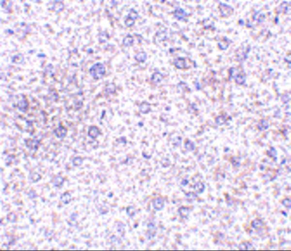
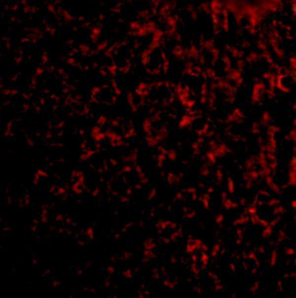
A08584-1 has mouse liver IHC and IF images (catalog image captions) and lists Human, Mouse, and Rat reactivity (catalog reactivity).
A08584-1 is listed for IHC-P and IF (catalog applications). Its IHC and IF images show mouse liver tissue at 2.5 μg/mL and 20 μg/mL, respectively (catalog image captions).
Which to pick: Choose A08584-1 for paraffin-section IHC: IHC-P is listed, and its own mouse liver IHC image uses 2.5 μg/mL (catalog applications; A08584-1 IHC image caption). The same SKU is listed for IF and has a mouse liver IF image at 20 μg/mL; ICC is not documented (catalog applications; A08584-1 IF image caption). It lists Human, Mouse, and Rat reactivity, but the pictured IHC and IF samples are mouse liver; the IHC caption does not report a fixative (catalog reactivity; A08584-1 image captions).