POFUT1 / GDP-fucose protein O-fucosyltransferase 1 · IHC design guide

Design Immunohistochemistry for POFUT1

Plan chromogenic IHC on paraffin sections with the IHC-validated antibody at 1:100–1:300 (datasheet: IHC). Use bronchial basal or duodenal glandular cells as positive staining references and parathyroid glandular cells as a negative comparator (HPA tissue IHC).

Evidence assembled Oct 2026 · For research use; verify linked source records and product datasheet before use
Immunohistochemistry protocol sheet for POFUT1 (IHC for POFUT1): expected localisation Cytoplasmic tissue staining (HPA tissue IHC); ER protein (UniProt), antibody A30700, validated IHC image, and IHC protocol steps
Printable POFUT1 IHC protocol sheet — expected localisation Cytoplasmic tissue staining (HPA tissue IHC); ER protein (UniProt), antibody A30700, controls and protocol steps. Open the full POFUT1 IHC guide →

POFUT1 Immunohistochemistry Experimental Design Guide

Expected localisation, validated protocols, controls and antibodies — the at-a-glance facts below, then the full design guide.

Must know before staining
Expected localisation Cytoplasmic tissue staining (HPA tissue IHC); ER protein (UniProt)
Staining pattern Cytoplasmic signal in bronchial basal and duodenal glandular cells (HPA tissue IHC)
Antigen retrieval Citrate pH 6.0 HIER, 95–98 °C, 20 min (rule: cytoplasmic / membrane antigen)
Positive control ⓘ Bronchus+4 more · see all
Negative control ⓘ Parathyroid gland+1 more · see all
Important caveats
Reasons your staining may differ from the expected pattern.
Fixation Keep fixation consistent across paraffin sections (standard IHC practice; not target-specific)
Caveat Staining and RNA expression show low consistency (HPA tissue IHC)
Regulation Low tissue specificity at RNA level (HPA tissue RNA)
Isoform / epitope 2 isoforms; signal peptide 1–26; epitope map unreported (UniProt)
Section 1

Recommended POFUT1 IHC & IF Protocols

The catalog antibody’s IHC-P protocol is accompanied by four published POFUT1 IHC methods (PMC6606927; PMC7581975; PMC6266312; PMC13529811).

Recommended immunohistochemistry (IHC-P) protocol parameters
SampleParaffin-embedded human lung carcinoma tissue; fixative not specified (datasheet A30700)
FixationImage fixative and duration unreported (datasheet A30700); verify before use.
Sectioning4–5 µm sections on charged slides (standard)
DeparaffinisationXylene, graded ethanol series to water (standard)
Antigen retrievalHeat-induced epitope retrieval in citrate buffer, pH 6.0, 20 min at 95–98 °C (standard rule: cytoplasmic / membrane antigen)
Peroxidase block3% H2O2, 10 min, room temperature (standard)
Blocking10% normal serum of the secondary host, 30 min, room temperature (standard)
Primary antibodyRabbit anti-POFUT1, 1:100 - 1:300 (datasheet A30700)
Primary incubationOvernight at 4 °C (standard)
DetectionHRP-polymer secondary, DAB chromogen 5–10 min (standard)
CounterstainHematoxylin, blue, dehydrate and mount (standard)
Expected resultPOFUT1-positive staining in basal cells of bronchus (HPA tissue IHC: High). HPA tissue profile: Cytoplasmic expression in several tissues. No signal in the no-primary control.
💡Decision noteStart with citrate pH 6.0 retrieval (page retrieval; PMC6606927). If staining is weak, consider the published pH 9 pressure-cooker method (PMC7581975).
Section 2

What Is the Expected POFUT1 Staining Pattern?

POFUT1 is an endoplasmic reticulum protein without a transmembrane segment (UniProt Q9H488 topology). In paraffin-section IHC, expect cytoplasmic staining, especially in bronchial basal cells, kidney tubular cells, and several glandular cell populations (HPA tissue IHC: High). HPA rates its tissue staining Approved but reports low consistency with RNA expression; treat the pattern as a guide to interpretation, not proof of antibody specificity (HPA tissue IHC).

What am I looking at on my slide?
Cytoplasmic staining in kidney tubular cells or bronchial basal cells, with identifiable tissue structure.This matches the reported compartment and High-staining cell populations (HPA tissue IHC). Judge signal in the named cells, since a positive tissue does not imply that every cell in its section should stain (HPA tissue IHC).
Predominantly nuclear or sharply plasma-membrane staining, without a convincing cytoplasmic component.That differs from the reported tissue-IHC cytoplasmic profile and the annotated endoplasmic reticulum location (HPA tissue IHC; UniProt Q9H488). Consider nonspecific staining or a detection artefact; location alone cannot identify its cause (general IHC practice).
Strong staining confined to an unexpected cell population while the reported positive cells are unstained.Question antibody cross-reactivity or endogenous detection activity (general IHC practice). Compare cell identity and distribution with the HPA examples; HPA's Approved rating has a low RNA-consistency caveat (HPA tissue IHC).
Diffuse chromogen over tissue, stroma, and empty regions, obscuring cell boundaries.A field-wide deposit does not establish the reported cell-selective cytoplasmic pattern (HPA tissue IHC). Review background controls and the detection workflow before assigning it to POFUT1 (general IHC practice).
No detectable staining in a kidney section's tubular cells or a bronchus section's basal cells.These are reported High-staining populations (HPA tissue IHC). An absent signal makes that run inconclusive: assess tissue preservation, controls, retrieval, and detection using general IHC practice before inferring biological absence.
💡Expected POFUT1 appearanceCall a result positive when identifiable bronchial basal, kidney tubular, or reported glandular cells show convincing cytoplasmic chromogen, potentially strong in HPA High populations; isolated nuclear, membrane-edge, or field-wide deposits are suspect (HPA tissue IHC; UniProt Q9H488; general IHC practice).
How each factor affects the staining
Tissue and cell selectionHPA reports High staining in bronchial basal cells, kidney tubular cells, duodenal and small-intestinal glandular cells, and other listed populations. Select and score the specified cells rather than treating an entire organ as uniformly positive (HPA tissue IHC).
Reported negative populationsParathyroid glandular cells and soft-tissue chondrocytes were Not detected (HPA tissue IHC). They can provide a distribution comparison, but the Approved rating and low RNA consistency limit claims that any one negative section proves specificity (HPA tissue IHC).
Subcellular annotationThe endoplasmic reticulum location and lack of a transmembrane segment support an intracellular expectation (UniProt Q9H488 topology). HPA describes cytoplasmic tissue-IHC staining; neither source establishes a required reticular pattern at routine chromogenic resolution (HPA tissue IHC).
Protein processing and isoformsUniProt annotates a signal peptide at residues 1–26, a chain at 27–388, two glycosylation sites, and two isoforms (UniProt Q9H488). Without an antibody epitope or isoform-specific validation in the payload, these facts cannot predict which form the IHC stain detects.
IHC antibody validationHPA054519 is Approved for IHC, whereas HPA059935 has no IHC status in the supplied record (HPA antibodies). HPA's tissue profile is also Approved with low consistency against RNA, so use morphology and controls when interpreting an individual stain (HPA tissue IHC).
IF/ICC Q: Should an IF image match the tissue-IHC pattern?A: HPA reports an approved centrosome location in ICC-IF, while its tissue-IHC profile is cytoplasmic (HPA subcellular; HPA tissue IHC). These are different assay observations; the ICC result does not redefine a positive chromogenic tissue section.
Why is my staining missing, weak or wrong?
SituationLikely causeNext action
A known High-staining cell population has no signal.Target-specific fixation effects are not established by the supplied assay evidence. Verify with a matched IHC source before attributing a result to fixation.Verify cell identity and tissue integrity, then check run controls, the documented retrieval conditions, antibody application, and detection reagents (general IHC practice).
Chromogen covers most cells and extracellular areas.Excess background or endogenous detection activity can obscure cell-specific staining (general IHC practice).Inspect the negative reagent control and background-blocking and detection steps; score only interpretable cellular signal (general IHC practice).
Only nuclei or crisp cell borders are stained.That distribution conflicts with the cytoplasmic tissue profile and ER annotation (HPA tissue IHC; UniProt Q9H488).Check a reported positive cell population and the control slide; treat compartment-mismatched staining as unconfirmed until the pattern is reproducible (general IHC practice).
An unexpected cell population stains more strongly than the expected one.Cross-reactivity or endogenous detection activity is possible; HPA tissue staining has low consistency with RNA expression (general IHC practice; HPA tissue IHC).Compare matched controls and cell morphology, and avoid assigning POFUT1 solely from intensity in that population (general IHC practice).
Parathyroid glandular cells or chondrocytes stain strongly.Those populations were Not detected in the supplied HPA tissue profile, although a single discordance cannot establish its cause (HPA tissue IHC).Confirm the cell identification and examine controls; record the discordance rather than using the stain as a validated negative comparison (general IHC practice).
An ICC-IF centrosome signal seems inconsistent with the IHC slide.HPA reports centrosome localisation for ICC-IF and cytoplasmic expression for tissue IHC (HPA subcellular; HPA tissue IHC).Interpret each assay against its own reported pattern and validation status; do not require a visible centrosome in chromogenic paraffin-section IHC (HPA subcellular; HPA tissue IHC).

Sample controls for POFUT1 IHC & IF

🧪Run kidney first: cells in tubules should stain (HPA: High in kidney tubule cells). Run parathyroid gland as the negative tissue (HPA: glandular cells Not detected); the supplied kidney row does not identify an internal negative cell type, so cells without specific chromogen staining can show slide background but should not be designated a biological negative (HPA: kidney row).
Positive control tissue: Bronchus (Basal cells, HPA High)
Negative control tissue: Parathyroid gland (HPA Not detected)
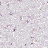
ICC-IF cell lines (HPA subcellular resource): HPA ICC-IF images show POFUT1 in CACO-2, SiHa, U2OS, with annotated localisation: Centrosome (approved) (HPA subcellular).
Technical controls: Include no-primary (secondary-only) and primary-host-matched isotype or nonimmune IgG controls, as appropriate to the antibody’s clonality (standard IHC practice). Use a POFUT1 knockout specimen if available or a peptide-block control (selected SKU A30700 tissue-IHC caption: peptide block); quench endogenous peroxidase and, if using avidin–biotin detection in kidney, control for endogenous biotin (standard IHC practice).
⚠️Feasibility: A target-specific fixation window or fixation effect is unreported, and the selected SKU A30700 paraffin-section caption does not state the fixative (selected SKU A30700 tissue-IHC caption). Antigen-retrieval dependency is unreported; optimize retrieval for paraffin IHC and check kidney tubule background, including endogenous biotin when applicable (standard IHC practice). The supplied evidence does not establish that frozen sections or IF are easier; IF/ICC has separate evidence showing centrosome localization, while UniProt places POFUT1 in the endoplasmic reticulum (HPA subcellular: centrosome; UniProt Q9H488: endoplasmic reticulum).

HPA tissue IHC evidence for POFUT1

Comprehensive Human Protein Atlas IHC scoring per tissue (reliability: Approved — Low consistency between antibody staining and RNA expression data.). Rows are taken directly from the HPA tissue chart — click any row's HPA link to view the source.

Positive expression · recommended positive controls

TissueCell typeLevelEvidenceSource
Bronchus Basal cells High Protein (IHC) HPA →
Duodenum Glandular cells High Protein (IHC) HPA →
Fallopian tube Ciliated cells (cell body) High Protein (IHC) HPA →
Kidney Cells in tubules High Protein (IHC) HPA →
Small intestine Glandular cells High Protein (IHC) HPA →

Undetected expression · recommended negative controls

TissueCell typeLevelEvidenceSource
Parathyroid gland Glandular cells Not detected Protein (IHC) HPA →
Soft tissue Chondrocytes Not detected Protein (IHC) HPA →
Section 3

Advanced POFUT1 IHC Tips

Troubleshoot POFUT1 staining in paraffin sections by checking retrieval, controls, cellular distribution and scoring before interpreting chromogenic signal.

What retrieval should I start with for weak POFUT1 staining in paraffin sections?
Start with citrate buffer at pH 6.0, heated to 95–98 °C for 20 min (page retrieval setting). Let sections cool in buffer before washing, then compare a test section with a section processed without retrieval (standard IHC practice). Keep antibody dilution and detection conditions identical so a change in signal can be attributed to retrieval. If staining remains weak, test a different retrieval condition on adjacent sections as a fallback, while checking whether tissue structure and background remain acceptable. Judge improvement in identifiable cells and intracellular signal rather than by overall brown color alone (standard IHC practice).
How should I troubleshoot fixation when POFUT1 staining is inconsistent?
The selected A30700 image identifies paraffin-embedded human lung carcinoma tissue but does not state its fixative (selected A30700 caption). Target-specific fixation sensitivity is therefore unknown; do not assign weak staining to a particular fixative without a controlled comparison. Record each specimen’s fixative, fixation duration, processing history and section age, then stain comparable sections together with the same retrieval and detection conditions (standard IHC practice). Check morphology alongside signal, because damaged tissue can make an apparent intensity difference difficult to interpret (standard IHC practice). If the history is unavailable, report that limitation and avoid treating staining differences as biological differences.
Which cellular staining pattern should support a POFUT1 IHC result?
Prioritize intracellular cytoplasmic staining in intact cells: POFUT1 is assigned to the endoplasmic reticulum (UniProt Q9H488 localisation), and tissue IHC describes cytoplasmic expression (HPA tissue IHC). The record lists a signal sequence at residues 1–26 and no transmembrane segment (UniProt Q9H488 processing and topology). Examine well-preserved cell bodies at high magnification before accepting a diffuse brown deposit as specific staining (standard IHC practice). The separate centrosome assignment comes from ICC/IF images and should not by itself redefine the expected chromogenic tissue pattern (HPA subcellular). A predominantly nuclear, extracellular or section-edge pattern warrants review of background and morphology before scoring.
Can the available evidence establish which POFUT1 isoform the antibody detects?
POFUT1 has 2 annotated isoforms, while the supplied tissue image does not identify an antibody epitope or establish isoform selectivity (UniProt Q9H488 isoforms; selected A30700 caption). Its precursor has a signal sequence at residues 1–26 and a reported chain spanning 27–388 (UniProt Q9H488 processing). Two glycosylation sites are annotated at residues 62 and 160, but their effect on staining by this antibody is unknown (UniProt Q9H488 glycosylation). Obtain the antibody’s immunogen or epitope information before making an isoform claim, and keep any peptide-block result specific to the tested reagent (standard antibody validation practice; selected A30700 caption).
How can IF help examine POFUT1 localisation alongside the IHC result?
Treat IF/ICC as a separate assay: the supplied subcellular images report a centrosome location, whereas tissue IHC reports cytoplasmic expression (HPA subcellular; HPA tissue IHC). Multiplex POFUT1 with an independently validated marker for the cell type being examined, then include single-stain controls to assess channel bleed-through (standard IF practice). Choose a fluorophore channel after inspecting unstained tissue autofluorescence, and keep exposure settings comparable across samples (standard IF practice). Because POFUT1 is assigned to the endoplasmic reticulum and lacks a transmembrane segment, test permeabilisation for access to an intracellular epitope; the epitope’s precise accessibility is not supplied (UniProt Q9H488 localisation and topology).
What should I check when POFUT1 sections show widespread brown background?
Compare staining in intact cells with deposits over folds, damaged regions and section edges before changing antibody concentration (standard IHC practice). Include a reagent control without primary antibody to assess detection-related background, and block endogenous peroxidase when using peroxidase and DAB detection (standard chromogenic IHC practice). The selected A30700 image includes a synthesized-peptide block, which provides a reagent-specific comparison for that image (selected A30700 caption). Persistent diffuse signal can be investigated by adjusting blocking and antibody dilution on adjacent sections while holding retrieval and DAB development constant (standard IHC practice). Do not score extracellular precipitate as POFUT1-positive cells.
How should I quantify POFUT1 staining across paraffin sections? ⚠ ANSWER MARKED FOR VERIFICATION
Define the cell population and tissue region before scoring, then record the percentage of positive cells and staining intensity in identifiable cell bodies (standard IHC scoring practice). An H-score can combine percentages at intensity levels 0–3 into a 0–300 summary; state the thresholds used (standard IHC scoring practice). For spatial comparisons, report positive-cell density per mm² of viable tissue and exclude folds, necrosis and blank area from the denominator (standard image-analysis practice). Normalize comparisons to the same cell type or tissue compartment, section area and staining batch. Keep cytoplasmic signal separate from edge deposits or other patterns that fail review (HPA tissue IHC; standard IHC practice).
How do I distinguish credible POFUT1 positivity from staining artefact?
Look for reproducible intracellular staining in intact cells that fits the cytoplasmic tissue profile and endoplasmic-reticulum assignment (HPA tissue IHC; UniProt Q9H488 localisation). HPA reports high staining in bronchial basal cells and kidney tubular cells, but rates its tissue IHC as Approved with low consistency between staining and RNA expression (HPA tissue IHC). Treat signal confined to an unexpected cell population, nuclei, extracellular material, necrotic areas or section edges as a reason to inspect morphology and controls before interpretation (standard IHC practice). Check a no-primary control for detection background, including endogenous enzyme signal, and compare any peptide-block result only within its tested antibody and conditions (standard IHC practice; selected A30700 caption).
Boster reagents

Best POFUT1 / GDP-fucose protein O-fucosyltransferase 1 IHC Antibodies

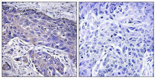
A30700 is listed for IHC, IF and ICC in human, mouse and rat (catalog: applications/reactivity). Its supplied IHC figure shows paraffin-embedded human lung carcinoma (catalog: IHC image caption).

Real IHC data Immunohistochemistry analysis of paraffin-embedded human lung carcinoma tissue, using POFUT1 Antibody. The picture on the right is blocked with the synthesized peptide.
Anti-POFUT1 Antibody
Cat # A30700

A30700 is listed for IHC, IF and ICC, with human, mouse and rat reactivity (catalog: applications/reactivity). Its supplied IHC image shows paraffin-embedded human lung carcinoma alongside a peptide-blocked comparison; no IF image is supplied (catalog: IHC image caption/IF image list).

Which to pick: For tissue IHC, choose A30700 at a listed dilution of 1:100–1:300 (catalog: IHC dilution); its image documents paraffin-embedded human lung carcinoma, but the fixative is unreported (catalog: IHC image caption). For IF/ICC, A30700 is the listed rabbit polyclonal option, with an IF dilution of 1:50 and no supplied IF image (catalog: host/clonality/applications/IF dilution/IF image list). For cross-species studies, A30700 lists human, mouse and rat reactivity, while its supplied tissue IHC image shows human tissue only (catalog: reactivity/IHC image caption).

Each figure is that product's own IHC / IF validation image from its datasheet.

References

  1. UniProt Consortium. UniProt entry Q9H488 (OFUT1_HUMAN, GDP-fucose protein O-fucosyltransferase 1).
  2. Human Protein Atlas. POFUT1 tissue IHC expression (reliability: Approved).
  3. Human Protein Atlas. POFUT1 subcellular location (ICC-IF): Localized to the centrosome..
  4. Human Protein Atlas. POFUT1 antibody validation summary (2 antibodies).
  5. poFUT1 promotes endometrial decidualization by enhancing the O-fucosylation of Notch1. EBioMedicine 2019 — PMC6606927.
  6. POFUT1 mRNA expression as an independent prognostic parameter in muscle-invasive bladder cancer. Translational oncology 2021 — PMC7581975.
  7. POFUT1 as a Promising Novel Biomarker of Colorectal Cancer. Cancers 2018 — PMC6266312.
  8. POFUT1 Serves as an Independent Prognostic Factor and Therapeutic Target by Activating the PI3K/AKT Pathway in Glioma. Brain and behavior 2026 — PMC13529811.
  9. PubMed PMID:11524432 — UniProt-cited evidence.
  10. PubMed PMID:8724849 — UniProt-cited evidence.
  11. PubMed PMID:12168954 — UniProt-cited evidence.