PPFIA1 / Liprin-alpha-1 · IHC design guide

Design Immunohistochemistry for PPFIA1

Plan PPFIA1 chromogenic IHC on paraffin sections using the catalog antibody at 2–5 μg/ml (datasheet A05735-1). Assess cytoplasmic staining against the reported tissue profile, while accounting for its uncertain reliability (HPA tissue IHC).

Evidence assembled Oct 2026 · For research use; verify linked source records and product datasheet before use
Immunohistochemistry protocol sheet for PPFIA1 (IHC for PPFIA1): expected localisation General cytoplasmic staining (HPA tissue IHC), antibody A05735-1, validated IHC image, and IHC protocol steps
Printable PPFIA1 IHC protocol sheet — expected localisation General cytoplasmic staining (HPA tissue IHC), antibody A05735-1, controls and protocol steps. Open the full PPFIA1 IHC guide →

PPFIA1 Immunohistochemistry Experimental Design Guide

Expected localisation, validated protocols, controls and antibodies — the at-a-glance facts below, then the full design guide.

Must know before staining
Expected localisation General cytoplasmic staining (HPA tissue IHC)
Staining pattern Cytoplasmic staining, including lung macrophages (HPA tissue IHC)
Antigen retrieval EDTA pH 8.0 HIER, heat-mediated (datasheet A05735-1)
Positive control ⓘ Bone marrow+4 more · see all
Negative control ⓘ Heart muscle+2 more · see all
Important caveats
Reasons your staining may differ from the expected pattern.
Fixation Keep paraffin-section fixation consistent (standard IHC practice; not target-specific)
Caveat Tissue staining awaits external verification (HPA tissue IHC)
Regulation No specific regulator reported (UniProt)
Isoform / epitope 2 isoforms; epitope coverage is unknown (UniProt)
Section 1

Recommended PPFIA1 IHC & IF Protocols

Compare the catalog antibody’s IHC-P protocol (datasheet: A05735-1) with two published PPFIA1 IHC workflows (PMC7227113; PMC10169376).

Recommended immunohistochemistry (IHC-P) protocol parameters
SampleParaffin-embedded human gastric signet ring cell carcinoma tissue; fixative not specified (datasheet A05735-1)
FixationImage fixative and duration unreported (datasheet A05735-1); verify before use.
Sectioning4–5 µm sections on charged slides (standard)
DeparaffinisationXylene, graded ethanol series to water (standard)
Antigen retrievalHeat retrieval: EDTA pH 8.0 (datasheet A05735-1); 20 min, 95–100 °C (standard)
Peroxidase block3% H2O2, 10 min, room temperature (standard)
Blocking10% goat serum (datasheet A05735-1)
Primary antibodyRabbit anti-PPFIA1, 2-5 μg/ml (datasheet A05735-1)
Primary incubationOvernight at 4 °C (datasheet A05735-1)
DetectionStreptavidin-biotin complex (SABC), DAB chromogen (datasheet A05735-1)
CounterstainHematoxylin, blue, dehydrate and mount (standard)
Expected resultPPFIA1-positive staining in hematopoietic cells of bone marrow (HPA tissue IHC: High). HPA tissue profile: General cytoplasmic expression. No signal in the no-primary control.
💡Decision noteStart with heat-mediated EDTA retrieval at pH 8.0 (datasheet: A05735-1). The esophageal study reports retrieval without specifying the solution (PMC10169376).
Section 2

What Is the Expected PPFIA1 Staining Pattern?

PPFIA1 is a cytoplasmic protein with cell cortex localization and no transmembrane segment (UniProt Q13136). In paraffin tissue sections, expect predominantly cytoplasmic staining, including in lung macrophages, skin fibroblasts, and endometrial glandular cells reported as high by HPA (HPA: tissue IHC). Treat the pattern as provisional: HPA rates its tissue IHC reliability Uncertain, with external verification pending (HPA: tissue IHC reliability).

What am I looking at on my slide?
Clear cytoplasmic staining in lung macrophages, skin fibroblasts, or endometrial glandular cells.This fits HPA's high staining in those cell types and its general cytoplasmic profile (HPA: tissue IHC). Compare cells within the same section; HPA's Uncertain reliability means agreement supports, but does not establish, antibody specificity (HPA: tissue IHC reliability).
Predominantly nuclear staining, or a crisp continuous outline around cells, with little cytoplasmic signal.This conflicts with the cytoplasmic and cell cortex localization and lack of a transmembrane segment (UniProt Q13136). Review staining and counterstain controls before assigning a new compartment; such a pattern may be artifact or nonspecific detection (general IHC practice).
Strong staining in cardiomyocytes, skeletal myocytes, or vaginal squamous epithelial cells.These cells were reported as not detected in HPA tissue IHC (HPA: tissue IHC). Check whether the signal follows tissue edges or endogenous detection activity; persistent cell-associated staining could reflect cross-reactivity. An HPA negative observation is not proof that every specimen must be negative (HPA: tissue IHC reliability; general IHC practice).
Broad, even chromogen across cells and extracellular areas, without recognizable cytoplasmic boundaries.That distribution does not match HPA's general cytoplasmic profile (HPA: tissue IHC). Excess antibody, incomplete blocking, residual detection reagent, or inadequate washing can produce background; compare a no-primary control and inspect whether the signal respects cell boundaries (general IHC practice).
No staining in a section expected to contain lung macrophages or skin fibroblasts.HPA reports high staining in these cells, so absence warrants a technical check, but their staining is not an assured positive control for every antibody and specimen (HPA: tissue IHC; HPA: tissue IHC reliability). Confirm the relevant cells are present, then review retrieval, antibody incubation, and detection controls (general IHC practice).
💡Expected PPFIA1 appearanceA convincing positive shows distinct, mainly cytoplasmic staining in HPA high cells such as lung macrophages or skin fibroblasts, with intensity above local background; dominant nuclear signal or equally strong staining in HPA not-detected cells argues for further specificity checks (HPA: tissue IHC; UniProt Q13136).
How each factor affects the staining
Tissue and cell selectionHPA reports high staining in bone marrow hematopoietic cells, kidney glomerular cells, and testis seminiferous duct cells, among others; cardiomyocytes and skeletal myocytes were not detected (HPA: tissue IHC). Select fields by the reported cell population, not tissue name alone (general IHC practice).
Antibody validationHPA labels tissue staining Uncertain and lists both HPA042271 and CAB017032 as IHC Uncertain; HPA042271 is ICC Supported (HPA: tissue IHC reliability; HPA: antibodies). Agreement with a reported tissue pattern therefore needs control-based interpretation rather than being treated as independent validation (general IHC practice).
Topology and compartmentPPFIA1 has no transmembrane segment and is annotated in the cytoplasm and cell cortex, near focal adhesions (UniProt Q13136). Interpret a diffuse intracellular pattern in that context; a purely extracellular deposit or continuous surface rim does not follow the annotated localization (UniProt Q13136).
Isoforms and epitope coverageUniProt lists two PPFIA1 isoforms and one annotated protein chain spanning residues 1–1202 (UniProt Q13136). The supplied record gives no antibody epitope or isoform coverage, so staining cannot be assigned to a particular isoform or assumed to detect both (UniProt Q13136; supplied antibody records).
IF/ICC Q&AWhat should IF/ICC show? HPA reports mainly cytosolic PPFIA1 with additional focal adhesion site localization, supported in its ICC-IF summary (HPA: subcellular). That cell-level pattern can inform compartment interpretation, while this section's tissue staining assessment remains based on IHC observations (HPA: tissue IHC).
Why is my staining missing, weak or wrong?
SituationLikely causeNext action
The expected high-staining cells are absent from the chosen field.A tissue label alone does not guarantee that the HPA-listed cell population appears in every field (HPA: tissue IHC; general IHC practice).Locate the relevant cells by morphology on the counterstained section and assess staining there; use an appropriate comparator section if they are absent (general IHC practice).
The relevant cells are present, but the section has no chromogenic signal.Antibody, retrieval, or detection failure is possible; HPA's reported high staining is observational and IHC reliability is Uncertain (HPA: tissue IHC; general IHC practice).Check the run's positive and no-primary controls, then review the catalog antibody's IHC-P retrieval, incubation, and detection steps before interpreting the tissue as negative (general IHC practice).
The slide has diffuse haze or staining between cells.Background from antibody concentration, blocking, washing, or detection chemistry can obscure the cytoplasmic profile (HPA: tissue IHC profile; general IHC practice).Compare with a no-primary control; optimize blocking and washes, and review antibody dilution against the catalog antibody's IHC-P instructions (general IHC practice).
Nuclei dominate while cytoplasm is weak.The pattern conflicts with UniProt's cytoplasm and cell cortex annotation and HPA's cytoplasmic tissue profile (UniProt Q13136; HPA: tissue IHC).Confirm compartment boundaries with the counterstain and controls; reassess specificity before scoring nuclear staining as PPFIA1 (general IHC practice).
HPA not-detected cell types stain as strongly as proposed positives.Cross-reactivity or endogenous detection activity may be contributing; HPA reports cardiomyocytes, skeletal myocytes, and vaginal squamous epithelial cells as not detected (HPA: tissue IHC; general IHC practice).Compare cell-associated signal with a no-primary control and examine its distribution; seek independent antibody or orthogonal support before calling those cells PPFIA1 positive (general IHC practice).
Results differ between sections or runs.Technical variation can alter IHC appearance, while HPA already reports only medium consistency between antibody staining and RNA expression (HPA: tissue IHC reliability; general IHC practice).Compare matched controls and the same cell populations across runs; document retrieval and detection settings, and avoid treating an isolated change in intensity as a biological difference (general IHC practice).

Sample controls for PPFIA1 IHC & IF

🧪Run lung first: macrophages must stain (HPA: High in lung macrophages). Use skeletal muscle myocytes as the negative tissue (HPA: Not detected in skeletal muscle myocytes); neighboring non-macrophage cells on the lung slide can show background, but their PPFIA1-negative status is unreported (HPA: lung row specifies macrophages only).
Positive control tissue: Bone marrow (Hematopoietic cells, HPA High)
Negative control tissue: Heart muscle (HPA Not detected)
ICC-IF cell lines (HPA subcellular resource): HPA ICC-IF images show PPFIA1 in A-431, U2OS, NIH 3T3, with annotated localisation: Cytosol (supported) (HPA subcellular).
Technical controls: Include no-primary or secondary-only and host-matched rabbit IgG isotype controls (hero caption: rabbit anti-PPFIA1), plus PPFIA1 knockout tissue as a biological negative if available (standard IHC practice). Block endogenous peroxidase and assess pigment in the lung section before interpreting DAB signal in macrophages (standard chromogenic IHC practice).
⚠️Feasibility: A target-specific fixation window or fixation effect is unreported, and the selected A05735-1 tissue-IHC caption does not state a fixative (hero caption: fixative not stated). The documented paraffin-section method uses heat retrieval in EDTA at pH 8.0; a retrieval dependency has not been established by comparison with unretrieved sections (hero caption: EDTA retrieval). Frozen sections or IF cannot be judged easier from the supplied methods, although ICC-IF images support cytosolic and focal-adhesion localization; lung macrophage pigment can complicate chromogenic scoring (HPA: supported subcellular locations; standard IHC practice).

HPA tissue IHC evidence for PPFIA1

Comprehensive Human Protein Atlas IHC scoring per tissue (reliability: Uncertain — Medium consistency between antibody staining and RNA expression data. Pending external verification.). Rows are taken directly from the HPA tissue chart — click any row's HPA link to view the source.

Positive expression · recommended positive controls

TissueCell typeLevelEvidenceSource
Bone marrow Hematopoietic cells High Protein (IHC) HPA →
Endometrium Glandular cells High Protein (IHC) HPA →
Kidney Cells in glomeruli High Protein (IHC) HPA →
Lung Macrophages High Protein (IHC) HPA →
Lymph node Non-germinal center cells High Protein (IHC) HPA →

Undetected expression · recommended negative controls

TissueCell typeLevelEvidenceSource
Heart muscle Cardiomyocytes Not detected Protein (IHC) HPA →
Skeletal muscle Myocytes Not detected Protein (IHC) HPA →
Vagina Squamous epithelial cells Not detected Protein (IHC) HPA →
Section 3

Advanced PPFIA1 IHC Tips

Troubleshoot PPFIA1 chromogenic IHC in paraffin sections by checking retrieval, controls, cellular localisation and scoring before interpreting differences between samples.

Which retrieval condition should I try first for PPFIA1 IHC?
Start with heat-mediated antigen retrieval in EDTA buffer at pH 8.0 (datasheet A05735-1). The selected image used this retrieval before 2 μg/ml primary antibody overnight at 4°C, so reproduce that sequence during initial optimisation (caption A05735-1). Include a known positive section and a no-primary control in each run to distinguish failed retrieval from detection background (standard IHC practice). If staining remains weak, compare heating durations on matched sections while holding antibody concentration and detection constant (standard IHC practice). Assess cytoplasmic staining first, given PPFIA1’s reported cytoplasmic and cortical localisation (UniProt Q13136 localisation).
Which fixation variables should I control when PPFIA1 staining varies between paraffin blocks?
No target-specific fixation sensitivity is established for PPFIA1 here, and the selected paraffin-section caption does not report its fixative (caption A05735-1). Record fixative identity, fixation interval, tissue thickness and processing history before attributing changes in DAB intensity to biology (standard IHC practice). Compare blocks using EDTA at pH 8.0, 2 μg/ml primary overnight at 4°C, and identical detection, with matched positive and no-primary controls (caption A05735-1; standard IHC practice). Repeat discordant samples on serial sections, but do not infer fixation tolerance from PPFIA1’s cytoplasmic localisation or phosphorylation sites because neither measures it (UniProt Q13136; standard IHC interpretation).
Where should convincing PPFIA1 staining appear in chromogenic paraffin sections?
Expect mainly cytoplasmic staining in interpretable cells, with possible cortical accentuation near focal adhesions (UniProt Q13136 localisation; HPA subcellular). PPFIA1 has no transmembrane segment, so uniform membrane outlining without cytoplasm deserves scrutiny before assigning a cellular score (UniProt Q13136 topology; standard IHC interpretation). Examine HPA-listed high compartments such as lung macrophages and kidney glomerular cells, while evaluating morphology and matched controls on your own section (HPA tissue IHC; standard IHC practice). Counterstain to distinguish nuclei from cytoplasm, and assess whether DAB follows intact cell contours instead of folds or tissue edges (standard IHC practice).
Could isoforms or epitope changes explain discordant PPFIA1 IHC results?
PPFIA1 has 2 annotated isoforms and three SAM domains spanning residues 878–1120, so epitope mapping matters when comparing antibodies (UniProt Q13136 isoforms and domains). The supplied caption documents staining with A05735-1 but gives no immunogen coordinates, leaving isoform coverage and the precise recognised epitope unresolved (caption A05735-1). If suitable antibodies are available, compare documented nonoverlapping epitopes on serial sections and seek concordant cytoplasmic patterns in the same cell populations (standard IHC validation; UniProt Q13136 localisation). Phosphorylation sites include serine 150 and threonine 230; interpret discordance as modification dependent only after establishing epitope overlap and specificity (UniProt Q13136 modified residues; standard IHC interpretation).
How can IF clarify PPFIA1 localisation alongside chromogenic IHC?
For follow-up IF, multiplex PPFIA1 with a validated macrophage marker in lung sections, where macrophages show high tissue staining (HPA tissue IHC; standard IF practice). Select spectrally separated fluorophores after assessing tissue autofluorescence, using unstained and single-label controls to distinguish it from specific signal (standard IF practice). Permeabilise gently after fixation for access to the cytosolic side, then optimise detergent exposure empirically; PPFIA1 is cytoplasmic and lacks a transmembrane segment (UniProt Q13136 localisation and topology; standard IF practice). HPA ICC/IF data support cytosolic and focal-adhesion localisation, while the selected paraffin caption supplies chromogenic IHC conditions only (HPA subcellular; caption A05735-1).
How do I distinguish PPFIA1 signal from DAB background?
Begin with no-primary and isotype controls, then inspect whether brown deposit persists in the same compartments or on damaged tissue (standard IHC practice). The selected paraffin workflow used 10% goat serum block, biotinylated goat anti-rabbit secondary, an avidin-biotin complex and DAB (caption A05735-1). Block endogenous peroxidase and consider endogenous biotin controls with this detection chemistry; adjust washes and primary concentration on matched sections (standard IHC practice). PPFIA1 staining should be predominantly cytoplasmic, whereas diffuse color across lumina, necrotic material or tissue edges warrants investigation (HPA tissue IHC; standard IHC interpretation).
How should I quantify PPFIA1 staining across heterogeneous sections? ⚠ ANSWER MARKED FOR VERIFICATION
Define the cell population and region before scoring, since HPA reports high staining in lung macrophages but no detection in cardiomyocytes (HPA tissue IHC). Compute an H-score of 0–300 from 0–3 intensity bins and cell percentages, or positive-cell density per mm² for sparse populations (standard IHC quantification). Normalize to viable, evaluable tissue area or eligible cell count, and keep segmentation and DAB thresholds fixed across sections (standard IHC quantification). Report cytoplasmic intensity separately from cortical accentuation, alongside replicate and control-slide performance; HPA rates its tissue staining reliability uncertain (UniProt Q13136 localisation; HPA tissue IHC; standard IHC practice).
When does unexpected PPFIA1 staining indicate artefact rather than biology?
Favor a reproducible cytoplasmic pattern with plausible cortical accentuation, since PPFIA1 is cytoplasmic and associates with focal adhesions (UniProt Q13136 localisation; HPA subcellular). Strong isolated nuclear or continuous membrane staining is atypical for its reported localisation and should prompt control review before biological interpretation (UniProt Q13136 localisation and topology; standard IHC interpretation). Check whether signal tracks tissue edges, necrosis or endogenous peroxidase activity, and whether it persists in no-primary controls (standard IHC practice). Compare identified cell types instead of whole-section color: macrophages can stain strongly while cardiomyocytes are reported undetected, and HPA rates its tissue evidence uncertain (HPA tissue IHC).
Boster reagents

Best PPFIA1 / Liprin-alpha-1 IHC Antibodies

The IHC-validated anti-PPFIA1 antibody has images from human paraffin-embedded tissue sections and IF data from A431 cells (catalog: A05735-1 image captions).

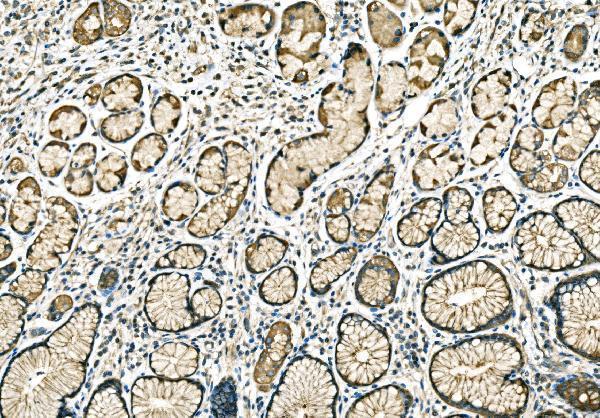
Real IHC data IHC analysis of PPFIA1 using anti-PPFIA1 antibody (A05735-1). PPFIA1 was detected in a paraffin-embedded section of human gastric signet ring cell carcinoma tissue. Heat mediated antigen retrieval was performed in EDTA buffer (pH 8.0, epitope retrieval solution). The tissue section was blocked with 10% goat serum. The tissue section was then incubated with 2 μg/ml rabbit anti-PPFIA1 Antibody (A05735-1) overnight at 4°C. Biotinylated goat anti-rabbit IgG was used as secondary antibody and incubated for 30 minutes at 37°C. The tissue section was developed using Strepavidin-Biotin-Complex (SABC) (Catalog # SA1022) with DAB as the chromogen.
Anti-PPFIA1 Antibody ®
Cat # A05735-1

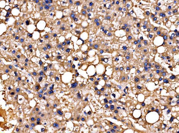
A05735-1 has IHC images from human gastric signet ring cell carcinoma, liver cancer and rectal differentiated adenocarcinoma paraffin sections (catalog: A05735-1 IHC captions). A05735-1 also has an IF image from A431 cells (catalog: A05735-1 IF caption).

Which to pick: Choose A05735-1 for paraffin-section IHC: its own captions document staining at 2 μg/ml after EDTA pH 8.0 heat retrieval, but do not report the fixative (catalog: A05735-1 IHC captions). Choose the same SKU for IF/ICC: IF and ICC are listed applications, and its A431 IF caption documents 5 μg/ml (catalog: A05735-1 applications and IF caption). No cross-species choice is supported: the listed reactivity is human, and clonality is unreported (catalog: A05735-1 reactivity and clone fields).

Each figure is that product's own IHC / IF validation image from its datasheet.

References

  1. UniProt Consortium. UniProt entry Q13136 (LIPA1_HUMAN, Liprin-alpha-1).
  2. Human Protein Atlas. PPFIA1 tissue IHC expression (reliability: Uncertain).
  3. Human Protein Atlas. PPFIA1 subcellular location (ICC-IF): Mainly localized to the cytosol. In addition localized to the focal adhesion sites..
  4. Human Protein Atlas. PPFIA1 antibody validation summary (2 antibodies).
  5. Cortactin expression predicts poor survival in laryngeal carcinoma. British journal of cancer 2008 — PMC2266851.
  6. Prognostic significance of TMEM16A, PPFIA1, and FADD expression in invasive ductal carcinoma of the breast. World journal of surgical oncology 2014 — PMC4028009.
  7. PPFIA1 expression associates with poor response to endocrine treatment in luminal breast cancer. BMC cancer 2020 — PMC7227113.
  8. High expression of PPFIA1 in human esophageal squamous cell carcinoma correlates with tumor metastasis and poor prognosis. BMC cancer 2023 — PMC10169376.
  9. PubMed PMID:7796809 — UniProt-cited evidence.
  10. PubMed PMID:16554811 — UniProt-cited evidence.
  11. PubMed PMID:15489334 — UniProt-cited evidence.