PPP1R8 / Nuclear inhibitor of protein phosphatase 1 · IHC design guide

Design Immunohistochemistry for PPP1R8

Plan PPP1R8 staining in paraffin sections around its widespread nuclear tissue pattern (HPA tissue IHC). The catalog antibody has an IHC working range of 2–5 μg/ml (datasheet A05396-2); compare nuclear staining across cells under consistent fixation.

Evidence assembled Oct 2026 · For research use; verify linked source records and product datasheet before use
Immunohistochemistry protocol sheet for PPP1R8 (IHC for PPP1R8): expected localisation Nuclear in tissue (HPA tissue IHC); cytoplasm also annotated (UniProt), antibody A05396-2, validated IHC image, and IHC protocol steps
Printable PPP1R8 IHC protocol sheet — expected localisation Nuclear in tissue (HPA tissue IHC); cytoplasm also annotated (UniProt), antibody A05396-2, controls and protocol steps. Open the full PPP1R8 IHC guide →

PPP1R8 Immunohistochemistry Experimental Design Guide

Expected localisation, validated protocols, controls and antibodies — the at-a-glance facts below, then the full design guide.

Must know before staining
Expected localisation Nuclear in tissue (HPA tissue IHC); cytoplasm also annotated (UniProt)
Staining pattern Nuclei across diverse tissue cell types (HPA tissue IHC)
Antigen retrieval EDTA pH 8.0 HIER, heat-mediated (datasheet A05396-2)
Positive control ⓘ Adrenal gland+4 more · see all
Negative control ⓘ None in HPA (detected in all 45 tissues); use no-primary + isotype controls
Important caveats
Reasons your staining may differ from the expected pattern.
Fixation Matched tissue-IHC evidence does not establish the fixation claim. Validate the specimen-specific method before use. (selected-SKU IHC image A05396-2)
Caveat Cell-type-dependent isoform ratios complicate comparisons (UniProt)
Regulation Heart and skeletal muscle highest; kidney lower (UniProt)
Isoform / epitope 3 isoforms; antibody epitope coverage is unknown (UniProt)
Section 1

Recommended PPP1R8 IHC & IF Protocols

The catalog antibody protocol is paired with one published mouse testis paraffin-section method (PMC5645368: Methods).

Recommended immunohistochemistry (IHC-P) protocol parameters
SampleParaffin-embedded human pancreas cancer tissue; fixative not specified (datasheet A05396-2)
FixationImage fixative and duration unreported (datasheet A05396-2); verify before use.
Sectioning4–5 µm sections on charged slides (standard)
DeparaffinisationXylene, graded ethanol series to water (standard)
Antigen retrievalHeat retrieval: EDTA pH 8.0 (datasheet A05396-2); 20 min, 95–100 °C (standard)
Peroxidase block3% H2O2, 10 min, room temperature (standard)
Blocking10% goat serum (datasheet A05396-2)
Primary antibodyRabbit anti-PPP1R8, 2-5 μg/ml (datasheet A05396-2)
Primary incubationOvernight at 4 °C (datasheet A05396-2)
DetectionHRP-conjugated secondary, DAB chromogen (datasheet A05396-2)
CounterstainHematoxylin, blue, dehydrate and mount (standard)
Expected resultPPP1R8-positive staining in glandular cells of adrenal gland (HPA tissue IHC: High). HPA tissue profile: Ubiquitous nuclear expression. No signal in the no-primary control.
💡Decision noteStart with heat-mediated EDTA retrieval at pH 8.0 (datasheet A05396-2); the published method does not specify retrieval (PMC5645368: Methods).
Section 2

What Is the Expected PPP1R8 Staining Pattern?

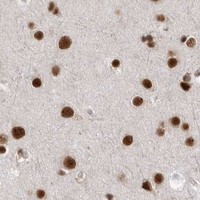
PPP1R8 should stain nuclei across many cell types, including glandular, hematopoietic, respiratory epithelial, neuronal and glial cells (HPA: ubiquitous nuclear expression; High staining in listed cells). HPA rates the tissue IHC profile Enhanced, with high consistency between antibody staining and RNA expression (HPA: tissue IHC reliability). Nuclear signal is the main expectation, although some cytoplasmic localization is possible (UniProt Q12972: primarily, but not exclusively, nuclear). PPP1R8 has no transmembrane segment (UniProt Q12972: topology).

What am I looking at on my slide?
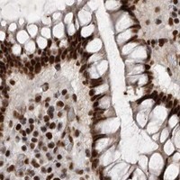
Nuclear chromogen is clear in breast glandular cells and bone marrow hematopoietic cells.This matches two reported High-staining populations (HPA: breast glandular cells; bone marrow hematopoietic cells). Judge signal against the counterstained nuclei (standard IHC practice).
Cytoplasmic staining dominates while nuclei remain pale.This conflicts with the expected predominant nuclear pattern (HPA: ubiquitous nuclear expression; UniProt Q12972: primarily nuclear). Some cytoplasmic signal is possible, so assess its strength relative to nuclei before calling the pattern aberrant (UniProt Q12972: cytoplasm).
Strong staining appears mainly in an unexpected cell population rather than the expected nuclei.Cross-reactivity or endogenous detection activity is possible (standard IHC practice). PPP1R8 is widely expressed, so staining in an unlisted cell type alone cannot prove an artefact (HPA: ubiquitous nuclear expression; UniProt Q12972: ubiquitous expression).
Chromogen is diffuse across tissue and obscures nuclear boundaries.The result cannot establish nuclear localization (standard IHC practice). Compare a no-primary control and assess blocking, detection background and antibody concentration (standard IHC practice).
Nuclei are unstained in adrenal gland glandular cells or bronchial respiratory epithelium.These are reported High-staining populations, making the negative result worth investigating (HPA: adrenal gland glandular cells; bronchus respiratory epithelial cells). Check controls and assay conditions before interpreting it as absent PPP1R8 (standard IHC practice).
💡Expected PPP1R8 appearanceCall the IHC result positive when distinct nuclear staining is strong in a reported High-staining cell population, such as breast glandular cells (HPA: High); dominant diffuse cytoplasmic or acellular chromogen is suspect (HPA: ubiquitous nuclear expression; standard IHC practice).
How each factor affects the staining
Expected compartmentNuclear staining is the main tissue IHC pattern (HPA: ubiquitous nuclear expression). UniProt also lists cytoplasm, so a minor cytoplasmic component need not invalidate clear nuclear staining (UniProt Q12972: subcellular location).
Choice of positive tissueAdrenal gland, appendix and breast glandular cells; bone marrow hematopoietic cells; and bronchial respiratory epithelial cells have High staining (HPA: tissue IHC). These provide documented populations for assessing nuclear signal.
Isoform compositionPPP1R8 has Alpha, Beta and Gamma isoforms, with cell-type-dependent ratios (UniProt Q12972: isoforms; tissue specificity). The supplied evidence gives no antibody epitope map, so it cannot predict isoform-specific IHC staining.
Protein topology and processingPPP1R8 has no transmembrane segment, signal peptide or propeptide, and its annotated chain spans residues 1–351 (UniProt Q12972: topology; processing). The supplied evidence gives no basis for a membrane or shed-protein pattern.
Antibody validationHPA027406, HPA027417 and HPA027452 each have Enhanced IHC status (HPA: antibody validation). That supports the reported pattern, but slide-specific background and detection controls remain necessary (standard IHC practice).
IF/ICC Q: What pattern should be expected?A: Nuclear speckles are the main location, with additional nucleoplasmic signal (HPA: subcellular ICC-IF, enhanced). This finer IF pattern should not be required to call chromogenic tissue IHC positive (standard IHC practice).
Why is my staining missing, weak or wrong?
SituationLikely causeNext action
High-staining reference tissue shows no nuclear signal.An assay or reagent failure is possible; HPA reports High staining in the selected reference cells (HPA: tissue IHC; standard IHC practice).Confirm the reference cell type, primary-antibody application and detection controls; then review retrieval and dilution against the antibody's IHC-P instructions (standard IHC practice).
Nuclear staining is weak but background is also low.The assay may be under-sensitive, or the examined field may contain fewer of the reported High-staining cells (standard IHC practice; HPA: tissue IHC).Recheck tissue morphology and the documented positive population; assess retrieval, antibody dilution and detection with a positive control (standard IHC practice).
Cytoplasm stains more strongly than nuclei.The balance departs from the predominant nuclear expectation, although UniProt allows some cytoplasmic localization (HPA: tissue IHC profile; UniProt Q12972: subcellular location).Compare an HPA-documented High-staining cell population and a no-primary control; reassess antibody dilution and detection background (standard IHC practice).
Unrelated cells or acellular areas carry prominent chromogen.Off-target binding or endogenous detection activity may contribute; the appearance alone cannot distinguish them (standard IHC practice).Inspect no-primary and detection controls, apply the relevant endogenous-activity block, and reassess whether signal follows nuclei (standard IHC practice).
Diffuse background makes nuclear scoring uncertain.Excess reagent signal or insufficient blocking can obscure compartment boundaries (standard IHC practice).Review blocking, washing and antibody dilution; use the nuclear counterstain to judge whether discrete nuclei remain readable (standard IHC practice).
A previously unlisted cell type stains clearly.The HPA list is a set of reported High-staining populations, while the overall profile is ubiquitous nuclear expression (HPA: tissue IHC).Assess nuclear localization and controls before assigning specificity; the supplied HPA record provides no negative cell population for exclusion (HPA: tissue IHC; standard IHC practice).

Sample controls for PPP1R8 IHC & IF

🧪Run bone marrow first and assess nuclear staining in hematopoietic cells, which HPA scores High (HPA: High in bone marrow hematopoietic cells; UniProt Q12972: primarily nuclear). HPA detects PPP1R8 in all 45 scored tissues, so use no-primary and isotype controls in place of a negative tissue; no cell population on the positive slide is established as a true internal negative, though non-target cells can be checked for diffuse background (HPA: no negative tissue rows; standard IHC practice).
Positive control tissue: Adrenal gland (Glandular cells, HPA High)
Negative control tissue: None in HPA: PPP1R8 is detected in all 45 scored tissues. Use a no-primary (secondary-only) and an isotype control instead.
ICC-IF cell lines (HPA subcellular resource): HPA ICC-IF images show PPP1R8 in A-431, U-251MG, U2OS, U2OS, siRNA 1 (10x), U2OS, siRNA 1 (40x), U2OS, siRNA 2 (10x), U2OS, siRNA 2 (40x), U2OS, scrambled (10x), U2OS, scrambled (40x), with annotated localisation: Nuclear speckles (enhanced) (HPA subcellular).
Technical controls: Include a no-primary, secondary-only section and a concentration-matched rabbit IgG isotype control, matching the captioned rabbit primary antibody (catalog antibody A05396-2 caption; standard IHC practice). Confirm specificity with PPP1R8 knockout material or immunizing-peptide blocking if available, and quench endogenous peroxidase in marrow before HRP/DAB detection because hematopoietic cells can contribute peroxidase activity (HPA: bone marrow hematopoietic cells; standard IHC practice).
⚠️Feasibility: A target-specific fixation window or fixation effect is unreported, and the selected A05396-2 paraffin-section caption does not state a fixative (catalog antibody A05396-2 caption). That caption uses heat retrieval in EDTA at pH 8.0, providing an IHC starting condition without establishing that retrieval is required across specimens (catalog antibody A05396-2 caption). Frozen sections are not established as easier; IF may help assess the reported nuclear-speckle pattern, but the supplied ICC-IF images are from cell lines rather than marrow, and endogenous peroxidase is a practical marrow concern for chromogenic detection (HPA: subcellular ICC-IF; HPA: bone marrow hematopoietic cells; standard IHC practice).

HPA tissue IHC evidence for PPP1R8

Comprehensive Human Protein Atlas IHC scoring per tissue (reliability: Enhanced — High consistency between antibody staining and RNA expression data.). Rows are taken directly from the HPA tissue chart — click any row's HPA link to view the source.

Positive expression · recommended positive controls

TissueCell typeLevelEvidenceSource
Adrenal gland Glandular cells High Protein (IHC) HPA →
Appendix Glandular cells High Protein (IHC) HPA →
Bone marrow Hematopoietic cells High Protein (IHC) HPA →
Breast Glandular cells High Protein (IHC) HPA →
Bronchus Respiratory epithelial cells High Protein (IHC) HPA →

Undetected expression · recommended negative controls

TissueCell typeLevelEvidenceSource
None in HPA: PPP1R8 is detected in all 45 scored tissues. Use a no-primary (secondary-only) and an isotype control instead.
Section 3

Advanced PPP1R8 IHC Tips

Use nuclear staining as the main readout for PPP1R8 in paraffin section IHC, while checking retrieval, controls and cell level scoring (HPA tissue IHC; UniProt Q12972).

How should I troubleshoot weak PPP1R8 staining after antigen retrieval?
Start with heat mediated retrieval in EDTA pH 8.0 for this paraffin section assay (datasheet A05396-2). The selected tissue image used that retrieval before staining a human pancreas cancer section with 2 μg/ml primary antibody overnight at 4°C (datasheet A05396-2). If nuclear staining remains weak, check heating consistency and compare retrieval durations on adjacent sections while keeping antibody concentration and detection constant (standard IHC practice; HPA tissue IHC: ubiquitous nuclear expression). If that fails, test an alternative retrieval buffer as a documented fallback, using matched controls to assess nuclear signal and tissue damage (standard IHC practice; UniProt Q12972: primarily nuclear).
Could fixation explain weak or patchy PPP1R8 staining?
Target specific fixation sensitivity is unknown: the selected paraffin section caption does not state a fixative, and the supplied evidence does not establish one (datasheet A05396-2). Record the fixative and fixation duration for each specimen, then compare similarly processed sections before changing antibody conditions (standard IHC practice). Check whether nuclear detail is preserved and whether staining drops preferentially in thick or poorly processed areas, since uneven processing can complicate IHC interpretation (standard IHC practice; HPA tissue IHC: ubiquitous nuclear expression). Keep the documented EDTA pH 8.0 retrieval and 2 μg/ml primary condition constant during that comparison (datasheet A05396-2).
Should I accept predominantly cytoplasmic PPP1R8 staining?
Treat nuclear staining as the principal IHC readout because tissue profiling reports ubiquitous nuclear expression (HPA tissue IHC). Nuclear speckles and nucleoplasm are reported by subcellular imaging, although chromogenic tissue sections may not resolve individual speckles (HPA subcellular; standard IHC practice). PPP1R8 is primarily, but not exclusively, nuclear, so a minor cytoplasmic component is biologically possible; diffuse cytoplasmic staining alone needs stronger validation (UniProt Q12972: subcellular location; standard IHC practice). Compare adjacent sections processed with EDTA pH 8.0 and include a no primary control before attributing cytoplasmic colour to PPP1R8 (datasheet A05396-2; standard IHC practice).
Can this antibody distinguish PPP1R8 isoforms or phosphorylation states in tissue?
Do not assign an isoform from this IHC stain without antibody epitope and isoform reactivity data; neither is supplied for the selected antibody (datasheet A05396-2). PPP1R8 has 3 isoforms, Alpha, Beta and Gamma, whose ratios vary by cell type (UniProt Q12972: isoforms and tissue specificity). Reported modified residues include phosphothreonine 161 and several phosphoserines, but the supplied evidence does not establish phosphorylation selective detection (UniProt Q12972: modified residues; datasheet A05396-2). For a suspected isoform or modification difference, document the question, use an independently validated reagent or orthogonal assay, and retain the IHC result as total antibody staining until specificity is established (standard IHC practice).
How can I adapt the PPP1R8 readout for multiplex IF?
For IF/ICC, pair PPP1R8 with a marker of the cell population under study and score signal within that marker defined population (standard IF practice; HPA tissue IHC: ubiquitous nuclear expression). Include a nuclear counterstain because PPP1R8 is mainly seen in nuclear speckles and also in nucleoplasm (HPA subcellular). Select fluorophores after checking the specimen's autofluorescence, and use single stain controls to assess channel bleed through (standard IF practice). PPP1R8 has no transmembrane segment and is primarily nuclear, so permeabilise sufficiently for antibody access to nuclear epitopes; the antibody's exact epitope is unspecified (UniProt Q12972: topology and subcellular location; standard IF practice; datasheet A05396-2).
What should I change when PPP1R8 IHC has high background?
First compare a no primary section with the stained section to identify colour arising from detection chemistry or tissue rather than primary antibody binding (standard IHC practice). The selected assay used 10% goat serum blocking, 2 μg/ml rabbit primary overnight at 4°C, and a peroxidase based DAB readout (datasheet A05396-2). Apply a peroxidase block before chromogen development and check wash quality, since residual enzyme activity or reagent can obscure nuclear interpretation (standard IHC practice). If background persists, titrate the primary around the documented condition while checking whether nuclear signal remains distinguishable from the no primary control (datasheet A05396-2; HPA tissue IHC: ubiquitous nuclear expression; standard IHC practice).
How should I score PPP1R8 across heterogeneous tissue sections? ⚠ ANSWER MARKED FOR VERIFICATION
Define the tissue region and cell population before scoring, then exclude folds, necrosis and section edges from the analysis (standard IHC practice). Score nuclear staining as percent positive cells or an H-score from 0–300, recording intensity categories and positivity thresholds consistently across slides (standard IHC practice; HPA tissue IHC: ubiquitous nuclear expression). If reporting positive cell density per mm², normalise to the analysed viable tissue area and state which cell population was counted (standard IHC practice). Compare matched processing and retrieval conditions, including the documented EDTA pH 8.0 step, because technical variation can distort apparent differences (datasheet A05396-2; standard IHC practice).
How do I separate true PPP1R8 signal from staining artefacts?
Look first for nuclear staining in intact cells, consistent with the reported tissue pattern, while allowing that PPP1R8 is not exclusively nuclear (HPA tissue IHC; UniProt Q12972: subcellular location). Check the identity of the stained cells: high staining has been reported in adrenal glandular cells and bone marrow hematopoietic cells, but those observations do not validate every specimen (HPA tissue IHC; standard IHC practice). Discount edge accentuation, necrotic deposits and colour in a no primary control when judging a positive result (standard IHC practice). If staining is predominantly in an unexpected compartment or cell population, repeat with matched retrieval and an independent specificity control before drawing a biological conclusion (datasheet A05396-2: EDTA pH 8.0; standard IHC practice).
Boster reagents

Best PPP1R8 / Nuclear inhibitor of protein phosphatase 1 IHC Antibodies

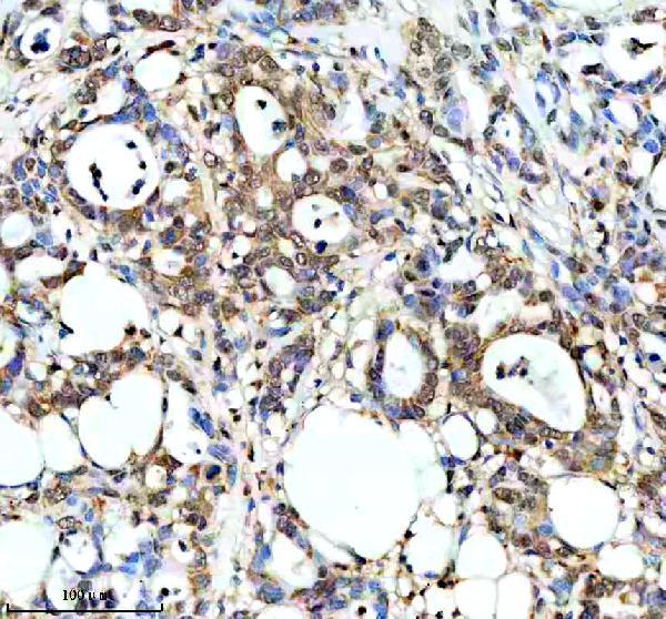
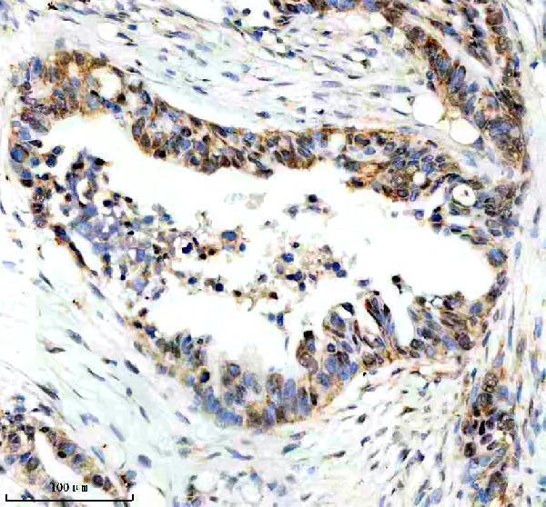
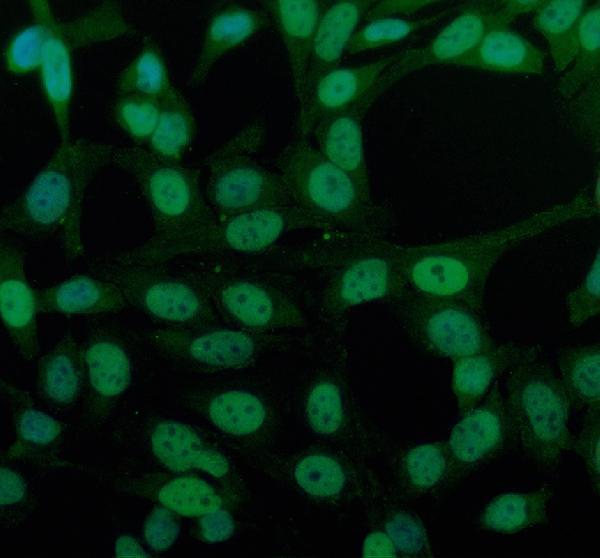
The catalog antibody has IHC data from human paraffin-embedded pancreatic cancer tissue and IF data from human pancreatic cancer tissue and HeLa cells (catalog image captions).

Real IHC data IHC analysis of PPP1R8 using anti-PPP1R8 antibody (A05396-2). PPP1R8 was detected in a paraffin-embedded section of human pancreas cancer tissue. Heat mediated antigen retrieval was performed in EDTA buffer (pH 8.0, epitope retrieval solution). The tissue section was blocked with 10% goat serum. The tissue section was then incubated with 2 μg/ml rabbit anti-PPP1R8 Antibody (A05396-2) overnight at 4°C. Peroxidase Conjugated Goat Anti-rabbit IgG was used as secondary antibody and incubated for 30 minutes at 37°C. The tissue section was developed using HRP Conjugated Rabbit IgG Super Vision Assay Kit (Catalog # SV0002) with DAB as the chromogen.
Anti-PPP1R8 Antibody ®
Cat # A05396-2

A05396-2 has IHC data from paraffin-embedded human pancreatic cancer tissue (IHC image caption). A05396-2 also has IF data from human pancreatic cancer tissue and HeLa cells (IF image captions).

Which to pick: Choose A05396-2 for tissue IHC: its own image shows staining of a paraffin-embedded human pancreatic cancer section after heat retrieval in EDTA, pH 8.0 (IHC image caption); the fixative is unreported (IHC image caption). Choose the same SKU for IF/ICC based on its IF images of HeLa cells and paraffin-embedded human pancreatic cancer tissue (IF image captions). For mouse or rat samples, A05396-2 lists reactivity with both species, but the supplied IHC and IF images show human samples only (catalog reactivity; image captions).

Each figure is that product's own IHC / IF validation image from its datasheet.