This website uses cookies to ensure you get the best experience on our website.


- Table of Contents
Plan PPP3CB chromogenic IHC in paraffin sections using the catalog antibody at 2–5 μg/ml (datasheet A07303-1). Compare cytoplasmic staining with HPA tissue observations, keeping its uncertain reliability in mind (UniProt; HPA tissue IHC).

Expected localisation, validated protocols, controls and antibodies — the at-a-glance facts below, then the full design guide.
| Expected localisation | Cytoplasmic (UniProt); extracellular signal in some tissues (HPA tissue IHC) | |
| Staining pattern | Macrophages and neurons: cytoplasmic; some extracellular signal (HPA tissue IHC) | |
| Antigen retrieval | EDTA pH 8.0 HIER, heat-mediated (datasheet A07303-1) | |
| Positive control | Breast+4 more · see all | |
| Negative control | Adrenal gland+2 more · see all |
| Fixation | Matched tissue-IHC evidence does not establish the fixation claim. Validate the specimen-specific method before use. (selected-SKU IHC image A07303-1) | |
| Caveat | Tissue staining has low concordance with RNA data (HPA tissue IHC) | |
| Regulation | Expression regulation unreported (UniProt) | |
| Isoform / epitope | Four isoforms; check epitope coverage (UniProt) |
The catalog antibody’s IHC-P protocol uses EDTA pH 8.0 retrieval (datasheet A07303-1). The published options cover glioma and pancreatic tissue microarrays (PMC10733678; PMC7785039).
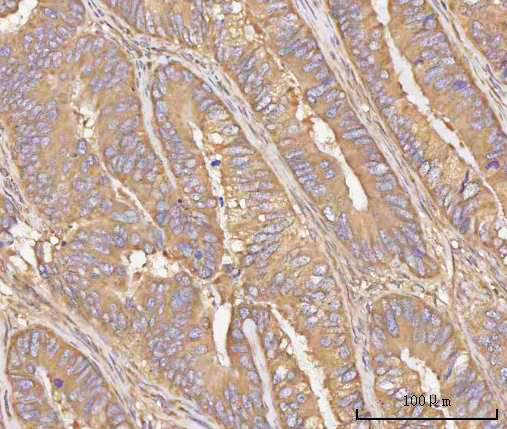
| Sample | Paraffin-embedded human rectum adenocarcinoma tissue; fixative not specified (datasheet A07303-1) |
| Fixation | Image fixative and duration unreported (datasheet A07303-1); verify before use. |
| Sectioning | 4–5 µm sections on charged slides (standard) |
| Deparaffinisation | Xylene, graded ethanol series to water (standard) |
| Antigen retrieval | Heat retrieval: EDTA pH 8.0 (datasheet A07303-1); 20 min, 95–100 °C (standard) |
| Peroxidase block | 3% H2O2, 10 min, room temperature (standard) |
| Blocking | 10% goat serum (datasheet A07303-1) |
| Primary antibody | Rabbit anti-PPP3CB, 2-5 μg/ml (datasheet A07303-1) |
| Primary incubation | Overnight at 4 °C (datasheet A07303-1) |
| Detection | HRP-conjugated secondary, DAB chromogen (datasheet A07303-1) |
| Counterstain | Hematoxylin, blue, dehydrate and mount (standard) |
| Expected result | PPP3CB-positive staining in glandular cells of breast (HPA tissue IHC: High). HPA tissue profile: General cytoplasmic expression with extracellular positivity in several tissues. No signal in the no-primary control. |
PPP3CB is expected mainly in the cytoplasm, consistent with its lack of a transmembrane segment (UniProt P16298: cytoplasm, topology). HPA reports high IHC staining in breast glandular cells, neurons in several brain regions, and lung macrophages (HPA: tissue IHC). Use these as candidate positive contexts, with caution: HPA rates the tissue staining pattern Uncertain because antibody staining and RNA expression have low consistency (HPA: reliability).
| Cytoplasmic staining in breast glandular cells, neurons, or lung macrophages, with distinguishable cellular boundaries. | This fits the expected compartment (UniProt P16298: cytoplasm) and cells reported as High by HPA (HPA: tissue IHC). Score intensity in the specified cells, not the whole section; the match is supportive, while HPA's Uncertain tissue reliability limits confirmation of specificity (HPA: reliability). |
| Signal is predominantly confined to nuclei or cell membranes, with little cytoplasmic staining. | This conflicts with the principal cytoplasmic assignment and lacks support from UniProt topology (UniProt P16298: cytoplasm, no transmembrane segment). Review morphology and controls before calling it PPP3CB. HPA's ICC-IF record lists nucleoplasm as an uncertain additional location, so occasional nuclear signal alone is inconclusive (HPA: subcellular). |
| Strong staining appears in an unexpected cell population within a section. | First identify the stained cells by morphology and compare them with the relevant HPA cell-specific entry (HPA: tissue IHC). A mismatch may reflect nonspecific binding or endogenous chromogenic detection activity (general IHC practice). It cannot establish a new PPP3CB cell distribution without independent validation, especially given HPA's Uncertain rating (HPA: reliability). |
| Broad haze obscures cells, or extracellular material stains more strongly than cell cytoplasm. | Treat diffuse haze as background until controls and tissue structure clarify it (general IHC practice). Extracellular positivity occurs in several HPA tissues, so extracellular signal is not automatically an artefact; it is also insufficient on its own to establish a cellular PPP3CB pattern (HPA: tissue IHC profile). |
| No visible staining occurs in a candidate positive tissue. | A blank result in a region reported as High by HPA calls for a technical check, including section integrity, antibody detection, and the assay's retrieval conditions (HPA: tissue IHC; general IHC practice). It does not prove absence of PPP3CB, and the HPA reference pattern itself remains Uncertain (HPA: reliability). |
| Compartment and topology | UniProt places PPP3CB in the cytoplasm and reports no transmembrane segment (UniProt P16298). Use cellular cytoplasm as the main localization criterion; the record gives no basis to expect a membrane-rim pattern. |
| Cell-specific reference patterns | HPA reports High staining in breast glandular cells, caudate, cortical and hippocampal neurons, and lung macrophages; it reports Medium staining in cerebellar Purkinje cells (HPA: tissue IHC). Compare like cell types when judging intensity, rather than treating all cells in a tissue as equivalent. |
| Strength of the reference evidence | HPA marks tissue IHC reliability Uncertain because staining and RNA expression show low consistency, with external verification pending; HPA008233 is also IHC Uncertain (HPA: reliability, antibodies). A pattern match supports interpretation but does not independently validate antibody specificity. |
| Isoforms and processing | UniProt lists four PPP3CB isoforms and a chain spanning residues 2–524, with no signal peptide or propeptide (UniProt P16298). The supplied record gives no antibody epitope, so it cannot establish which isoforms an IHC antibody detects or whether isoform differences alter staining. |
| IF/ICC Q&A: should the same localization be expected? | Mainly cytosolic staining is supported in HPA ICC-IF; nucleoplasm is an uncertain additional location and calyx is approved (HPA: subcellular). HPA cautions that this annotation uses antibodies targeting proteins from multiple genes, so it should not override the uncertain tissue IHC assessment (HPA: subcellular, reliability). |
| Situation | Likely cause | Next action |
|---|---|---|
| No staining in an HPA High cell population. | The result may reflect an assay failure or absent detectable signal; HPA's Uncertain reference cannot resolve which (HPA: tissue IHC, reliability). | Check section morphology and a working detection control, then review the antibody's IHC dilution and retrieval conditions against its supplied instructions (general IHC practice). Do not infer a PPP3CB-specific fixation effect from this result. |
| All cell types or the entire section show a uniform chromogenic tint. | Widespread tint may arise from background binding or endogenous detection activity (general IHC practice). | Compare a no-primary control; review blocking, washes, and detection chemistry, then reassess whether cytoplasmic signal remains in the expected cells (general IHC practice; UniProt P16298: cytoplasm; HPA: tissue IHC). |
| Predominantly nuclear or membrane-rim staining persists. | That pattern conflicts with the main cytoplasmic assignment; HPA lists nucleoplasm only as an uncertain additional ICC-IF location (UniProt P16298: cytoplasm, topology; HPA: subcellular). | Recheck cell boundaries and counterstain, compare controls, and seek independent specificity evidence before scoring the pattern as PPP3CB (general IHC practice; HPA: reliability Uncertain). |
| Extracellular staining dominates the section. | HPA reports extracellular positivity in several tissues, but does not establish that this alone identifies the source of PPP3CB signal (HPA: tissue IHC profile). | Score intracellular staining separately from extracellular deposits; inspect a no-primary control and report the extracellular observation without treating it as cell-specific evidence (general IHC practice; HPA: tissue IHC profile). |
| A nominally negative comparator stains strongly. | HPA lists adrenal gland glandular cells, ovarian follicle cells, and parathyroid gland glandular cells as Not detected, yet rates the overall tissue pattern Uncertain (HPA: tissue IHC, reliability). | Confirm the actual stained cell type, review the no-primary control, and seek orthogonal validation before interpreting unexpected staining as PPP3CB expression (general IHC practice; HPA: reliability). |
| The expected cytoplasmic cells stain, but the signal is hard to separate from background. | Weak contrast may limit cell-level scoring, while HPA's Uncertain rating limits how much the reference pattern can settle specificity (general IHC practice; HPA: reliability). | Compare matched controls and cell morphology; optimize general IHC conditions within the antibody's validated instructions, then document the cell-specific result and any remaining uncertainty (general IHC practice; HPA: tissue IHC). |
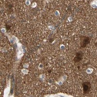


Comprehensive Human Protein Atlas IHC scoring per tissue (reliability: Uncertain — Low consistency between antibody staining and RNA expression data. Pending external verification.). Rows are taken directly from the HPA tissue chart — click any row's HPA link to view the source.
| Tissue | Cell type | Level | Evidence | Source |
|---|---|---|---|---|
| Breast | Glandular cells | High | Protein (IHC) | HPA → |
| Caudate | Neuronal cells | High | Protein (IHC) | HPA → |
| Cerebral cortex | Neuronal cells | High | Protein (IHC) | HPA → |
| Hippocampus | Neuronal cells | High | Protein (IHC) | HPA → |
| Lung | Macrophages | High | Protein (IHC) | HPA → |
Troubleshoot PPP3CB staining in paraffin sections by checking retrieval, cell type, compartment and controls before comparing chromogenic IHC signals (UniProt P16298; HPA tissue IHC).
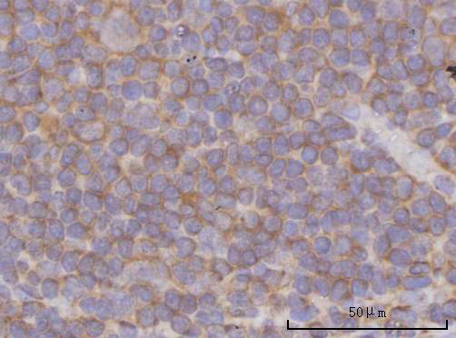
The IHC-validated anti-PPP3CB antibody has paraffin-section images from human rectum adenocarcinoma, tonsil and urothelial carcinoma (catalog IHC captions); its listed reactivity also includes monkey, mouse and rat (catalog reactivity).
A07303-1 is the sole SKU shown, with IHC images from paraffin sections of human rectum adenocarcinoma, tonsil and urothelial carcinoma (catalog IHC captions). Its application list includes IHC but not IF/ICC, and its listed reactivity is human, monkey, mouse and rat (catalog applications and reactivity).
Which to pick: Choose A07303-1 for human tissue IHC on paraffin sections; its captions document 2 μg/ml antibody after heat retrieval in EDTA, pH 8.0, but do not report the fixative (A07303-1 IHC captions). There is no IF/ICC-validated SKU in this payload (catalog applications; catalog IF images: none). For work in monkey, mouse or rat, A07303-1 lists reactivity with those species, while the supplied IHC images document human tissue only (catalog reactivity; A07303-1 IHC captions).