PRKRA / Interferon-inducible double-stranded RNA-dependent protein kinase activator A · IHC design guide

Design Immunohistochemistry for PRKRA

Plan PRKRA paraffin IHC around cytoplasmic staining in selected brain cells (HPA tissue IHC). Use matched tissue controls, consistent fixation, and cell-level scoring to interpret the result.

Evidence assembled Oct 2026 · For research use; verify linked source records and product datasheet before use
Immunohistochemistry protocol sheet for PRKRA (IHC for PRKRA): expected localisation Cytoplasmic tissue staining (HPA tissue IHC), antibody A02744-2, validated IHC image, and IHC protocol steps
Printable PRKRA IHC protocol sheet — expected localisation Cytoplasmic tissue staining (HPA tissue IHC), antibody A02744-2, controls and protocol steps. Open the full PRKRA IHC guide →

PRKRA Immunohistochemistry Experimental Design Guide

Expected localisation, validated protocols, controls and antibodies — the at-a-glance facts below, then the full design guide.

Must know before staining
Expected localisation Cytoplasmic tissue staining (HPA tissue IHC)
Staining pattern Cytoplasmic staining in brain neuronal and glial cells (HPA tissue IHC)
Antigen retrieval EDTA pH 8.0 HIER, heat-mediated (datasheet A02744-2)
Positive control ⓘ Caudate+4 more · see all
Negative control ⓘ Adipose tissue+4 more · see all
Important caveats
Reasons your staining may differ from the expected pattern.
Fixation Keep fixation consistent across paraffin sections (standard IHC practice; not target-specific)
Caveat Strong brain staining may not generalize to other tissues (HPA tissue IHC)
Regulation No expression regulator annotated (UniProt)
Isoform / epitope 3 isoforms; verify epitope coverage (UniProt)
Section 1

Recommended PRKRA IHC & IF Protocols

The catalog antibody’s IHC-P protocol (datasheet: A02744-2) is followed by published PRKRA staining details from three articles (PMC2688376; PMC10361375; PMC11185611).

Recommended immunohistochemistry (IHC-P) protocol parameters
SampleParaffin-embedded mouse brain tissue; fixative not specified (datasheet A02744-2)
FixationImage fixative and duration unreported (datasheet A02744-2); verify before use.
Sectioning4–5 µm sections on charged slides (standard)
DeparaffinisationXylene, graded ethanol series to water (standard)
Antigen retrievalHeat retrieval: EDTA pH 8.0 (datasheet A02744-2); 20 min, 95–100 °C (standard)
Peroxidase block3% H2O2, 10 min, room temperature (standard)
Blocking10% goat serum (datasheet A02744-2)
Primary antibodyRabbit anti-PRKRA, 2-5 μg/ml (datasheet A02744-2)
Primary incubationOvernight at 4 °C (datasheet A02744-2)
DetectionHRP-conjugated secondary, DAB chromogen (datasheet A02744-2)
CounterstainHematoxylin, blue, dehydrate and mount (standard)
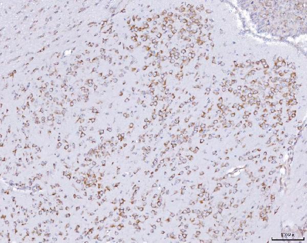
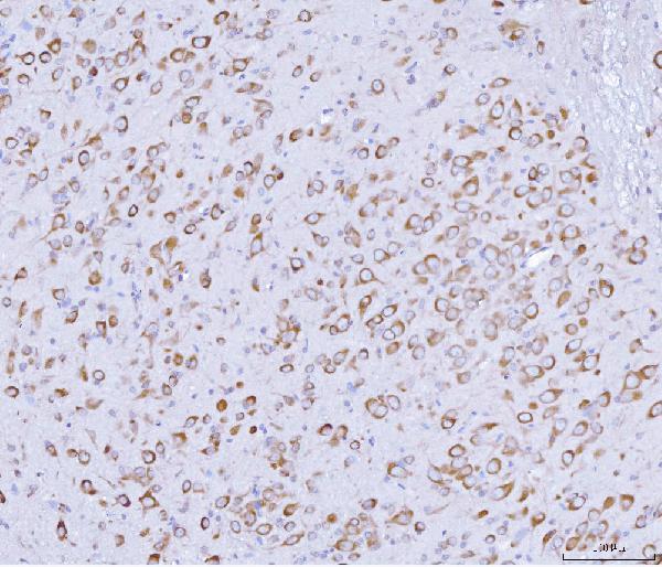
Expected resultPRKRA-positive staining in neuronal cells of caudate (HPA tissue IHC: High). HPA tissue profile: Cytoplasmic expression in several different tissues. No signal in the no-primary control.
💡Decision noteStart with heat-mediated EDTA pH 8.0 retrieval for the catalog antibody (datasheet: A02744-2); the published excerpts do not specify retrieval conditions (PMC2688376; PMC10361375; PMC11185611).
Section 2

What Is the Expected PRKRA Staining Pattern?

PRKRA is a cytoplasmic protein with perinuclear localization and no transmembrane segment (UniProt O75569). In paraffin section IHC, expect cytoplasmic staining in the specific cells reported by HPA, including caudate neurons, cortical glia, and colon glandular cells (HPA: tissue IHC). HPA rates its tissue staining profile Approved, pending external verification (HPA: reliability).

What am I looking at on my slide?
Cytoplasmic signal in caudate neurons or cerebral cortex glia.This fits HPA's High staining in those cell types and the cytoplasmic localization annotated by UniProt (HPA: tissue IHC; UniProt O75569). Score the named cells rather than treating every cell in the section as an expected positive.
Predominantly nuclear, membranous, or extracellular signal in an otherwise positive section.A dominant signal in these compartments does not match the expected tissue IHC pattern (HPA: cytoplasmic expression; UniProt O75569). Check morphology, counterstain, detection controls, and antibody conditions before interpreting it as PRKRA.
Strong staining in adipocytes or bone marrow hematopoietic cells.HPA reports PRKRA as Not detected in those cells (HPA: adipose tissue; bone marrow). Consider cross-reactivity or endogenous detection activity; compare matched control sections before assigning the signal to PRKRA.
Uniform haze across tissue, stroma, and empty slide areas.That distribution obscures cell boundaries and cannot establish the HPA cell-specific pattern (HPA: tissue IHC). Assess nonspecific binding, residual detection reagent, and background in a control lacking primary antibody (general IHC practice).
No signal in caudate neurons or hippocampal neurons.Both are reported High by HPA (HPA: caudate; hippocampus). An absent result warrants checking tissue preservation, retrieval conditions, antibody dilution, and detection controls (general IHC practice); HPA does not establish PRKRA-specific fixation sensitivity.
💡Expected PRKRA appearanceCall a section positive when the named HPA cell population shows predominantly cytoplasmic chromogen, with High signal in caudate neurons or cortical glia and Medium signal in colon glandular cells (HPA: tissue IHC); diffuse slide haze or dominant nuclear staining is suspect (HPA: cytoplasmic expression; general IHC practice).
How each factor affects the staining
Which compartment and cells should guide IHC scoring?Use cytoplasmic signal in the named cell population: HPA reports High staining in caudate neurons, cerebellar molecular-layer cells, cortical glia, and hippocampal neurons; Medium staining in colon, duodenum, epididymis, and fallopian tube glandular cells (HPA: tissue IHC).
How should low or absent staining be interpreted?HPA reports Low staining in several epithelial populations, including stomach glandular cells, and Not detected in selected populations, including adipocytes and liver cholangiocytes (HPA: tissue IHC). These are cell-specific observations; a negative population is a comparison, not proof that an entire organ lacks PRKRA.
What does antibody validation establish?Three listed antibodies have IHC Approved status: HPA034996, HPA034997, and CAB004648 (HPA: antibody validation). The supplied statuses do not say IHC Enhanced; HPA also describes the tissue profile as pending external verification (HPA: reliability).
Do isoforms or modified residues change the expected stain?UniProt lists 3 isoforms and phosphoserines at residues 18, 167, 246, and 287 (UniProt O75569). Without an antibody epitope or isoform-specific IHC evidence, these annotations cannot predict a different compartment or staining intensity.
What is expected on the separate IF/ICC guide page?HPA reports mainly cytosolic IF signal, with additional nucleoplasmic localization, in its ICC-IF assessment (HPA: subcellular localization). That supported nucleoplasmic observation should not be used to recast dominant nuclear chromogen in tissue IHC as the expected tissue pattern.
Why is my staining missing, weak or wrong?
SituationLikely causeNext action
Known-positive neuronal cells show no chromogen.A failed staining run or unsuitable assay conditions are possibilities; HPA reports High staining in caudate and hippocampal neurons (HPA: tissue IHC).Check section quality, retrieval, antibody dilution, and detection with a concurrently stained positive section (general IHC practice). The supplied sources do not specify a PRKRA retrieval condition.
All compartments stain equally, including nuclei.The pattern conflicts with cytoplasmic tissue IHC localization (HPA: tissue IHC; UniProt O75569); background or ambiguous chromogen localization may explain it.Inspect cell boundaries and counterstain, compare a control lacking primary antibody, and optimize blocking or washing if background persists (general IHC practice).
Signal appears in HPA Not detected cells.Cross-reactivity or endogenous detection activity may contribute; HPA reports adipocytes and bone marrow hematopoietic cells as Not detected (HPA: tissue IHC).Compare the suspect cells with a known-positive cell population and a control lacking primary antibody; review endogenous enzyme blocking where relevant to the chromogenic system (general IHC practice).
Weak staining appears in stomach glandular cells.A weak result may fit HPA's Low category for that cell population (HPA: stomach). HPA categories do not by themselves establish a technical failure.Score the correct cell population and compare with a concurrently stained High reference, such as caudate neurons, under the same assay conditions (HPA: caudate; general IHC practice).
Colon glands stain, but the whole section is called uniformly positive.HPA reports Medium staining specifically in colon glandular cells (HPA: colon); whole-section scoring can hide cell-specific distribution.Record the percentage and intensity in glandular cells separately from surrounding tissue, and note any background that prevents confident cellular assignment (general IHC practice).
A strong nuclear signal is defended using IF/ICC images.HPA supports additional nucleoplasmic localization in ICC-IF, while its tissue IHC profile describes cytoplasmic expression (HPA: subcellular localization; tissue IHC).Interpret the paraffin section against tissue IHC morphology and controls. Reserve the ICC-IF observation for the separate IF/ICC guide; it does not supply an IHC nuclear scoring rule.

Sample controls for PRKRA IHC & IF

🧪Run caudate first and assess staining in neuronal cells, where PRKRA is High (HPA: caudate neuronal cells, High). Use adipose tissue as the negative tissue, focusing on adipocytes where PRKRA is Not detected (HPA: adipose tissue adipocytes, Not detected); on the caudate slide, unstained non-neuronal cells can serve as background comparators, but their PRKRA-negative status is unestablished by the supplied HPA row.
Positive control tissue: Caudate (Neuronal cells, HPA High)
Negative control tissue: Adipose tissue (HPA Not detected)
ICC-IF cell lines (HPA subcellular resource): HPA ICC-IF images show PRKRA in A-431, PC-3, U2OS, NIH 3T3, with annotated localisation: Cytosol (supported) (HPA subcellular).
Technical controls: Include a no-primary, secondary-only control, a species- and immunoglobulin-matched rabbit IgG isotype control, and PRKRA knockout material if available as a biological specificity control (selected A02744-2 caption: rabbit primary; standard IHC practice). For HRP/DAB staining of caudate, block endogenous peroxidase; for IF, assess brain autofluorescence (selected A02744-2 caption: HRP/DAB; standard IHC/IF practice).
⚠️Feasibility: A target-specific fixation window or fixation effect is unreported, and the selected A02744-2 paraffin-section caption does not state the fixative (selected A02744-2 caption). That caption reports heat retrieval in EDTA at pH 8.0 for mouse brain IHC, but does not establish that retrieval is required for PRKRA staining (selected A02744-2 caption). The supplied evidence does not establish whether frozen sections or IF are easier; brain autofluorescence may complicate IF interpretation (HPA: ICC-IF cell-line images; standard IF practice).

HPA tissue IHC evidence for PRKRA

Comprehensive Human Protein Atlas IHC scoring per tissue (reliability: Approved — Pending external verification.). Rows are taken directly from the HPA tissue chart — click any row's HPA link to view the source.

Positive expression · recommended positive controls

TissueCell typeLevelEvidenceSource
Caudate Neuronal cells High Protein (IHC) HPA →
Cerebellum Cells in molecular layer High Protein (IHC) HPA →
Cerebral cortex Glial cells High Protein (IHC) HPA →
Hippocampus Neuronal cells High Protein (IHC) HPA →
Colon Glandular cells Medium Protein (IHC) HPA →

Undetected expression · recommended negative controls

TissueCell typeLevelEvidenceSource
Adipose tissue Adipocytes Not detected Protein (IHC) HPA →
Adrenal gland Glandular cells Not detected Protein (IHC) HPA →
Bone marrow Hematopoietic cells Not detected Protein (IHC) HPA →
Cervix Glandular cells Not detected Protein (IHC) HPA →
Heart muscle Cardiomyocytes Not detected Protein (IHC) HPA →
Section 3

Advanced PRKRA IHC Tips

Troubleshoot PRKRA staining in paraffin section chromogenic IHC using the selected antibody’s tissue protocol, cellular localisation, and tissue expression evidence.

How should I optimize retrieval when PRKRA staining is weak in paraffin sections?
Start with heat mediated retrieval in EDTA at pH 8.0 (datasheet A02744-2). The selected antibody was used after that retrieval on a paraffin embedded mouse brain section, followed by 10% goat serum blocking and 2 μg/ml primary antibody overnight at 4°C (datasheet A02744-2). If staining remains weak, adjust heating duration in small steps while holding antibody concentration, detection, and development constant, and inspect tissue integrity after each change (standard IHC practice). Compare matched sections with a no primary control so increased DAB background is not mistaken for improved cytoplasmic signal (standard IHC practice; UniProt O75569 localisation).
Does the available evidence identify an optimal fixative for PRKRA IHC?
Target specific sensitivity to fixation is unknown: the selected paraffin section caption does not state a fixative (datasheet A02744-2). Record the actual fixative and fixation interval for each specimen, then compare similarly processed sections before changing retrieval or antibody concentration (standard IHC practice). If signal varies across a batch, assess fixation history and section quality alongside a tissue control, because processing differences can change antigen accessibility in paraffin IHC (standard IHC practice). Do not attribute a weak result to PRKRA phosphorylation or its cytoplasmic localisation without an experiment that isolates fixation as the variable (UniProt O75569 modified residues and localisation; standard IHC practice).
Which cellular pattern should count as plausible PRKRA staining?
Prioritise cytoplasmic staining, including a possible perinuclear pattern, when reading chromogenic sections (UniProt O75569 localisation). Cytosolic localisation is supported by cell imaging, while nucleoplasmic localisation is also reported; a nuclear signal therefore needs separate validation rather than automatic rejection (HPA subcellular). PRKRA has no transmembrane segment, so a crisp, exclusive cell membrane rim is inconsistent with its annotated topology and warrants a background check (UniProt O75569 topology). Compare cellular staining against adjacent no primary sections and morphology, especially where dense DAB or counterstain makes a perinuclear deposit appear nuclear (standard IHC practice).
Can this IHC result distinguish PRKRA isoforms or phosphorylation states?
Do not assign staining to a single splice form without an epitope map and isoform specific validation: the record lists 3 isoforms (UniProt O75569 isoforms). PRKRA contains double stranded RNA binding domains at residues 34–101, 126–194, and 240–308, and annotated phosphoserines include residues 18, 167, 246, and 287 (UniProt O75569 domains and modified residues). The selected tissue caption specifies antibody concentration and retrieval, but no epitope or phospho specificity (datasheet A02744-2). Treat DAB intensity as antibody reactivity until an appropriate isoform or modification specific control supports a narrower interpretation (standard IHC practice).
How can I check a PRKRA IHC pattern with multiplex IF?
On the separate IF/ICC workflow, pair PRKRA with a validated cell type marker to test whether signal falls in the expected population; neuronal cells in caudate and hippocampus and glial cells in cerebral cortex show high tissue IHC staining (HPA tissue IHC). Select fluorophores after inspecting unstained tissue for autofluorescence, and keep single stain controls for spectral bleed through (standard IF practice). Permeabilise for an intracellular epitope because PRKRA is cytoplasmic and has no transmembrane segment, then titrate permeabilisation to preserve morphology (UniProt O75569 localisation and topology; standard IF practice). Do not transfer the selected paraffin section retrieval or primary incubation conditions to IF without testing them there (datasheet A02744-2; standard IF practice).
What should I change when PRKRA DAB staining is widespread or granular?
First compare a no primary section and assess whether DAB appears in the same structures, then check the peroxidase block and development time (standard IHC practice). The selected tissue protocol used 10% goat serum blocking, 2 μg/ml primary antibody overnight at 4°C, a peroxidase conjugated secondary, and DAB detection (datasheet A02744-2). If background persists, titrate primary concentration and shorten chromogen development while retaining a known positive control section (standard IHC practice). Judge residual signal against the expected cytoplasmic pattern and local tissue morphology, since diffuse edge staining or pigment can obscure the cellular distribution (UniProt O75569 localisation; standard IHC practice).
How should I score PRKRA across regions with different cell populations? ⚠ ANSWER MARKED FOR VERIFICATION
Define the cell population and region before scoring, then report the percentage of positive cells and intensity separately or combine them as an H score on a 0–300 scale (standard IHC practice). If cell counts vary, normalise positive counts to the number of eligible cells; for spatial comparisons, report positive cell density per mm² of evaluable tissue (standard IHC practice). Apply the same segmentation, counterstain, DAB threshold, and exclusion rules to every section (standard IHC practice). Stratify neuronal and glial populations where morphology permits, because reported high staining occurs in neuronal cells in caudate and hippocampus and glial cells in cerebral cortex (HPA tissue IHC).
How can I separate true PRKRA staining from tissue artefacts?
A convincing result has cellular cytoplasmic signal in a plausible cell population, with a clean no primary control and preserved morphology (UniProt O75569 localisation; HPA tissue IHC; standard IHC practice). High staining in caudate neuronal cells or cerebral cortex glial cells can support assay assessment, but the tissue IHC profile is approved pending external verification (HPA tissue IHC). Investigate exclusive membrane rims, edge restricted deposits, necrotic areas, and staining that persists without primary antibody before calling cells positive (UniProt O75569 topology; standard IHC practice). Check endogenous peroxidase and chromogen deposition as possible causes of DAB signal, and confirm any unexpected nuclear pattern independently because nucleoplasmic localisation is also reported (standard IHC practice; HPA subcellular).
Boster reagents

Best PRKRA / Interferon-inducible double-stranded RNA-dependent protein kinase activator A IHC Antibodies

A02744-2 has paraffin-section IHC images from mouse and rat brain and human ovarian cancer (A02744-2 image captions). M02744 lists IHC use and human, mouse, and rat reactivity (catalog).

Real IHC data IHC analysis of PRKRA using anti-PRKRA antibody (A02744-2). PRKRA was detected in a paraffin-embedded section of mouse brain tissue. Heat mediated antigen retrieval was performed in EDTA buffer (pH 8.0, epitope retrieval solution). The tissue section was blocked with 10% goat serum. The tissue section was then incubated with 2 μg/ml rabbit anti-PRKRA Antibody (A02744-2) overnight at 4°C. Peroxidase Conjugated Goat Anti-rabbit IgG was used as secondary antibody and incubated for 30 minutes at 37°C. The tissue section was developed using HRP Conjugated Rabbit IgG Super Vision Assay Kit (Catalog # SV0002) with DAB as the chromogen.
Anti-PRKRA Antibody ®
Cat # A02744-2

A02744-2 has IHC images from paraffin sections of mouse and rat brain and human ovarian cancer (A02744-2 image captions). M02744 lists IHC use and human, mouse, and rat reactivity, but has no IHC image in the payload (catalog).

Which to pick: Choose A02744-2 for tissue IHC: its own captions document paraffin-section staining in all three listed species, though the fixative is unreported (A02744-2 image captions). M02744 is a rabbit monoclonal with listed IHC use and human, mouse, and rat reactivity, but no IHC image; A02744-2 has the stronger cross-species image evidence (catalog; A02744-2 image captions). Neither SKU lists IF/ICC use or provides IF images, so there is no supported IF/ICC pick (catalog).

Each figure is that product's own IHC / IF validation image from its datasheet.

References

  1. UniProt Consortium. UniProt entry O75569 (PRKRA_HUMAN, Interferon-inducible double-stranded RNA-dependent protein kinase activator A).
  2. Human Protein Atlas. PRKRA tissue IHC expression (reliability: Approved).
  3. Human Protein Atlas. PRKRA subcellular location (ICC-IF): Mainly localized to the cytosol. In addition localized to the nucleoplasm..
  4. Human Protein Atlas. PRKRA antibody validation summary (3 antibodies).
  5. DICER1 and PRKRA in Colon Adenocarcinoma. Biomarker insights 2008 — PMC2688376.
  6. PRKRA promotes pancreatic cancer progression by upregulating MMP1 transcription via the NF-κB pathway. Heliyon 2023 — PMC10361375.
  7. A frameshift mutation in the murine Prkra gene causes dystonia and exhibits abnormal cerebellar development and reduced eIF2α phosphorylation. bioRxiv : the preprint server for biology 2024 — PMC11185611.
  8. PRKRA/PACT Expression Promotes Chemoresistance of Mucinous Ovarian Cancer. Molecular cancer therapeutics 2019 — PMC6318044.
  9. PubMed PMID:9687506 — UniProt-cited evidence.
  10. PubMed PMID:10336432 — UniProt-cited evidence.
  11. PubMed PMID:11230166 — UniProt-cited evidence.