This website uses cookies to ensure you get the best experience on our website.


- Table of Contents
Plan PRODH staining in paraffin sections with the catalog antibody’s 2–5 μg/ml IHC range (datasheet A03231-3). Compare the observed cytoplasmic pattern with tissue evidence, allowing for low consistency between antibody staining and RNA expression (HPA tissue IHC).

Expected localisation, validated protocols, controls and antibodies — the at-a-glance facts below, then the full design guide.
| Expected localisation | Cytoplasmic tissue staining (HPA tissue IHC); mitochondrial matrix localization (UniProt) | |
| Staining pattern | Cytoplasmic staining in glandular and epithelial cells (HPA tissue IHC) | |
| Antigen retrieval | EDTA pH 8.0 HIER, heat-mediated (datasheet A03231-3) | |
| Positive control | Adrenal gland+4 more · see all | |
| Negative control | Spleen |
| Fixation | Keep fixation consistent across sections (standard IHC practice; not target-specific) | |
| Caveat | Staining may detect proteins from more than one gene (HPA tissue IHC) | |
| Regulation | Expression varies by tissue (UniProt) | |
| Isoform / epitope | 3 isoforms; epitope coverage is unspecified (UniProt) |
The catalog antibody protocol is followed by 3 published PRODH IHC protocols from lung cancer, breast cancer xenograft, and glioma studies (PMC10815008; PMC9261841; PMC10814259).
| Sample | Paraffin-embedded human lung adenocarcinoma tissue; fixative not specified (datasheet A03231-3) |
| Fixation | Image fixative and duration unreported (datasheet A03231-3); verify before use. |
| Sectioning | 4–5 µm sections on charged slides (standard) |
| Deparaffinisation | Xylene, graded ethanol series to water (standard) |
| Antigen retrieval | Heat retrieval: EDTA pH 8.0 (datasheet A03231-3); 20 min, 95–100 °C (standard) |
| Peroxidase block | 3% H2O2, 10 min, room temperature (standard) |
| Blocking | 10% goat serum (datasheet A03231-3) |
| Primary antibody | Rabbit anti-PRODH, 2-5 μg/ml (datasheet A03231-3) |
| Primary incubation | Overnight at 4 °C (datasheet A03231-3) |
| Detection | HRP-conjugated secondary, DAB chromogen (datasheet A03231-3) |
| Counterstain | Hematoxylin, blue, dehydrate and mount (standard) |
| Expected result | PRODH-positive staining in glandular cells of adrenal gland (HPA tissue IHC: High). HPA tissue profile: General cytoplasmic expression. No signal in the no-primary control. |
PRODH is a mitochondrial matrix protein with no annotated transmembrane segment (UniProt O43272: location and topology). In paraffin IHC, expect a cytoplasmic pattern in epithelial and glandular cells, with high staining reported in several tissues (HPA tissue IHC: general cytoplasmic expression; High in listed cells). Interpret intensity cautiously: HPA rates the tissue pattern Approved but reports low agreement with RNA and cautions that antibodies may target proteins from multiple genes (HPA tissue IHC: reliability).
| Granular cytoplasmic staining in bronchial respiratory epithelium or colonic glandular cells. | This fits the expected compartment and cells: PRODH is in the mitochondrial matrix (UniProt O43272: location), and HPA reports High staining in both cell types (HPA tissue IHC). Chromogenic granules alone cannot prove mitochondrial identity (general IHC practice). |
| Predominantly nuclear staining, with little cytoplasmic staining, in an expected positive cell population. | Treat this as discordant with the principal location (UniProt O43272: mitochondrial matrix; HPA subcellular: mitochondria approved). HPA calls nucleoplasmic localization uncertain in ICC-IF, so nuclear IHC alone should not establish it (HPA subcellular: nucleoplasm uncertain). |
| Strong signal in spleen red pulp cells while expected positive epithelial cells stain weakly. | HPA reports PRODH staining as Not detected in red pulp cells and High in several glandular and epithelial populations (HPA tissue IHC). Check specificity and endogenous chromogen activity; an unexpected positive is not proof of PRODH expression (general IHC practice). |
| Similar faint color across tissue, empty spaces, and unrelated cell populations. | A uniform haze is less persuasive than cell-associated cytoplasmic staining (HPA tissue IHC: general cytoplasmic expression; general IHC practice). Background may arise from detection reagents, insufficient blocking, or incomplete washing; compare with an appropriate control slide (general IHC practice). |
| No signal in bronchial respiratory epithelium or colonic glandular cells. | These are reported High by HPA, so a blank result warrants a run check (HPA tissue IHC). Assess tissue preservation, retrieval conditions, antibody and detection performance using the catalog antibody's IHC-P instructions and run controls (general IHC practice). |
| Compartment and optical resolution | The mitochondrial matrix assignment predicts intracellular cytoplasmic signal (UniProt O43272: location). HPA describes tissue IHC broadly as cytoplasmic and ICC-IF as mainly mitochondrial (HPA tissue IHC; HPA subcellular). Chromogenic IHC morphology can support this pattern but does not resolve the matrix itself (general IHC practice). |
| Tissue and cell selection | Adrenal, appendix, cervical, colonic, duodenal, and gallbladder glandular cells; bronchial respiratory epithelium; and esophageal squamous epithelium are reported High (HPA tissue IHC). Spleen red pulp cells are reported Not detected (HPA tissue IHC). Choose the named cell population when comparing controls, since whole-section color can hide where the signal occurs (general IHC practice). |
| Interpretation confidence | HPA labels tissue staining Approved while noting low consistency with RNA and possible targeting of proteins from multiple genes (HPA tissue IHC: reliability). Its listed antibody, HPA020361, has IHC Approved status, whereas the antibody record does not claim IHC Enhanced validation (HPA antibodies). Treat a plausible pattern as supporting evidence, especially when staining conflicts with the listed cells (HPA tissue IHC). |
| Isoforms and antibody coverage | UniProt lists three PRODH isoforms, but the supplied record does not map the catalog antibody's epitope to them (UniProt O43272: isoforms; supplied antibody record). Do not infer which isoform a positive cell contains or explain a negative cell by isoform loss without epitope information (general IHC interpretation). |
| IF/ICC Q: Which location can corroborate IHC? | A: HPA rates mitochondrial ICC-IF localization approved; nucleoplasm and vesicles are uncertain, with a multiple-gene targeting caution (HPA subcellular). Use that finding to assess compartment plausibility, while interpreting the paraffin chromogenic result on its own IHC evidence (HPA tissue IHC: general cytoplasmic expression). |
| Situation | Likely cause | Next action |
|---|---|---|
| Expected positive cells are blank throughout the run. | A failed stain is possible when cells reported High show no color (HPA tissue IHC: bronchial respiratory epithelium and colonic glandular cells). | Check that a run control developed; review the catalog antibody's IHC-P instructions for retrieval, dilution, and detection, then repeat with the named positive tissue (general IHC practice). Do not assign PRODH absence from a failed control. |
| The positive tissue stains, but the pattern is mostly nuclear. | The predominant location conflicts with mitochondrial matrix PRODH (UniProt O43272: location); HPA's nucleoplasmic ICC-IF signal is uncertain (HPA subcellular). | Review staining and counterstain controls, inspect cytoplasmic signal in the same cells, and seek an independent specificity check before scoring nuclear staining as PRODH (general IHC practice; HPA tissue IHC: multiple-gene caution). |
| Spleen red pulp cells show strong chromogenic signal. | That population is reported Not detected (HPA tissue IHC). Endogenous enzyme activity or nonspecific reagent staining can resemble a positive reaction (general IHC practice). | Compare an appropriate detection control, confirm the endogenous activity block for the chosen chromogen system, and review whether the color is confined to cells (general IHC practice). |
| Color spreads across cells and surrounding tissue. | Diffuse color lacks the defined cytoplasmic pattern reported by HPA (HPA tissue IHC: general cytoplasmic expression); excess reagent or inadequate washing can raise background (general IHC practice). | Compare the control slide, verify blocking and wash steps, and adjust antibody concentration only within the catalog IHC-P instructions (general IHC practice). Reassess named positive cell populations after background falls (HPA tissue IHC). |
| Brain or skeletal muscle RNA enrichment is used to predict the strongest IHC control. | HPA lists RNA as tissue enhanced in brain, choroid plexus, and skeletal muscle, but reports low RNA–staining consistency (HPA tissue IHC: RNA specificity and reliability). | Base the initial IHC control on an HPA High staining cell population, such as bronchial respiratory epithelium, and score its cells directly (HPA tissue IHC; general IHC practice). |
| Two plausible tissues give different staining strengths. | HPA reports High staining in specific epithelial and glandular populations, while UniProt describes a broader tissue expression distribution (HPA tissue IHC; UniProt O43272: tissue specificity). | Compare equivalent cell types and run controls before attributing the difference to PRODH abundance (general IHC practice). Record the compartment and background separately from intensity; HPA cautions about low RNA–staining agreement and multiple-gene targeting (HPA tissue IHC: reliability). |


Comprehensive Human Protein Atlas IHC scoring per tissue (reliability: Approved — Low consistency between antibody staining and RNA expression data. Caution, targets protein from more than one gene.). Rows are taken directly from the HPA tissue chart — click any row's HPA link to view the source.
| Tissue | Cell type | Level | Evidence | Source |
|---|---|---|---|---|
| Adrenal gland | Glandular cells | High | Protein (IHC) | HPA → |
| Appendix | Glandular cells | High | Protein (IHC) | HPA → |
| Bronchus | Respiratory epithelial cells | High | Protein (IHC) | HPA → |
| Cervix | Glandular cells | High | Protein (IHC) | HPA → |
| Colon | Glandular cells | High | Protein (IHC) | HPA → |
| Tissue | Cell type | Level | Evidence | Source |
|---|---|---|---|---|
| Spleen | Cells in red pulp | Not detected | Protein (IHC) | HPA → |
Troubleshoot PRODH chromogenic IHC by checking retrieval, compartment, cell type, controls and scoring before interpreting staining intensity.
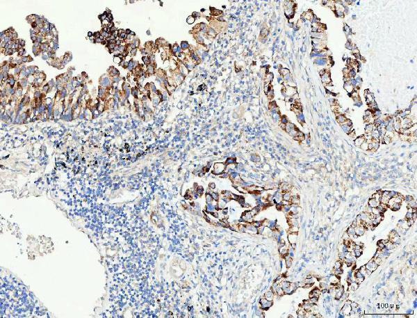
A03231-3 has real IHC data from a paraffin-embedded human lung adenocarcinoma section (A03231-3 IHC image caption). Human, mouse, and rat reactivity is listed (catalog reactivity).
A03231-3 is the only card and lists IHC as an application, with human, mouse, and rat reactivity (catalog applications; catalog reactivity). Its image documents IHC on a paraffin-embedded human lung adenocarcinoma section (A03231-3 IHC image caption).
Which to pick: For tissue IHC, choose A03231-3: its paraffin-section image documents 2 μg/ml primary antibody after EDTA retrieval at pH 8.0 (A03231-3 IHC image caption). No SKU is listed for IF/ICC validation or has an IF image, so there is no validated IF/ICC choice here (catalog applications; IF image alts). For cross-species work, A03231-3 lists human, mouse, and rat reactivity, but its IHC image documents human tissue only; the fixative is unreported (catalog reactivity; A03231-3 IHC image caption).