This website uses cookies to ensure you get the best experience on our website.
- Table of Contents
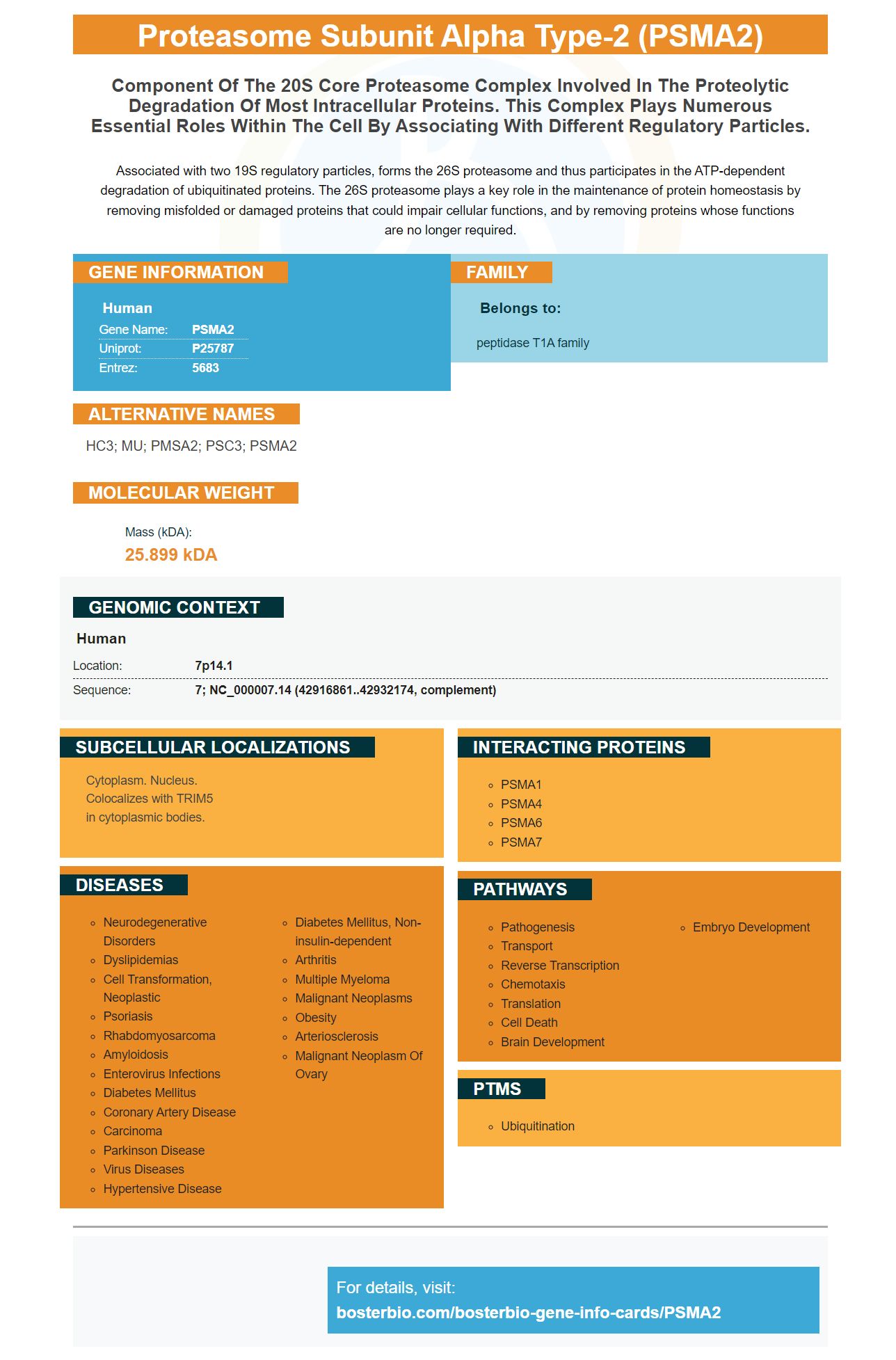
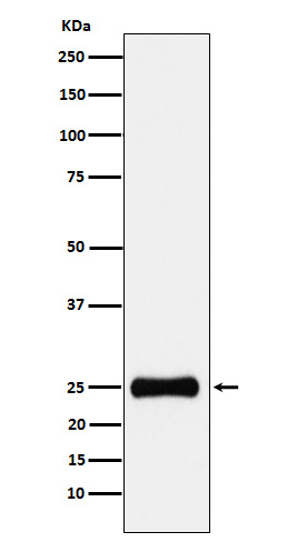
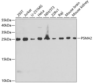
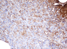
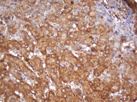
Facts about Proteasome subunit alpha type-2.
Associated with two 19S regulatory particles, forms the 26S proteasome and thus participates in the ATP-dependent degradation of ubiquitinated proteins. The 26S proteasome plays a key role in the maintenance of protein homeostasis by removing misfolded or damaged proteins that could impair cellular functions, and by removing proteins whose functions are no longer required.
Human | |
---|---|
Gene Name: | PSMA2 |
Uniprot: | P25787 |
Entrez: | 5683 |
Belongs to: |
---|
peptidase T1A family |
HC3; MU; PMSA2; PSC3; PSMA2
Mass (kDA):
25.899 kDA
Human | |
---|---|
Location: | 7p14.1 |
Sequence: | 7; NC_000007.14 (42916861..42932174, complement) |
Cytoplasm. Nucleus. Colocalizes with TRIM5 in cytoplasmic bodies.
Boster Bio offers the Anti-Proteasome 20S alpha 2/PSMA2 Marker. Let's take a look at the company's history, the development of the marker, and the validation on IHC/WB before you purchase this antibody. We will also discuss the use this marker to detect PSMA2 ligands. We will be discussing the history of the company as well the development of PSMA2 Marker and its uses.
The PSMA2 protein encodes the protein that forms an alpha subunit in the proteasome. The proteasome is a multi-catalytic proteinase complex composed of a ring-shaped 20S core containing twenty-eight nonidentical subunits. Proteasomes can be found in high amounts in eukaryotic cell cells. They cleave proteins using ATP and ubiquitin. The immunoproteasome has a critical role in the processing of class I MHC peptides.
Boster Bio Antiteasomal 20S beta 2/PSMA2 marker inhibits ubiquitination. It is a critical process in the enzymological processes. This enzyme is involved with the biosynthesis cholesterol and fatty acid. It is important to distinguish between these two types of ubiquitin.
If you're interested in learning about Steve Boster's past, you're not alone. There are many public records available online. These include Steve Boster’s previous and current addresses as well as mobile phone numbers. Email addresses. You can also find public records about Steve Boster’s relatives. You can search the internet for Steve Boster by age and state, or by location.
Steve Boster was an active Staunton resident, Virginia resident, in addition to his scholarly writings. He was the son James and Evelyn Meier Boster. He worked as a manager for many decades in retail sales. He was also a Concordia Hall, Staunton, Virginia member. His family includes Natosha Poeck and Crystal Boster as well as six grandchildren. His siblings are Jack Boster and Sandra Blanton brothers, as well as his daughter Jonathan Boster and sister Tammy.
As an initial antibody screen, WB is recommended. To determine specificity, it is necessary to perform immunoblotting. To determine the phospho-specificity and viability of the antibody, immunoblotting should be done on tissue samples or cell pellets treated with siRNA or phosphatase. Further validation is recommended for IHC or ICC if multiple bands are found in the WB.
Western blot is typically the first validation step in antibody testing. If an antibody recognizes denatured antigens, it is a reasonable initial validation step. A single band of the same molecular weight indicates specificity. Multiple bands representing the same target could be represented at different levels of post-translational modification or splice variants. These findings should cause concern for further experiments.
Immunohistochemistry with antibodies against PSMA2 markers requires that they recognize a particular protein epitope. To validate an antibody against PSMA2 using WB, the target protein should be quantified with an appropriate quantitative method. ImageJ or A3UA are two options to measure the protein expression in TMA areas. It should show strong correlation. In this study we used a monoclonal anti-phospho-4EBP1 antibody (clone 1-D5). Nuclear staining is observed on FFPE lung carcinoma cells using the phospho-4EBP1 antibody.
Company 1 has the smallest antibody validation. While the antibody was commercially available, it provided scant information on the validation process. The datasheet of this antibody provided little or no background information about its target. Three of the four companies provided the complete sequence, while the fourth identified the area surrounding the phosphorylation spot. A lack of examples or references also prevented validation of antibodies on WB with the PSMA2 marker and IHC with the PSMA2 mark.
The group's recommendations provide a good example of how an antibody should be validated. Companies 2-5 have data sheets that show representative samples of their antigens and their applications. But Company 6 has immunocytochemistry results from a positive-control cell line lysate. It is important that you are aware of the limitations and limitations of antibodies that have not been validated.
There are two mAbs that target PSMA, 3F11 and 1A11. They recognize linear epitopes spanning residues 226-243 of the human protein. In addition, they recognize PSMA orthologs from other species. The 5D3 mAbs and 5B1 mAbs can recognize surface-exposed conformational epitopes, making them suitable for in vivo imaging.
The currently-disclosed antibody exhibits high binding specificity as well as biological activity to PSMA epitopes. It also inhibits cell growth. The present invention also includes pharmaceutical applications that can be used with the antibodies. The disclosed subject matter also contains humanizing antibodies, which can be used to reduce their immunogenicity as well as toxicity. Humanized antibodies are produced from non-human cells, which have their constant regions altered to mimic those of human antibodies.
PMID: 2025653 by Tamura T., et al. Molecular cloning and sequence analysis of cDNAs for five major subunits of human proteasomes (multi-catalytic proteinase complexes).
PMID: 7811265 by Kristensen P., et al. Human proteasome subunits from 2-dimensional gels identified by partial sequencing.