PSMD8 / 26S proteasome non-ATPase regulatory subunit 8 · IHC design guide

Design Immunohistochemistry for PSMD8

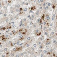
Plan PSMD8 paraffin IHC using the catalog antibody’s documented protocol (datasheet A12465-1). Compare staining with the mainly cytoplasmic tissue profile and score Purkinje cell nuclear signal separately (HPA tissue IHC).

Evidence assembled Oct 2026 · For research use; verify linked source records and product datasheet before use
Immunohistochemistry protocol sheet for PSMD8 (IHC for PSMD8): expected localisation Mainly cytoplasmic staining in most tissues (HPA tissue IHC), antibody A12465-1, validated IHC image, and IHC protocol steps
Printable PSMD8 IHC protocol sheet — expected localisation Mainly cytoplasmic staining in most tissues (HPA tissue IHC), antibody A12465-1, controls and protocol steps. Open the full PSMD8 IHC guide →

PSMD8 Immunohistochemistry Experimental Design Guide

Expected localisation, validated protocols, controls and antibodies — the at-a-glance facts below, then the full design guide.

Must know before staining
Expected localisation Mainly cytoplasmic staining in most tissues (HPA tissue IHC)
Staining pattern Mostly cytoplasmic; Purkinje cell nuclei stain strongly (HPA tissue IHC)
Antigen retrieval EDTA pH 8.0 HIER, heat-mediated (datasheet A12465-1)
Positive control ⓘ Cerebellum+4 more · see all
Negative control ⓘ Bone marrow+2 more · see all
Important caveats
Reasons your staining may differ from the expected pattern.
Fixation Keep fixation consistent across samples (standard IHC practice; not target-specific); Matched tissue-IHC evidence does not establish the fixation claim. Validate the specimen-specific method before use. (selected-SKU IHC image A12465-1)
Caveat Purkinje cell nuclei may stain despite the cytoplasmic trend (HPA tissue IHC)
Regulation Expression regulation is unreported (UniProt)
Isoform / epitope No annotated isoforms; one chain spans residues 1–350 (UniProt)
Section 1

Recommended PSMD8 IHC & IF Protocols

The catalog antibody’s IHC-P protocol (datasheet: A12465-1) is accompanied by published PSMD8 protocols for ovarian and bladder cancer tissue (PMC10286491; PMC12704262).

Recommended immunohistochemistry (IHC-P) protocol parameters
SampleParaffin-embedded mouse brain tissue; fixative not specified (datasheet A12465-1)
FixationImage fixative and duration unreported (datasheet A12465-1); verify before use.
Sectioning4–5 µm sections on charged slides (standard)
DeparaffinisationXylene, graded ethanol series to water (standard)
Antigen retrievalHeat retrieval: EDTA pH 8.0 (datasheet A12465-1); 20 min, 95–100 °C (standard)
Peroxidase block3% H2O2, 10 min, room temperature (standard)
Blocking10% goat serum (datasheet A12465-1)
Primary antibodyRabbit anti-PSMD8, 2-5 μg/ml (datasheet A12465-1)
Primary incubationOvernight at 4 °C (datasheet A12465-1)
DetectionStreptavidin-biotin complex (SABC), DAB chromogen (datasheet A12465-1)
CounterstainHematoxylin, blue, dehydrate and mount (standard)
Expected resultPSMD8-positive staining in purkinje cells - nucleus of cerebellum (HPA tissue IHC: High). HPA tissue profile: Mainly cytoplasmic expression in most tissues. No signal in the no-primary control.
💡Decision noteStart with heat-mediated EDTA retrieval at pH 8.0 (datasheet: A12465-1). The ovarian study used heat retrieval without specifying a buffer (PMC10286491).
Section 2

What Is the Expected PSMD8 Staining Pattern?

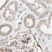
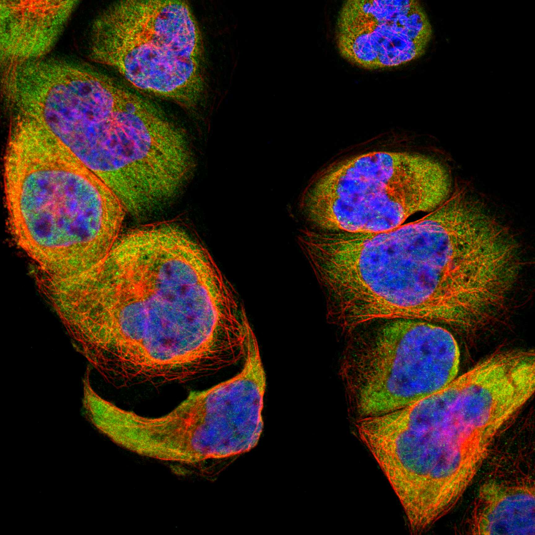
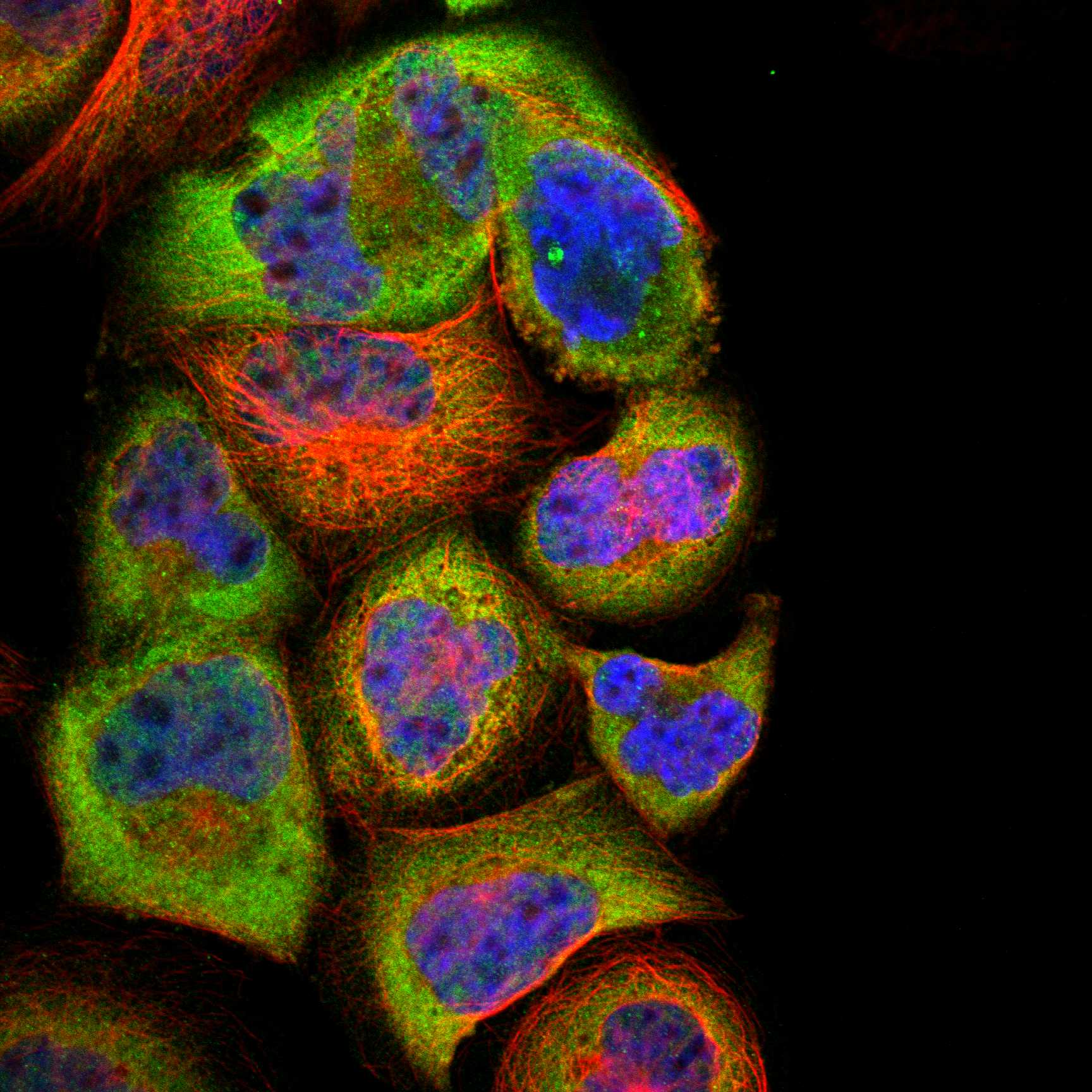
Expect mainly cytoplasmic PSMD8 staining across most tissues, with intensity varying by cell type (HPA tissue IHC: Supported; medium consistency with RNA). Cerebral cortex neuronal cells stain strongly, while cerebellar Purkinje cells show high nuclear staining (HPA tissue IHC). PSMD8 has no annotated transmembrane segment (UniProt P48556 topology), so a membrane-restricted pattern is unexpected. ICC-IF places PSMD8 mainly in the cytosol, with additional nuclear speckles (HPA subcellular: approved).

What am I looking at on my slide?
Cytoplasmic staining in many cells, with stronger neuronal staining in cerebral cortex and nuclear staining in Purkinje cells.This fits the reported IHC pattern: mainly cytoplasmic expression in most tissues, high staining in cortical neuronal cells, and high nuclear staining in cerebellar Purkinje cells (HPA tissue IHC). Judge the compartment in its tissue context; nuclear Purkinje staining is an observed exception to the broad cytoplasmic profile.
A sharp plasma-membrane outline dominates the stain, with little cytoplasmic signal.Treat a membrane-restricted result as suspect because PSMD8 has no annotated transmembrane segment (UniProt P48556 topology), and HPA reports mainly cytoplasmic tissue staining (HPA tissue IHC). Review morphology, controls, and detection conditions before interpreting the outline as PSMD8.
Strong staining appears in bone-marrow hematopoietic cells or oral or vaginal squamous epithelium.HPA lists these cell populations as not detected in its tissue IHC record (HPA tissue IHC). Investigate cross-reactivity or endogenous chromogen-generating activity (general IHC practice). An HPA negative observation is a comparison point, not proof that every specimen must be negative.
Color coats the section broadly, including tissue spaces or many unrelated structures.Diffuse background does not resolve the cell and compartment differences reported for PSMD8 (HPA tissue IHC). Check a no-primary control, blocking, washes, and detection reagent conditions to distinguish nonspecific staining from cellular signal (general IHC practice).
No staining is visible in cerebral cortex neuronal cells or cerebellar Purkinje cells.These are useful positive comparisons because HPA reports high staining in cortical neuronal cells and Purkinje-cell nuclei (HPA tissue IHC). Confirm tissue integrity and a working detection control, then review retrieval and antibody conditions (general IHC practice); absence alone cannot identify the failed step.
💡Expected PSMD8 appearanceCall an IHC result consistent when cellular staining is mainly cytoplasmic, with high signal in cortical neuronal cells or Purkinje-cell nuclei (HPA tissue IHC); an isolated membrane rim or widespread structure-free color is suspect (UniProt P48556 topology; general IHC practice).
How each factor affects the staining
Strength of tissue evidenceHPA rates the IHC pattern Supported and describes medium consistency between antibody staining and RNA data (HPA tissue IHC). Use the listed cells as reference patterns while allowing for specimen and assay differences (general IHC practice).
Cell-type variationHPA reports medium staining in adipocytes and several glandular or epithelial populations, low staining in selected glial and glandular cells, and not detected staining in listed marrow and squamous cells (HPA tissue IHC). Score identified cell populations separately.
Compartment and protein contextPSMD8 is a component of the 19S proteasome regulatory-particle lid and lacks an annotated transmembrane segment (UniProt P48556). HPA's tissue profile is mainly cytoplasmic, while ICC-IF also resolves nuclear speckles (HPA tissue IHC; HPA subcellular).
RNA versus stainSkeletal muscle is tissue-enhanced at the RNA level (HPA tissue IHC: RNA specificity). That label does not specify an IHC intensity or establish a positive control in skeletal muscle; select an observed protein-staining example when assessing the slide (HPA tissue IHC).
Antibody evidenceHPA lists two antibodies with IHC status Supported; one also has ICC status Approved (HPA antibodies: HPA006702, CAB021101). These statuses support their reported applications but do not validate an unlisted antibody or a particular laboratory's conditions.
Why is my staining missing, weak or wrong?
SituationLikely causeNext action
Known-positive neuronal tissue has no visible signal.The assay may have failed at detection, retrieval, or antibody incubation (general IHC practice); HPA reports high staining in cortical neuronal cells (HPA tissue IHC).Check a working detection control and section quality, then review the antibody's IHC-P instructions for retrieval and incubation conditions (general IHC practice).
The whole section has diffuse brown background.Nonspecific binding, incomplete washing, or endogenous detection activity can obscure cell boundaries (general IHC practice).Compare a no-primary control; review blocking, washes, and the detection system's endogenous-activity controls (general IHC practice). Reassess whether a cellular pattern remains (HPA tissue IHC).
Signal is confined to cell membranes.That compartment conflicts with PSMD8's lack of an annotated transmembrane segment and HPA's mainly cytoplasmic IHC profile (UniProt P48556 topology; HPA tissue IHC).Inspect a positive reference and no-primary control, then review primary-antibody and detection conditions before assigning the rim to PSMD8 (general IHC practice).
Bone-marrow hematopoietic cells stain strongly.HPA reports these cells as not detected; cross-reactivity or endogenous detection activity is possible (HPA tissue IHC; general IHC practice).Run a no-primary control and compare cell-specific staining with a known-positive tissue; interpret persistent signal cautiously (general IHC practice; HPA tissue IHC).
Every nuclear signal is being scored as an artifact.The rule is too broad: HPA reports high nuclear staining in Purkinje cells and additional nuclear-speckle localization by ICC-IF (HPA tissue IHC; HPA subcellular).Score nuclei by identified cell type and staining pattern; investigate unexpected nuclear staining elsewhere against controls and the mainly cytoplasmic tissue profile (HPA tissue IHC; general IHC practice).
Q: Must an IF/ICC image reproduce the chromogenic IHC pattern exactly?The assays report related but different observations: HPA describes mainly cytoplasmic tissue IHC and cytosol plus nuclear speckles in ICC-IF (HPA tissue IHC; HPA subcellular).A: Use the ICC-IF localization as context for compartment interpretation, and judge the paraffin-section result against tissue IHC cell types and controls (HPA tissue IHC; HPA subcellular; general IHC practice).

Sample controls for PSMD8 IHC & IF

🧪Run cerebellum first and require staining in Purkinje cell nuclei (HPA: High in Purkinje cells - nucleus). Use bone marrow hematopoietic cells as the negative tissue (HPA: Not detected); on the cerebellum slide, treat cells with only background signal as internal negative references if present, since the supplied HPA rows do not identify a negative cerebellar cell type.
Positive control tissue: Cerebellum (Purkinje cells - nucleus, HPA High)
Negative control tissue: Bone marrow (HPA Not detected)
ICC-IF cell lines (HPA subcellular resource): HPA ICC-IF images show PSMD8 in A-431, U-251MG, U2OS, ASC52telo, hTERT-RPE1 (serum starved), Sperm, KOLF2.1J, with annotated localisation: Cytosol (approved) (HPA subcellular).
Technical controls: Include a no-primary, secondary-only control and a rabbit immunoglobulin isotype control matched to the primary antibody’s class and clonality where applicable (caption: rabbit anti-PSMD8); use PSMD8 knockout material as a biological negative if available (UniProt P48556: PSMD8). For the cerebellum slide, quench endogenous peroxidase and check for endogenous biotin background with the biotin-based detection system (caption: SABC with DAB).
⚠️Feasibility: No target-specific fixation window or fixation effect is reported in the supplied evidence, and the selected A12465-1 paraffin-section caption leaves the fixative unreported. Heat retrieval in EDTA at pH 8.0 is a documented starting condition, but retrieval dependency is unreported (caption: mouse brain IHC). The evidence does not establish whether frozen sections or tissue IF are easier; for IF, assess brain autofluorescence separately from PSMD8 signal (HPA: cerebellum High; standard IF practice).

HPA tissue IHC evidence for PSMD8

Comprehensive Human Protein Atlas IHC scoring per tissue (reliability: Supported — Medium consistency between antibody staining and RNA expression data.). Rows are taken directly from the HPA tissue chart — click any row's HPA link to view the source.

Positive expression · recommended positive controls

TissueCell typeLevelEvidenceSource
Cerebellum Purkinje cells - nucleus High Protein (IHC) HPA →
Cerebral cortex Neuronal cells High Protein (IHC) HPA →
Adipose tissue Adipocytes Medium Protein (IHC) HPA →
Adrenal gland Glandular cells Medium Protein (IHC) HPA →
Appendix Glandular cells Medium Protein (IHC) HPA →

Undetected expression · recommended negative controls

TissueCell typeLevelEvidenceSource
Bone marrow Hematopoietic cells Not detected Protein (IHC) HPA →
Oral mucosa Squamous epithelial cells Not detected Protein (IHC) HPA →
Vagina Squamous epithelial cells Not detected Protein (IHC) HPA →
Section 3

Advanced PSMD8 IHC Tips

Troubleshoot PSMD8 staining in paraffin sections by checking retrieval, compartment, controls, and scoring before interpreting differences between samples.

How should I retrieve PSMD8 when staining is weak?
Use heat-mediated retrieval in EDTA at pH 8.0 for PSMD8 paraffin sections (datasheet A12465-1). The selected antibody stained a paraffin-embedded mouse brain section after this retrieval, with 2 μg/ml primary antibody incubated overnight at 4°C (datasheet A12465-1). If staining remains weak, compare retrieval heating and cooling conditions on adjacent sections while holding antibody concentration and detection constant (standard IHC practice). Include a section with detectable neuronal staining as a process control, since neuronal cells in cerebral cortex are reported as high and the antibody’s tissue staining has Supported reliability (HPA tissue IHC).
Could fixation explain inconsistent PSMD8 staining across blocks?
Target-specific PSMD8 sensitivity to fixation is unknown: the selected paraffin-section caption does not state a fixative (datasheet A12465-1). Record each block’s fixative and fixation duration, then compare similarly processed sections using the same EDTA pH 8.0 retrieval and detection run (datasheet A12465-1; standard IHC practice). If signal differs between blocks, inspect morphology and staining of an internal process control before attributing the difference to PSMD8 abundance (standard IHC practice). Do not infer a fixation effect from the reported cytoplasmic tissue pattern or from PSMD8’s lack of a transmembrane segment (HPA tissue IHC; UniProt P48556 topology).
Should PSMD8 stain nuclei, cytoplasm, or both in tissue sections?
Expect predominantly cytoplasmic tissue staining, while allowing nuclear signal where cell context supports it (HPA tissue IHC: mainly cytoplasmic in most tissues; Purkinje cell nuclei high). Cell imaging places PSMD8 mainly in cytosol and additionally in nuclear speckles, so nuclear staining alone does not establish an artefact (HPA subcellular: approved locations). Inspect signal within intact cell boundaries and compare the same compartment across adjacent sections stained together (standard IHC practice). Diffuse extracellular DAB deposits or staining confined to section edges warrant investigation because PSMD8 is a component of the intracellular 19S regulatory particle and has no transmembrane segment (UniProt P48556; standard IHC practice).
How can epitope uncertainty affect PSMD8 IHC interpretation?
The supplied record lists 0 isoforms and a 350-residue PSMD8 chain, so it provides no basis for assigning different IHC patterns to splice variants (UniProt P48556). It identifies a PCI domain at residues 162–331 and phosphoserine at residue 106, but the selected antibody’s binding epitope is not specified here (UniProt P48556; datasheet A12465-1). Avoid attributing weak staining to either feature without epitope information or direct validation (standard IHC interpretation). Compare staining after the documented EDTA pH 8.0 retrieval with an independently validated PSMD8 reagent or a target-depleted control, if available, to assess specificity (datasheet A12465-1; standard IHC practice).
How should IF help resolve ambiguous PSMD8 staining in tissue?
Use IF as a separate localisation check, pairing PSMD8 with a validated marker of the expected cell type; Purkinje cells are a useful candidate because their nuclei show high tissue-IHC staining (HPA tissue IHC). Choose spectrally separated fluorophores and favor a far-red PSMD8 channel when tissue autofluorescence obscures shorter wavelengths (standard IF practice). PSMD8 has no transmembrane segment and is reported mainly in cytosol, with additional nuclear-speckle localisation; permeabilise cells sufficiently to expose intracellular epitopes while checking that morphology remains intact (UniProt P48556 topology; HPA subcellular; standard IF practice). Include single-label and no-primary controls to distinguish bleed-through and autofluorescence from PSMD8 signal (standard IF practice).
What should I check when PSMD8 DAB staining looks diffuse?
First compare a no-primary section with the stained section to locate signal arising from the detection system (standard IHC practice). The selected workflow uses a biotinylated secondary, streptavidin–biotin complex, and DAB; tissue-associated background may therefore require checking endogenous biotin and peroxidase blocking (datasheet A12465-1; standard IHC practice). Also review the 10% goat-serum block, 2 μg/ml primary concentration, and overnight incubation at 4°C against the documented workflow (datasheet A12465-1). Titrate primary antibody or shorten chromogen development on matched sections if background persists, while retaining a section with expected cellular staining to monitor sensitivity (standard IHC practice; HPA tissue IHC).
How should I score PSMD8 staining across tissue samples? ⚠ ANSWER MARKED FOR VERIFICATION
Define the cell population and compartment before scoring, since PSMD8 is mainly cytoplasmic in tissues but Purkinje cell nuclei can stain strongly (HPA tissue IHC). For comparable regions, record the percentage of positive cells and intensity categories, then calculate an H-score or report positive-cell density per mm² (standard IHC quantification). Normalize counts to viable cells or analysed tissue area, and use the same exposure, DAB development, and positivity threshold across the batch (standard IHC practice). Keep nuclear and cytoplasmic scores separate where both occur, and exclude folds, edges, and necrotic areas using prespecified criteria (HPA tissue IHC; standard IHC practice).
When does PSMD8 staining support a biological conclusion?
Look for reproducible intracellular staining in intact cells: PSMD8 belongs to the 19S proteasome regulatory particle, and tissue IHC is mainly cytoplasmic (UniProt P48556; HPA tissue IHC). Nuclear staining can be plausible in Purkinje cells, whereas an extracellular rim, edge-only signal, or staining concentrated in necrotic tissue should prompt an artefact check (HPA tissue IHC: Purkinje cell nuclei high; standard IHC practice). Compare no-primary and peroxidase-block controls when DAB staining may reflect the detection chemistry rather than antibody binding (standard IHC practice). Interpret differences cautiously because HPA rates tissue-IHC reliability Supported with medium consistency between staining and RNA expression (HPA tissue IHC).
Boster reagents

Best PSMD8 / 26S proteasome non-ATPase regulatory subunit 8 IHC Antibodies

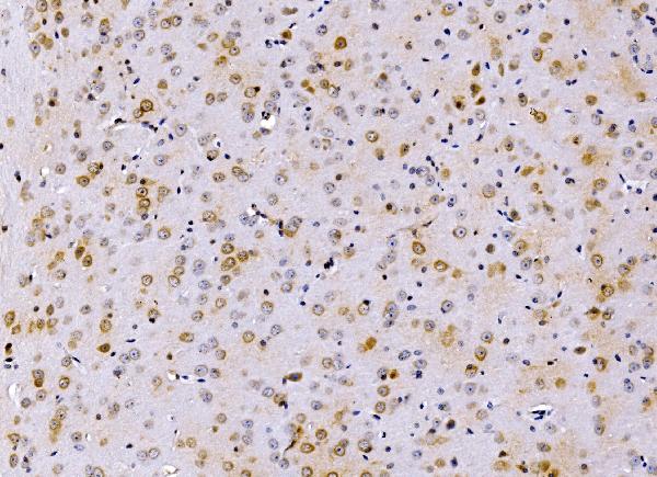
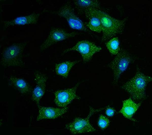
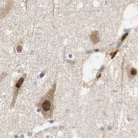
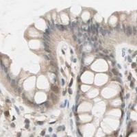
A12465-1 has paraffin-section IHC images from mouse and rat brain and human breast cancer, plus IF/ICC data in A549 cells (catalog image captions).

Real IHC data IHC analysis of PSMD8 using anti-PSMD8 antibody (A12465-1). PSMD8 was detected in a paraffin-embedded section of mouse brain tissue. Heat mediated antigen retrieval was performed in EDTA buffer (pH 8.0, epitope retrieval solution). The tissue section was blocked with 10% goat serum. The tissue section was then incubated with 2 μg/ml rabbit anti-PSMD8 Antibody (A12465-1) overnight at 4°C. Biotinylated goat anti-rabbit IgG was used as secondary antibody and incubated for 30 minutes at 37°C. The tissue section was developed using Strepavidin-Biotin-Complex (SABC) (Catalog # SA1022) with DAB as the chromogen.
Anti-PSMD8 Antibody ®
Cat # A12465-1

A12465-1 is listed for IHC and IF/ICC, with human, mouse and rat reactivity (catalog: applications/reactivity). Its IHC captions show paraffin sections of mouse and rat brain and human breast cancer; its IF caption shows A549 cells (catalog image captions).

Which to pick: Choose A12465-1 for paraffin-section tissue IHC: its captions show EDTA pH 8.0 retrieval and 2 μg/ml primary antibody in mouse brain, rat brain and human breast cancer sections; the fixative is unreported (catalog IHC captions). For IF/ICC, A12465-1 has an A549-cell example at 5 μg/ml (catalog IF caption). It is the listed cross-species choice for tissue IHC because those captions cover human, mouse and rat; the antibody is rabbit-hosted, and clonality is unreported (catalog IHC captions; catalog: host/clone).

Each figure is that product's own IHC / IF validation image from its datasheet.

References

  1. UniProt Consortium. UniProt entry P48556 (PSMD8_HUMAN, 26S proteasome non-ATPase regulatory subunit 8).
  2. Human Protein Atlas. PSMD8 tissue IHC expression (reliability: Supported).
  3. Human Protein Atlas. PSMD8 subcellular location (ICC-IF): Mainly localized to the cytosol. In addition localized to the nuclear speckles..
  4. Human Protein Atlas. PSMD8 antibody validation summary (2 antibodies).
  5. PSMD8 can serve as potential biomarker and therapeutic target of the PSMD family in ovarian cancer: based on bioinformatics analysis and in vitro validation. BMC cancer 2023 — PMC10286491.
  6. PSMD8 cooperates with USP14 to promote bladder cancer progression by inhibiting ferroptosis. iScience 2025 — PMC12704262.
  7. PSMD8 promotes breast cancer progression and attenuates paclitaxel sensitivity by stabilizing KIF10. Cancer cell international 2026 — PMC13563711.
  8. PubMed PMID:14702039 — UniProt-cited evidence.
  9. PubMed PMID:15057824 — UniProt-cited evidence.
  10. PubMed PMID:15489334 — UniProt-cited evidence.