RAB1B / Ras-related protein Rab-1B · IHC design guide

Design Immunohistochemistry for RAB1B

Plan RAB1B paraffin IHC using the general cytoplasmic pattern and high staining in selected glandular cells (HPA tissue IHC). Compare test sections with controls and interpret staining in light of the antibody’s potential to detect proteins from more than one gene (HPA tissue IHC).

Evidence assembled Oct 2026 · For research use; verify linked source records and product datasheet before use
Immunohistochemistry protocol sheet for RAB1B (IHC for RAB1B): expected localisation General cytoplasmic staining (HPA tissue IHC), antibody A04589-1, validated IHC image, and IHC protocol steps
Printable RAB1B IHC protocol sheet — expected localisation General cytoplasmic staining (HPA tissue IHC), antibody A04589-1, controls and protocol steps. Open the full RAB1B IHC guide →

RAB1B Immunohistochemistry Experimental Design Guide

Expected localisation, validated protocols, controls and antibodies — the at-a-glance facts below, then the full design guide.

Must know before staining
Expected localisation General cytoplasmic staining (HPA tissue IHC)
Staining pattern Cytoplasmic staining; high in some glandular cells (HPA tissue IHC)
Antigen retrieval EDTA pH 8.0 HIER, heat-mediated (datasheet A04589-1)
Positive control ⓘ Duodenum+4 more · see all
Negative control ⓘ Adipose tissue+4 more · see all
Important caveats
Reasons your staining may differ from the expected pattern.
Fixation Matched tissue-IHC evidence does not establish the fixation claim. Validate the specimen-specific method before use. (selected-SKU IHC image A04589-1)
Caveat Antibody may detect proteins from more than one gene (HPA tissue IHC)
Regulation No specific regulator annotated (UniProt)
Isoform / epitope 0 annotated isoforms; one 1–201 chain (UniProt)
Section 1

Recommended RAB1B IHC & IF Protocols

Start with the catalog antibody’s IHC-P protocol (datasheet: A04589-1), then compare the published colorectal tissue and xenograft IHC conditions below (PMC5391239; PMC9980309).

Recommended immunohistochemistry (IHC-P) protocol parameters
SampleParaffin-embedded human prostate cancer tissue; fixative not specified (datasheet A04589-1)
FixationImage fixative and duration unreported (datasheet A04589-1); verify before use.
Sectioning4–5 µm sections on charged slides (standard)
DeparaffinisationXylene, graded ethanol series to water (standard)
Antigen retrievalHeat retrieval: EDTA pH 8.0 (datasheet A04589-1); 20 min, 95–100 °C (standard)
Peroxidase block3% H2O2, 10 min, room temperature (standard)
Blocking10% goat serum (datasheet A04589-1)
Primary antibodyRabbit anti-RAB1B, 2μg/ml (datasheet A04589-1)
Primary incubationOvernight at 4 °C (datasheet A04589-1)
DetectionStreptavidin-biotin complex (SABC), DAB chromogen (datasheet A04589-1)
CounterstainHematoxylin, blue, dehydrate and mount (standard)
Expected resultRAB1B-positive staining in glandular cells of duodenum (HPA tissue IHC: High). HPA tissue profile: General cytoplasmic expression. No signal in the no-primary control.
💡Decision noteTry heat-mediated EDTA at pH 8.0 first (datasheet: A04589-1). Sodium citrate is a published alternative, though its pH was not reported (PMC9980309).
Section 2

What Is the Expected RAB1B Staining Pattern?

RAB1B is a cytoplasmic, membrane-associated protein concentrated around the ER–Golgi pathway and perinuclear region; it has no transmembrane segment (UniProt Q9H0U4: localization and topology). In paraffin-section IHC, expect cytoplasmic staining in glandular cells of duodenum and small intestine, follicle cells of ovary, and decidual cells of placenta (HPA: High). HPA describes general cytoplasmic expression, but rates tissue IHC only Approved and cautions that the antibodies target protein from more than one gene (HPA: tissue IHC).

What am I looking at on my slide?
Cytoplasmic staining in glandular cells of duodenum or small intestine, with some perinuclear concentration.This fits the reported High tissue pattern and RAB1B's ER–Golgi and perinuclear localization (HPA: tissue IHC; UniProt Q9H0U4). Chromogenic IHC cannot by itself identify an individual organelle (standard IHC practice).
Predominantly nuclear staining, with little cytoplasmic signal in an otherwise positive tissue.A nuclear-dominant pattern conflicts with the reported cytoplasmic and membrane localization (UniProt Q9H0U4; HPA: tissue IHC). Check morphology, counterstain and detection controls before assigning that signal to RAB1B (standard IHC practice).
Strong staining in adipocytes or esophageal squamous epithelial cells.HPA reports these cell populations as Not detected; unexpected signal warrants checks for cross-reactivity or endogenous detection activity (HPA: tissue IHC; standard IHC practice). An HPA Not detected result is a comparison point, not proof that every specimen must be negative.
Color is spread evenly across cells, stroma and blank areas, obscuring cell boundaries.Diffuse, tissue-independent color is difficult to score as specific cytoplasmic staining (standard IHC practice). Evaluate background and detection controls; HPA's general cytoplasmic profile does not justify calling uniform haze a positive result (HPA: tissue IHC).
No cytoplasmic signal in glandular cells of duodenum or small intestine.That conflicts with HPA's High observations and makes the run inconclusive until section quality, detection and control performance are checked (HPA: tissue IHC; standard IHC practice). HPA's cross-gene caution also limits how confidently its reference staining identifies RAB1B alone (HPA: reliability description).
💡Expected RAB1B appearanceCall positive when cytoplasmic, often perinuclear color is visible in the expected cell population—such as High-staining duodenal glandular cells—while nuclear-dominant color or uniform haze prompts review (HPA: tissue IHC; UniProt Q9H0U4; standard IHC practice).
How each factor affects the staining
Cellular compartmentRAB1B cycles between cytoplasmic and membrane-associated states and is reported near the ER and Golgi (UniProt Q9H0U4). Expect a cytoplasmic IHC pattern; exact organelle assignment requires greater spatial resolution (standard IHC practice).
Tissue and cell contextHPA reports High staining in duodenal and small-intestinal glandular cells, ovarian follicle cells and placental decidual cells; adipocytes and skeletal myocytes are Not detected (HPA: tissue IHC). Compare the named cell population, not the whole tissue.
Antibody evidenceHPA lists two antibodies with Approved IHC status, yet describes only medium consistency with RNA data and cautions that they target protein from more than one gene (HPA: tissue IHC and antibody validation). Treat staining as supportive rather than gene-specific proof.
Isoforms and processingThe supplied UniProt record lists one chain, residues 1–201, no signal peptide, no propeptide and no annotated isoforms (UniProt Q9H0U4). These annotations give no basis for predicting a separate secreted or shed staining pattern.
Fixation and retrievalTarget-specific fixation sensitivity and epitope access are unreported in the supplied UniProt and HPA records (UniProt Q9H0U4; HPA: tissue IHC). If a run fails, review the laboratory's validated retrieval and staining controls without inferring a RAB1B-specific fixation effect (standard IHC practice).
IF/ICC Q: Where should signal localize?A: Mainly to the Golgi apparatus, with additional ER localization in the HPA cell-image data (HPA: subcellular ICC-IF, Supported). This helps interpret compartment, but does not turn paraffin-section chromogenic IHC into an organelle-level assay (standard IHC practice).
Why is my staining missing, weak or wrong?
SituationLikely causeNext action
Expected glandular cells show no color.The run may have failed, or the examined section may lack evaluable target cells (standard IHC practice); HPA reports High glandular-cell staining in duodenum and small intestine (HPA: tissue IHC).Check tissue identity and morphology, then review the laboratory's positive and detection controls and validated IHC conditions before interpreting absence (standard IHC practice).
Nuclei appear to be the main positive compartment.Nuclear-dominant signal is inconsistent with the reported cytoplasmic localization (UniProt Q9H0U4; HPA: tissue IHC). Counterstain overlap or nonspecific color can complicate visual assignment (standard IHC practice).Inspect a detection control and assess color separately from the nuclear counterstain; score only convincingly cellular staining in the expected compartment (standard IHC practice).
Adipocytes or esophageal squamous cells stain strongly.Those populations are Not detected in HPA's tissue profile; cross-reactivity or endogenous detection activity is possible (HPA: tissue IHC; standard IHC practice). The HPA antibody warning makes specificity checks especially relevant (HPA: reliability description).Compare with the expected positive cell population and examine appropriate detection controls; seek independent specificity evidence before calling unexpected cells RAB1B-positive (standard IHC practice).
Background obscures the cytoplasmic pattern.Widespread color can arise from nonspecific binding or endogenous detection activity (standard IHC practice). It does not match HPA's cell-associated cytoplasmic profile (HPA: tissue IHC).Inspect controls and review blocking, washing and detection conditions used in the validated IHC workflow; interpret only signal distinguishable from background (standard IHC practice).
The slide appears diffusely cytoplasmic, without a distinct Golgi focus.HPA reports general cytoplasmic tissue IHC, whereas its supported Golgi and ER localization comes from ICC-IF images (HPA: tissue IHC; HPA: subcellular ICC-IF). Chromogenic section IHC has limited organelle resolution (standard IHC practice).Judge the relevant cell type and cytoplasmic compartment first; reserve a Golgi-level claim for appropriately resolved imaging (HPA: tissue IHC; standard IHC practice).
A High-staining HPA reference is used as sole proof of antibody specificity.HPA rates tissue IHC Approved with medium RNA concordance and warns that the antibodies target protein from more than one gene (HPA: reliability description).Report the observed cell pattern and validation limit separately; use independent specificity evidence before attributing all color exclusively to RAB1B (standard IHC practice).

Sample controls for RAB1B IHC & IF

🧪Run duodenum first and expect staining in its glandular cells (HPA: duodenal glandular cells High). Use adipose tissue adipocytes as the biological negative (HPA: adipocytes Not detected); on the duodenum slide, any unstained non-glandular cells can provide a background reference, but HPA does not identify a verified negative cell type there (HPA: duodenal glandular cells High).
Positive control tissue: Duodenum (Glandular cells, HPA High)
Negative control tissue: Adipose tissue (HPA Not detected)
ICC-IF cell lines (HPA subcellular resource): HPA ICC-IF images show RAB1B in A-431, U-251MG, U2OS, with annotated localisation: Golgi apparatus (supported) (HPA subcellular).
Technical controls: Include no-primary (secondary-only) and host-matched rabbit IgG isotype controls, and confirm specificity with RAB1B knockout material where available (caption: rabbit anti-RAB1B antibody). For duodenal SABC/DAB staining, block endogenous peroxidase and biotin to limit false signal (HPA: duodenum; caption: SABC/DAB detection).
⚠️Feasibility: No target-specific fixation window or fixation effect is reported, and the selected A04589-1 paraffin-section caption does not state a fixative (caption: fixative not stated). Heat-mediated EDTA retrieval at pH 8.0 is documented for that antibody in a paraffin-embedded prostate cancer section; its dependence on retrieval in duodenum remains unreported (caption: EDTA pH 8.0 retrieval). Frozen sections are not established as easier, while IF/ICC offers supported Golgi and endoplasmic reticulum localization; in duodenal chromogenic sections, endogenous biotin can interfere with the caption’s SABC detection (HPA: supported Golgi and endoplasmic reticulum ICC-IF; caption: SABC/DAB detection).

HPA tissue IHC evidence for RAB1B

Comprehensive Human Protein Atlas IHC scoring per tissue (reliability: Approved — Medium consistency between antibody staining and RNA expression data. Caution, targets protein from more than one gene.). Rows are taken directly from the HPA tissue chart — click any row's HPA link to view the source.

Positive expression · recommended positive controls

TissueCell typeLevelEvidenceSource
Duodenum Glandular cells High Protein (IHC) HPA →
Epididymis Glandular cells High Protein (IHC) HPA →
Ovary Follicle cells High Protein (IHC) HPA →
Placenta Decidual cells High Protein (IHC) HPA →
Small intestine Glandular cells High Protein (IHC) HPA →

Undetected expression · recommended negative controls

TissueCell typeLevelEvidenceSource
Adipose tissue Adipocytes Not detected Protein (IHC) HPA →
Bone marrow Hematopoietic cells Not detected Protein (IHC) HPA →
Caudate Glial cells Not detected Protein (IHC) HPA →
Esophagus Squamous epithelial cells Not detected Protein (IHC) HPA →
Parathyroid gland Glandular cells Not detected Protein (IHC) HPA →
Section 3

Advanced RAB1B IHC Tips

These questions focus on interpreting RAB1B staining in paraffin sections while keeping antibody evidence separate from general IHC practice.

Where should I start if RAB1B staining is weak after antigen retrieval?
Start with heat-mediated retrieval in EDTA at pH 8.0 for paraffin sections (datasheet A04589-1). The selected tissue-IHC image used that retrieval with 2 μg/ml primary antibody overnight at 4°C, providing a starting condition for this SKU (caption A04589-1). If staining remains weak, compare retrieval duration across matched sections while holding antibody concentration and detection conditions constant (standard IHC practice). Include a positive control section and inspect tissue integrity, because excessive heating can damage morphology and complicate scoring (standard IHC practice). Record any retrieval change alongside its control result before attributing improved signal to RAB1B (standard IHC practice).
Can I infer an optimal fixative for RAB1B from the available IHC evidence?
No target-specific fixation sensitivity is established here: the selected paraffin-section caption does not state a fixative (caption A04589-1). HPA tissue staining patterns describe detection across tissues, but do not establish how RAB1B responds to fixation (HPA: tissue IHC). For a new specimen series, document the fixative and processing history, then compare similarly processed positive controls under the same retrieval and detection conditions (standard IHC practice). Keep section thickness, retrieval, and antibody incubation consistent when assessing a fixation-related loss of signal (standard IHC practice). Do not infer fixation tolerance from RAB1B localisation or its annotated modifications (UniProt Q9H0U4).
Which staining pattern should I expect in chromogenic RAB1B IHC?
Expect predominantly cytoplasmic staining at tissue scale, with a possible perinuclear emphasis (HPA: general cytoplasmic expression; UniProt Q9H0U4 localisation). Subcellular evidence places RAB1B mainly at the Golgi apparatus and additionally at the endoplasmic reticulum (HPA: supported subcellular locations). A chromogenic section may not resolve those organelles, so score a reproducible cytoplasmic pattern without claiming organelle identity from DAB alone (standard IHC practice). Compare the same cell type across matched sections and note whether staining is diffuse, perinuclear, or granular (standard IHC practice). Treat isolated nuclear signal as a reason to check controls and morphology rather than as an expected RAB1B compartment (UniProt Q9H0U4 localisation).
How should epitope uncertainty affect my interpretation of RAB1B staining?
The record lists one 201-aa chain and no annotated isoforms, so isoform-specific staining cannot be inferred from this evidence (UniProt Q9H0U4). RAB1B has no annotated transmembrane segment, but that alone does not identify the antibody epitope or establish its accessibility after processing (UniProt Q9H0U4 topology). The record includes modifications at several residues, including the C-terminal cysteine at position 201; their effect on this antibody is unreported (UniProt Q9H0U4 modifications). Check the catalog antibody’s stated immunogen or epitope, if available, before making claims about modification-sensitive staining (standard IHC practice). Use an independent specificity control to investigate unexpected patterns (standard IHC practice).
How can IF help assess a questionable RAB1B IHC pattern?
Use IF as a separate localisation check: HPA reports supported Golgi localisation with additional endoplasmic-reticulum localisation for RAB1B (HPA: subcellular). For multiplexing, pair RAB1B with a validated marker for the expected cell type, such as the glandular cells highlighted in several HPA tissue entries (HPA: tissue IHC). Choose fluorophores and controls that distinguish specific signal from tissue autofluorescence, especially when a broad cytoplasmic pattern is being evaluated (standard IF practice). Plan permeabilisation around the antibody’s actual epitope accessibility; RAB1B lacks a transmembrane segment, but the epitope side is not specified here (UniProt Q9H0U4 topology). Compare IF and DAB patterns by compartment and cell type, allowing for their different resolution (standard microscopy practice).
What should I check when DAB staining obscures RAB1B-positive cells?
First compare a no-primary control with the stained section to identify signal contributed by detection reagents or tissue background (standard IHC practice). The selected image used a biotinylated secondary antibody, a streptavidin-biotin complex, and DAB, so that detection chain deserves its own controls (caption A04589-1). Apply a peroxidase block and assess whether endogenous biotin contributes staining when using this detection approach (standard chromogenic IHC practice). The caption reports a 10% goat-serum block; adjust blocking and washes systematically if nonspecific staining persists (caption A04589-1; standard IHC practice). Judge any improvement against preserved cytoplasmic cell staining and tissue morphology (HPA: general cytoplasmic expression; standard IHC practice).
How should I score RAB1B IHC across specimens with different cell composition? ⚠ ANSWER MARKED FOR VERIFICATION
Define the cell population and analysis region before scoring, then apply the same thresholds to every specimen (standard IHC practice). For glandular compartments, report the percentage of positive cells and an intensity-weighted H-score, normalised to the number of evaluable cells in that compartment (standard IHC practice). If counting discrete positive cells, report density per mm² of viable tissue and document excluded areas (standard IHC practice). HPA reports high staining in glandular cells of duodenum, epididymis, and small intestine, making cell composition relevant to comparisons (HPA: tissue IHC). Keep retrieval, exposure to DAB, and image-analysis settings consistent across batches and include control sections (standard IHC practice).
When is a strong RAB1B signal more likely to be artefactual?
A plausible result follows a reproducible cytoplasmic or perinuclear pattern in intact cells, consistent with reported tissue and subcellular localisation (HPA: tissue IHC; UniProt Q9H0U4 localisation). Check an unexpected nuclear-only pattern, staining confined to section edges, or signal concentrated in necrotic areas against morphology and controls (standard IHC practice). Because the selected method uses DAB with a biotin-based detection chain, assess endogenous enzyme and biotin contributions before assigning diffuse brown signal to RAB1B (caption A04589-1; standard IHC practice). HPA rates its tissue antibody data Approved while cautioning that the antibody targets protein from more than one gene (HPA: reliability description). Confirm consequential cell-type claims with an independent specificity strategy (standard IHC practice).
Boster reagents

Best RAB1B / Ras-related protein Rab-1B IHC Antibodies

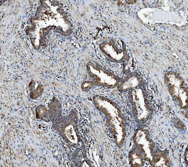
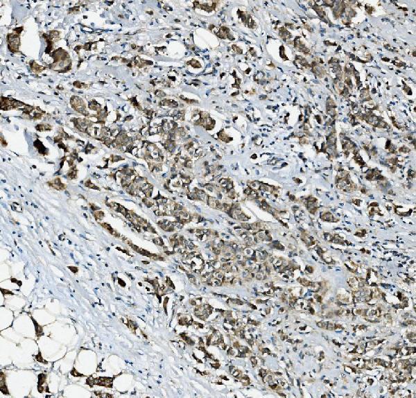
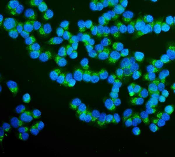
A04589-1 has IHC data from human paraffin sections and IF/ICC data from T-47D cells (catalog image captions); the catalog lists Human, Mouse, and Rat reactivity (catalog applications/reactivity).

Real IHC data IHC analysis of RAB1B using anti-RAB1B antibody (A04589-1). RAB1B was detected in paraffin-embedded section of human prostate cancer tissue. Heat mediated antigen retrieval was performed in EDTA buffer (pH8.0, epitope retrieval solution). The tissue section was blocked with 10% goat serum. The tissue section was then incubated with 2μg/ml rabbit anti-RAB1B Antibody (A04589-1) overnight at 4°C. Biotinylated goat anti-rabbit IgG was used as secondary antibody and incubated for 30 minutes at 37°C. The tissue section was developed using Strepavidin-Biotin-Complex (SABC) (Catalog # SA1022) with DAB as the chromogen.
Anti-RAB1B Antibody ®
Cat # A04589-1

A04589-1 will render with IHC data from paraffin sections of human prostate, breast, and ovarian cancer and placenta (catalog IHC captions). Its IF/ICC data show T-47D cells (catalog IF caption).

Which to pick: Choose A04589-1 for tissue IHC in human paraffin sections; its IHC captions report EDTA retrieval at pH 8.0, with the fixative unreported (catalog IHC captions). Choose the same SKU for IF/ICC in T-47D cells (catalog IF caption). For other species, the catalog lists Mouse and Rat reactivity, while its IHC captions document human tissue only (catalog reactivity; catalog IHC captions).

Each figure is that product's own IHC / IF validation image from its datasheet.

References

  1. UniProt Consortium. UniProt entry Q9H0U4 (RAB1B_HUMAN, Ras-related protein Rab-1B).
  2. Human Protein Atlas. RAB1B tissue IHC expression (reliability: Approved).
  3. Human Protein Atlas. RAB1B subcellular location (ICC-IF): Mainly localized to the Golgi apparatus. In addition localized to the endoplasmic reticulum..
  4. Human Protein Atlas. RAB1B antibody validation summary (2 antibodies).
  5. Identification of Endoplasmic Reticulum Stress-Related Genes in Osteoporosis Pathogenesis. Mediators of inflammation 2025 — PMC12413945.
  6. Overexpression of Rab1B and MMP9 predicts poor survival and good response to chemotherapy in patients with colorectal cancer. Aging 2017 — PMC5391239.
  7. LINC00173 facilitates tumor progression by stimulating RAB1B-mediated PA2G4 and SDF4 secretion in nasopharyngeal carcinoma. Molecular oncology 2023 — PMC9980309.
  8. Vibrio MARTX toxin processing and degradation of cellular Rab GTPases by the cytotoxic effector Makes Caterpillars Floppy. Proceedings of the National Academy of Sciences of the United States of America 2024 — PMC11194500.
  9. PubMed PMID:11230166 — UniProt-cited evidence.
  10. PubMed PMID:14702039 — UniProt-cited evidence.
  11. PubMed PMID:15489334 — UniProt-cited evidence.