This website uses cookies to ensure you get the best experience on our website.


- Table of Contents
Plan paraffin-section RAB3IP IHC around the reported cytoplasmic tissue pattern (HPA tissue IHC). Compare staining with appropriate controls because the tissue IHC profile has uncertain reliability (HPA tissue IHC).

Expected localisation, validated protocols, controls and antibodies — the at-a-glance facts below, then the full design guide.
| Expected localisation | Predominantly cytoplasmic tissue staining (HPA tissue IHC) | |
| Staining pattern | Cytoplasmic staining across several tissue types (HPA tissue IHC) | |
| Antigen retrieval | EDTA pH 8.0 HIER, heat-mediated (datasheet A09085-3) | |
| Positive control | Appendix+4 more · see all | |
| Negative control | Adipose tissue+4 more · see all |
| Fixation | Keep fixation consistent across sections (standard IHC practice; not target-specific). Selected-image fixative and duration unreported (datasheet A09085-3); verify before use. | |
| Caveat | Low consistency between IHC staining and RNA expression (HPA tissue IHC) | |
| Regulation | PKC redistributes RAB3IP to lamellipodia (UniProt) | |
| Isoform / epitope | 8 isoforms; check epitope coverage (UniProt) |
The catalog antibody’s IHC-P protocol (datasheet A09085-3) is paired with a published RAB3IP IHC protocol for human gastric cancer tissue (PMC6277663).
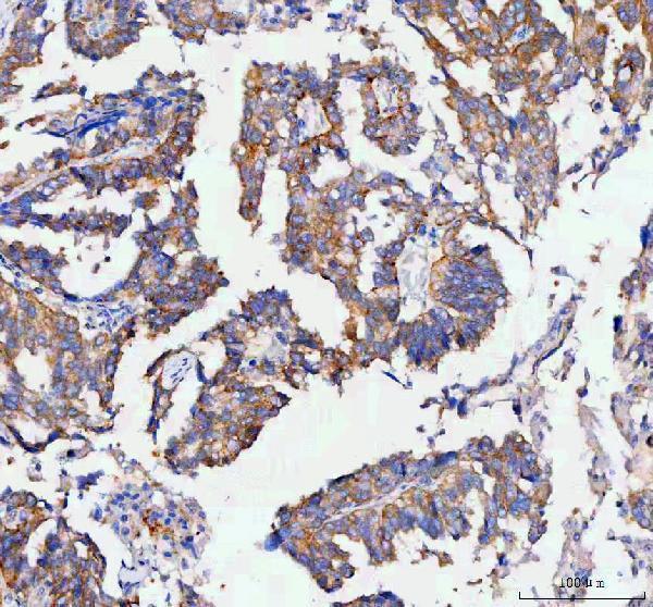
| Sample | Paraffin-embedded human ovarian serous adenocarcinoma tissue; fixative not specified (datasheet A09085-3) |
| Fixation | Image fixative and duration unreported (datasheet A09085-3); verify before use. |
| Sectioning | 4–5 µm sections on charged slides (standard) |
| Deparaffinisation | Xylene, graded ethanol series to water (standard) |
| Antigen retrieval | Heat retrieval: EDTA pH 8.0 (datasheet A09085-3); 20 min, 95–100 °C (standard) |
| Peroxidase block | 3% H2O2, 10 min, room temperature (standard) |
| Blocking | 10% goat serum (datasheet A09085-3) |
| Primary antibody | Rabbit anti-RAB3IP, 2-5 μg/ml (datasheet A09085-3) |
| Primary incubation | Overnight at 4 °C (datasheet A09085-3) |
| Detection | HRP-conjugated secondary, DAB chromogen (datasheet A09085-3) |
| Counterstain | Hematoxylin, blue, dehydrate and mount (standard) |
| Expected result | RAB3IP-positive staining in glandular cells of appendix (HPA tissue IHC: High). HPA tissue profile: Cytoplasmic expression in several different tissue types. No signal in the no-primary control. |
In paraffin-section IHC, expect predominantly cytoplasmic RAB3IP staining in selected glandular cells and kidney tubule cells (HPA: tissue IHC; UniProt Q96QF0: predominantly cytoplasmic). RAB3IP has no transmembrane segment (UniProt Q96QF0: topology). Treat this as a provisional pattern: HPA rates its tissue IHC reliability Uncertain because antibody staining has low consistency with RNA expression data and awaits external verification (HPA: tissue IHC reliability).
| Cytoplasmic staining in appendix, breast, or endometrial glandular cells, or kidney tubule cells. | This matches cell populations scored High by HPA and its broader cytoplasmic tissue profile (HPA: tissue IHC). Judge the signal by the named cell type, not by staining anywhere in the organ. HPA's Uncertain reliability means agreement is supportive, not definitive proof of specificity (HPA: tissue IHC reliability). |
| Predominantly sharp plasma-membrane or extracellular staining, without convincing cytoplasmic staining. | This is discordant with the expected tissue pattern (HPA: cytoplasmic tissue expression) and with a protein lacking a transmembrane segment (UniProt Q96QF0: topology). Consider nonspecific deposition or detection artefact. A small nuclear component alone is not disqualifying: UniProt describes predominantly cytoplasmic RAB3IP with a small nuclear proportion (UniProt Q96QF0: subcellular location). |
| Strong staining mainly in a cell population HPA scores as not detected, such as adipocytes. | Adipocytes are scored Not detected (HPA: adipose-tissue IHC). Cross-reactivity or endogenous detection activity is possible; compare staining with a no-primary control and the expected positive cell population. A negative HPA score is a comparison point, not absolute proof of absence, given the uncertain tissue IHC reliability (HPA: tissue IHC reliability). |
| Diffuse chromogen across cells, stroma, or section edges, obscuring cell boundaries. | An uninterpretable distribution cannot establish RAB3IP localization. Check no-primary and detection-only controls for nonspecific or endogenous detection activity, then reassess blocking and washing as general IHC practice. The expected tissue profile is cellular and cytoplasmic (HPA: tissue IHC); diffuse background should not be scored as target-positive. |
| No convincing signal in appendix glandular cells or kidney tubule cells. | Both populations are scored High (HPA: appendix and kidney IHC), so an absent signal warrants a run check. Confirm tissue preservation, control performance, the antibody's IHC-P instructions, and detection reagents before interpreting the specimen as negative. HPA's Uncertain reliability limits any single tissue's value as a definitive positive control (HPA: tissue IHC reliability). |
| Tissue and cell selection | HPA scores glandular cells in appendix, breast, endometrium, epididymis, fallopian tube, and gallbladder, kidney tubule cells, and liver cholangiocytes as High (HPA: tissue IHC). It scores adipocytes and heart cardiomyocytes as Not detected (HPA: tissue IHC). Select the specific cell population when comparing slides; an organ-wide positive or negative label loses that distinction. |
| Source disagreement and IHC confidence | UniProt lists expression in heart and says RAB3IP is not detected in liver (UniProt Q96QF0: tissue specificity), while HPA scores cardiomyocytes Not detected and liver cholangiocytes High by IHC (HPA: tissue IHC). These statements concern different evidence and cell populations. Record the observed cell type and avoid resolving the discrepancy by assumption; HPA rates tissue IHC Uncertain (HPA: tissue IHC reliability). |
| Subcellular interpretation | UniProt describes cytoplasmic, cytoskeletal, vesicular, centrosomal, lamellipodial, and minor nuclear localization (UniProt Q96QF0: subcellular location). HPA's tissue IHC summary is cytoplasmic (HPA: tissue IHC), so that is the principal paraffin-section comparison. Fine structures may be difficult to resolve in chromogenic sections; do not require a centrosomal or vesicular pattern for an IHC positive call. |
| Antibody evidence | HPA lists tissue IHC for HPA039794 as Uncertain and ICC for HPA057728 as Supported (HPA: antibody validation). Those ratings apply to different antibodies and applications. ICC support does not independently validate an IHC-P staining pattern. No target-specific fixation sensitivity or antigen-retrieval effect is supplied by UniProt or HPA; follow the IHC-validated antibody's documented workflow without claiming a RAB3IP-specific retrieval requirement. |
| IF/ICC Q&A: should the IHC compartment rule carry over? | No single compartment rule should be transferred. HPA reports mainly nucleoplasmic staining, with additional cytosol and centrosome localization, in ICC-IF (HPA: subcellular ICC-IF); its tissue IHC summary is cytoplasmic (HPA: tissue IHC). Use the separate IF/ICC guide for that application. The HPA ICC rating is Supported for HPA057728 (HPA: antibody validation). |
| Situation | Likely cause | Next action |
|---|---|---|
| Expected glandular or tubule cells are blank. | The run may have insufficient detection, or the sampled cells may be absent; HPA scores these specified populations High (HPA: tissue IHC). | Locate the named cells on a counterstained section, verify a run control, and check the IHC-validated antibody's instructions and detection reagents. Treat a persistent negative cautiously because HPA tissue IHC reliability is Uncertain (HPA: tissue IHC reliability). |
| Most structures, including stroma, carry similar color. | Diffuse background may reflect nonspecific reagent binding or endogenous detection activity; it does not match HPA's cellular cytoplasmic summary (HPA: tissue IHC). | Compare no-primary and detection-only controls. Reassess blocking, washes, and detection chemistry as general IHC practice, then score only signal that remains cell-associated and interpretable. |
| Only membrane rims or extracellular deposits stain. | That distribution conflicts with cytoplasmic tissue IHC and the lack of a transmembrane segment (HPA: tissue IHC; UniProt Q96QF0: topology). | Inspect the no-primary control and compare a specified HPA-positive cell population. Do not assign extracellular or membrane-only color to RAB3IP without independent support. |
| Nuclear staining accompanies cytoplasmic staining. | A small nuclear fraction is described by UniProt (UniProt Q96QF0: subcellular location), while HPA tissue IHC emphasizes cytoplasm (HPA: tissue IHC). | Record both compartments and the cell type. Check controls if nuclear staining dominates; do not automatically reject a limited nuclear component or import the mainly nucleoplasmic ICC-IF result as the paraffin IHC standard (HPA: subcellular ICC-IF). |
| Liver cholangiocytes stain, but a broad liver expectation suggests negativity. | HPA scores cholangiocytes High by IHC (HPA: liver IHC), whereas UniProt says liver expression was not detected (UniProt Q96QF0: tissue specificity). | Document that the signal is in cholangiocytes, compare controls, and report the source discrepancy. Avoid calling either result a universal liver pattern; HPA tissue IHC remains Uncertain (HPA: tissue IHC reliability). |
| A strongly stained cell population lies outside the expected HPA pattern. | Cross-reactivity or endogenous detection activity is possible; HPA's tissue profile is cell-type specific and rated Uncertain (HPA: tissue IHC and reliability). | Identify the stained cell type, compare no-primary and detection-only controls, and inspect a specified HPA-positive population in the same run. Report an unresolved signal as unverified rather than scoring it as RAB3IP. |
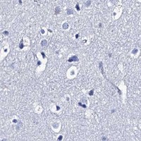
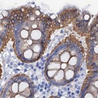


Comprehensive Human Protein Atlas IHC scoring per tissue (reliability: Uncertain — Low consistency between antibody staining and RNA expression data. Pending external verification.). Rows are taken directly from the HPA tissue chart — click any row's HPA link to view the source.
| Tissue | Cell type | Level | Evidence | Source |
|---|---|---|---|---|
| Appendix | Glandular cells | High | Protein (IHC) | HPA → |
| Breast | Glandular cells | High | Protein (IHC) | HPA → |
| Endometrium | Glandular cells | High | Protein (IHC) | HPA → |
| Epididymis | Glandular cells | High | Protein (IHC) | HPA → |
| Fallopian tube | Glandular cells | High | Protein (IHC) | HPA → |
| Tissue | Cell type | Level | Evidence | Source |
|---|---|---|---|---|
| Adipose tissue | Adipocytes | Not detected | Protein (IHC) | HPA → |
| Adrenal gland | Glandular cells | Not detected | Protein (IHC) | HPA → |
| Bone marrow | Hematopoietic cells | Not detected | Protein (IHC) | HPA → |
| Caudate | Glial cells | Not detected | Protein (IHC) | HPA → |
| Cerebellum | Cells in granular layer | Not detected | Protein (IHC) | HPA → |
Troubleshoot RAB3IP staining in paraffin sections by checking retrieval, cellular compartment, tissue context and controls before scoring chromogenic signal.
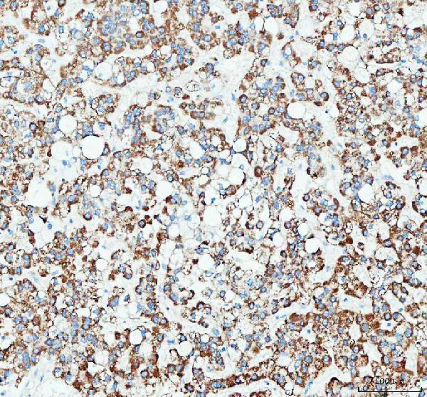
A09085-3 has IHC data from paraffin sections of human ovarian serous adenocarcinoma and liver cancer, plus mouse and rat kidney (catalog image captions); no IF/ICC data are supplied (catalog applications and images).
A09085-3 is listed for IHC in human, mouse and rat samples (catalog applications and reactivity). Its IHC captions show paraffin sections of human ovarian serous adenocarcinoma, human liver cancer, mouse kidney and rat kidney (catalog image captions).
Which to pick: Choose A09085-3 for paraffin-section IHC; its ovarian tissue caption documents EDTA pH 8.0 retrieval and 2 μg/ml primary antibody (A09085-3 IHC image caption). The fixative is unreported (catalog image captions), and IF/ICC is not listed or illustrated, so there is no payload-supported IF/ICC pick (catalog applications and images). For human, mouse or rat IHC, A09085-3 is the listed option with species-specific tissue captions; its host is rabbit and clonality is unreported (catalog reactivity, image captions and host/clone fields).