This website uses cookies to ensure you get the best experience on our website.
- Table of Contents
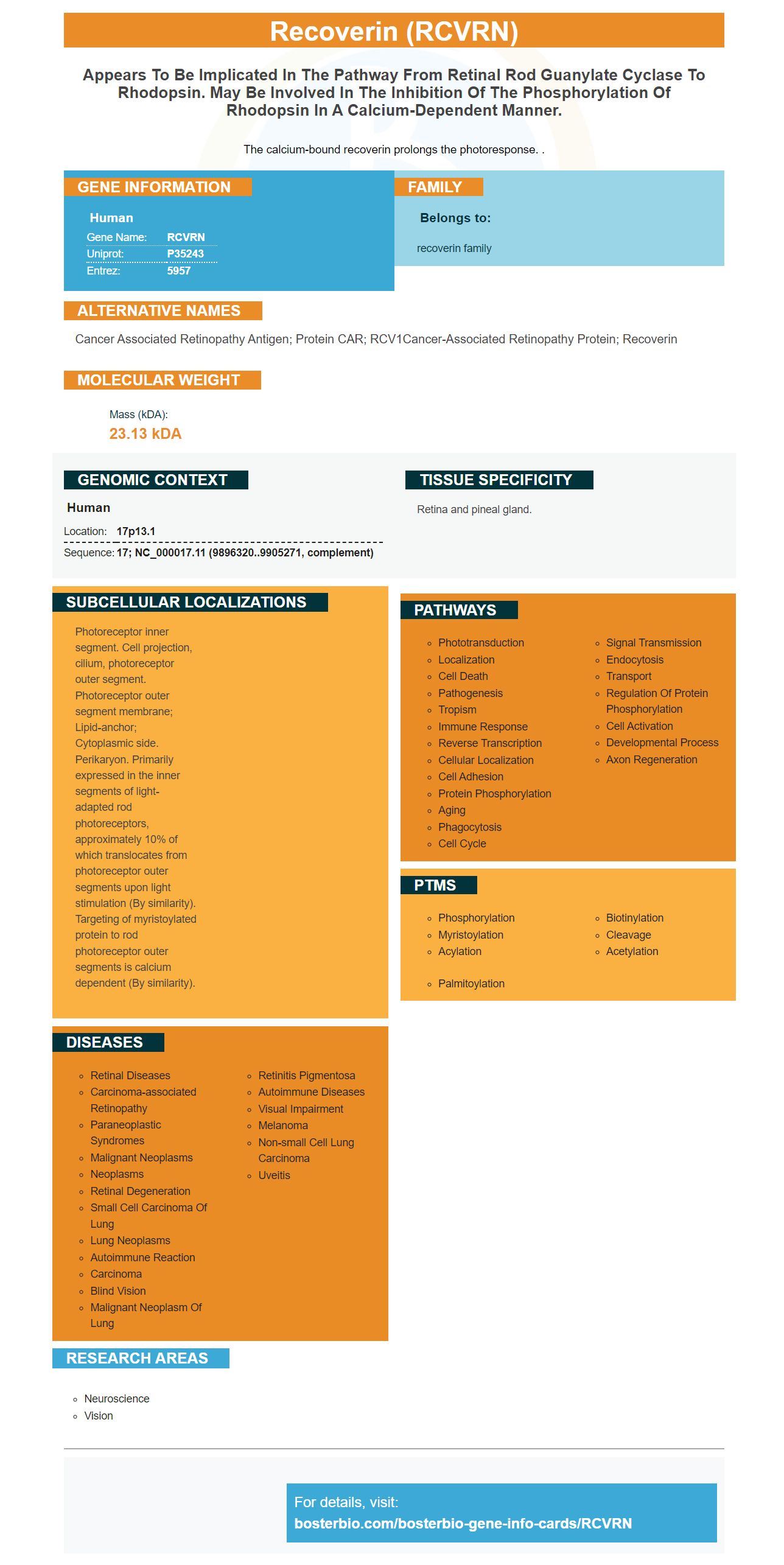
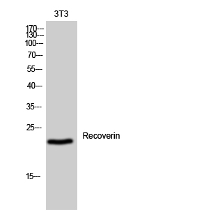
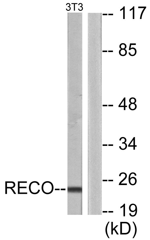
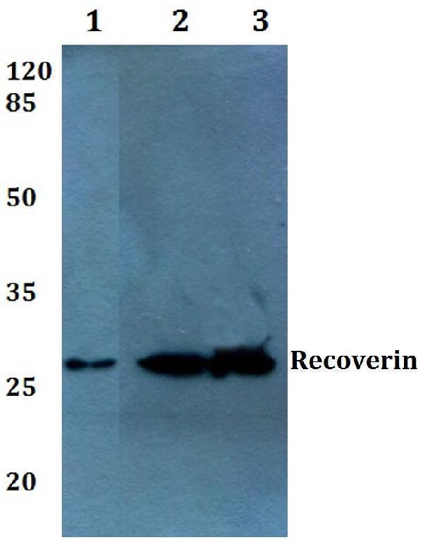
Facts about Recoverin.
The calcium-bound recoverin prolongs the photoresponse. .
Human | |
---|---|
Gene Name: | RCVRN |
Uniprot: | P35243 |
Entrez: | 5957 |
Belongs to: |
---|
recoverin family |
cancer associated retinopathy antigen; Protein CAR; RCV1Cancer-associated retinopathy protein; recoverin
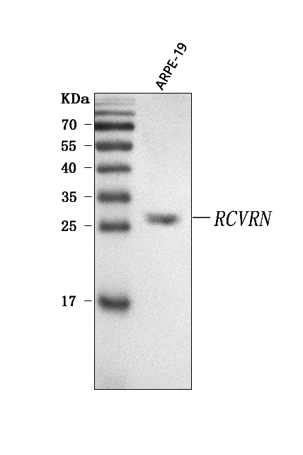
Mass (kDA):
23.13 kDA
Human | |
---|---|
Location: | 17p13.1 |
Sequence: | 17; NC_000017.11 (9896320..9905271, complement) |
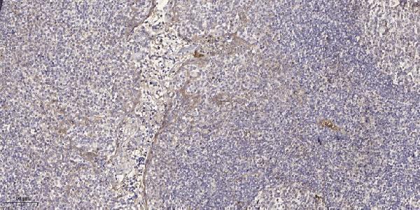
Retina and pineal gland.
Photoreceptor inner segment. Cell projection, cilium, photoreceptor outer segment. Photoreceptor outer segment membrane; Lipid-anchor; Cytoplasmic side. Perikaryon. Primarily expressed in the inner segments of light-adapted rod photoreceptors, approximately 10% of which translocates from photoreceptor outer segments upon light stimulation (By similarity). Targeting of myristoylated protein to rod photoreceptor outer segments is calcium dependent (By similarity).
The Anti-Recoverin RCVRN Marker from Boster Bio is a high affinity primary antibody that has been cited in many research publications for over 25 years. Boster antibodies are trusted and used by the research community and have been validated in Western Blotting, Immunohistochemistry, and ELISA. Here are the best uses for the RCVRN Marker.
Anti-Recoverin RCVRN markers are a new class anti-recoverin antibodies that target retinal protein. They have been shown not to increase the growth of tumor cells. These antibodies are not only present in cancer patients, but also in healthy individuals. The anti-recoverin RCVRN antibodies are rare in humans, 5% of CAR patients have antibodies to recoverin. A paraneoplastic syndrome can be diagnosed even if there are no anti-recoverin antibody. Anti-recoverin antibodies are associated with over 30 different retinal antigens.
Originally discovered as a human autoantigen, recoveryin is now an internationally recognized calcium sensor protein. It binds two Ca(2+), ions and is immunogenic. It has been shown to form complexes of rhodopsin-kinase. In tumor cells, aberrant hypomethylation has affected the promoter region up-stream of the first exon.
By labeling a target protein with a fluorescent label (such as the RCVRN), a high-affinity antigen is created. The antibody must recognize the epitope on the tissue. This may not always be possible. The affinity constant of specific antibodies may not be known. To solve this problem, high-affinity primary antibodies using the RCVRN marker were developed.
A high-affinity IgM antibody is a type of a monoclonal antibody that recognizes specific targets and molecules. It is made by B cells. High-affinity IgM antibodies are produced by cells with an increased ratio of InsA(1)/InsA(4). This is called affinity maturation. In addition, different days in the life cycle were compared to determine their direct binding affinity. WT mice reach high insulin-IgM levels at day 52. Mice with IgD deficiency don't reach this level until day 72. Both groups showed signs of diabetics.
This study shows high-affinity primary antibodies that use the RCVRNA mark are capable of recognizing dsDNA, and nuclear structures. This partially polyreactive IgM may be part of an effective primary immune response. A B cell must recognize its cognate antibody in order to produce an efficient secondary response. However, it is unclear whether or not this polyreactivity will affect the production of high-affinity IgG antibodies.
As discussed previously, antibodies produced using the RCVRN markers are able to recognize high-affinity antigens. This is not always desirable. High affinity antibodies may not be easy to separate from antigens. They may also be difficult for elution from affinity columns unless harsher methods used. High affinity antibodies are still useful. They are still viable options for high-affinity prim antibodies.
The antibody's ability to bind to one molecule to a target is measured by its affinity. In a bimolecular interaction, the rate at which an antibody binds to a target is proportional the concentration of the reactants. KD values are proportional to the affinity. The antibody's efficiency is also higher if the KD value is higher.
The affinity maturation results are obtained by dividing InsA(1) by InsA(4). These experiments result in relative units of both antigens. Professor Nobuo Saguchi discovered the RCVRN genes. Professor Nobuo Sakaguchi discovered the RCVRN gene. It encodes recoverin, a member EF-hand of neuronal calcium sensor protein proteins. It is composed of four EFhand domains. One mediates high and lower-affinity calcium binding and the other mediates high affinity.
In this study, the subclasses immunoglobulin IgG and M were created. After multiple freeze-thaw cycles, the serum of immunized mouse was purified of antibodies. The manufacturer's protocol required that the serum be dialyzed using HiTrap IgM cells. Coomassie staining was used to detect antibodies in the serum. SDS-PAGE was used to confirm their presence.
The use of RCVRN as a biomarker in clinical trials is a novel approach to study retinal degeneration. The biomarker, which can easily be extracted from human cells and used as a therapeutic agent for the disease, is very easy to create. RCVRN’s eGFP reporter is very efficient in obtaining photoreceptor-cell numbers and its purification effects are very favorable. The RCVRN eGFP reader is useful for cell transfer in animal models with RD.
To study RCVRN in-vivo, researchers first prepared embryoid bodies from PGP1 HiPSCs. Then they stained them with an anti-bIII tubulin and anti–rhodopsin (RCVRN) antibodies. The cells then developed distinct photoreceptor layers and were distinguished by the markers. Similarly, RCVRN expression was high in both the groups. To visualize the sequential growth of these retinal types, the antibodies to RCVRN ISL-1 and ROD were used.
A RCVRN-eGFP reporter strain was also successfully established, and it is capable of differentiation into human ROs. This line also showed high expression under a variety phenotypic conditions, such as adiponectin. RCVRN-eGFP reporter lines reexamines RCVRN's ability in vivo to identify specific types of photoreceptors.
In addition to its immunoreactivity, RCVRN-eGFP labeled postmitotic photoreceptor precursors in retinal organoids. The RCVRN-eGFP-eGFP-eGFP+ cells did not express the proliferation marker Ki67. Furthermore, RCVRN-eGFP-positive cells expressed the photoreceptor precursor marker CRX, cone-specific recovery protein RXRr, and rod-specific phototransduction protein NRL. RCVRN eGFP++ cells also labeled photoreceptor precursors. Additionally, they also expressed a subunit CNGB3 of the cone specific phototransduction protein.
The RCVRN-eGFP reporter hiPSC line was able to efficiently generate photoreceptors and monitor endogenous expression of human recoverin. These reporter ROs can serve as stable enrichment tools for photoreceptor cells and may eventually be used for photoreceptor cell therapy. This study identified potential CD Biomarkers that could prove to be of great value for clinical use. There is much more to this novel biomarker than just clinical trials, and these will be useful for the field.
Using an animal genetic editing method, eGFP reporters hiPSCs were created. To generate these reporter hiPSCs, the viral P2A-eGFP fusion was inserted before the RCVRN exon 3 TGA stop codon. After this, the clones of reporter hiPSCs were expanded and characterized with standard hiPSC anatomy and normal karyotype.
PMID: 1387789 by Murakami A., et al. Isolation of human retinal genes: recoverin cDNA and gene.
PMID: 8500558 by Wiechmann A.F., et al. Molecular cloning and nucleotide sequence of a cDNA encoding recoverin from human retina.