This website uses cookies to ensure you get the best experience on our website.


- Table of Contents
Plan paraffin-section REST IHC around its general nuclear tissue pattern (HPA tissue IHC) and the catalog antibody’s 2–5 μg/ml IHC range (datasheet A03852-3). Score nuclear staining by cell type, accounting for hypoxia-associated nuclear increases (UniProt).

Expected localisation, validated protocols, controls and antibodies — the at-a-glance facts below, then the full design guide.
| Expected localisation | Nuclear tissue staining (HPA tissue IHC); cytoplasm also annotated (UniProt) | |
| Staining pattern | General nuclear staining across tissues (HPA tissue IHC) | |
| Antigen retrieval | EDTA pH 8.0 HIER, heat-mediated (datasheet A03852-3) | |
| Positive control | Bronchus+4 more · see all | |
| Negative control | None in HPA (detected in all 45 tissues); use no-primary + isotype controls |
| Fixation | Matched tissue-IHC evidence does not establish the fixation claim. Validate the specimen-specific method before use. (selected-SKU IHC image A03852-3) | |
| Caveat | Neuronal nuclear REST can decrease in dementia (UniProt) | |
| Regulation | Hypoxia increases nuclear REST (UniProt) | |
| Isoform / epitope | Four isoforms; validate antibody coverage of each (UniProt) |
The catalog antibody protocol uses EDTA pH 8.0 retrieval (datasheet A03852-3). Published IHC protocols cover Merkel cell carcinoma (PMC6226622) and brain sections (PMC5797046).
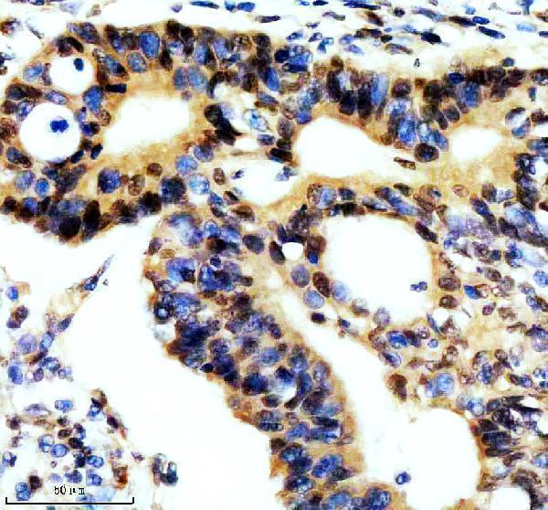
| Sample | Paraffin-embedded human colon cancer tissue; fixative not specified (datasheet A03852-3) |
| Fixation | Image fixative and duration unreported (datasheet A03852-3); verify before use. |
| Sectioning | 4–5 µm sections on charged slides (standard) |
| Deparaffinisation | Xylene, graded ethanol series to water (standard) |
| Antigen retrieval | Heat retrieval: EDTA pH 8.0 (datasheet A03852-3); 20 min, 95–100 °C (standard) |
| Peroxidase block | 3% H2O2, 10 min, room temperature (standard) |
| Blocking | 10% goat serum (datasheet A03852-3) |
| Primary antibody | Rabbit anti-REST, 2-5 μg/ml (datasheet A03852-3) |
| Primary incubation | Overnight at 4 °C (datasheet A03852-3) |
| Detection | HRP-conjugated secondary, DAB chromogen (datasheet A03852-3) |
| Counterstain | Hematoxylin, blue, dehydrate and mount (standard) |
| Expected result | REST-positive staining in respiratory epithelial cells of bronchus (HPA tissue IHC: High). HPA tissue profile: General nuclear expression. No signal in the no-primary control. |
REST is mainly nuclear, with additional cytoplasmic localization (UniProt Q13127; HPA: general nuclear IHC expression; HPA: nucleoplasm and cytosol in ICC-IF). In paraffin sections, look for nuclear staining in respiratory epithelial, squamous epithelial, glandular and alveolar type I cells listed as high by HPA (HPA tissue IHC). REST has no transmembrane segment, so a membrane outline is unexpected (UniProt Q13127 topology). HPA rates its tissue IHC evidence Supported, with medium consistency and external verification pending (HPA tissue IHC).
| Distinct nuclear chromogen in bronchial respiratory epithelium, with nuclei identifiable beneath the counterstain. | This fits a high-staining cell population and REST’s principal nuclear location (HPA: bronchus respiratory epithelial cells, High; HPA: general nuclear IHC expression; UniProt Q13127). Score the stained nuclei within that cell population rather than treating all structures in the section as equivalent. |
| Nuclear staining in endometrial glandular cells; weaker cytoplasmic color is also visible. | The nuclear component fits the expected pattern (HPA: endometrium glandular cells, High; UniProt Q13127: nucleus). Some cytoplasmic REST is plausible (UniProt Q13127: cytoplasm; HPA ICC-IF: additional cytosol). Record the two compartments separately; cytoplasmic color alone does not demonstrate the expected nuclear pattern. |
| A crisp cell-surface rim or extracellular deposit dominates while nuclei lack signal. | A dominant membrane or extracellular pattern is inconsistent with REST’s reported localization and lack of a transmembrane segment (UniProt Q13127; HPA: general nuclear IHC expression). Treat it as suspect staining and review morphology and detection controls before assigning it to REST. |
| Strong signal dominates a population HPA lists as low, such as cerebral cortex glial cells. | This disagrees with the supplied tissue profile (HPA: cerebral cortex glial cells, Low), but is not proof of cross-reactivity: REST is reported as ubiquitous (UniProt Q13127). Check cell identification and nuclear localization; use detection controls to assess endogenous activity before interpreting the difference. |
| The chromogen is diffuse across cells and tissue spaces, or absent in a high-staining reference area. | Diffuse color does not resolve the expected nuclear pattern (HPA: general nuclear IHC expression). Absence from a listed high-staining population, such as bronchial respiratory epithelium, makes the run inconclusive for that specimen (HPA: bronchus, High). Compare controls and section quality before calling biological loss. |
| Cell population and tissue | HPA reports high staining in bronchial respiratory, cervical and esophageal squamous, several glandular, and lung alveolar type I cells, but low staining in specified glial and other populations (HPA tissue IHC). Identify the cells before comparing intensity across sections. |
| Intracellular distribution | REST is reported in nucleus and cytoplasm (UniProt Q13127), while ICC-IF places it mainly in nucleoplasm with additional cytosol (HPA ICC-IF). Nuclear enrichment can increase in hypoxia and aging neurons (UniProt Q13127); interpret a changed nuclear-to-cytoplasmic balance in its specimen context. |
| Isoform coverage | Four REST isoforms are listed (UniProt Q13127). The supplied evidence does not map the IHC antibody’s epitope across them, so a staining difference cannot be assigned to one isoform without additional antibody information. |
| Strength of tissue evidence | HPA labels tissue IHC Supported, describes medium agreement with RNA, and says external verification is pending (HPA tissue IHC). Use its high and low entries as comparison points, not absolute positive or negative guarantees for every specimen. |
| Antigen retrieval and detection | Target-specific fixation effects are not established by the supplied assay evidence. Verify with a matched IHC source before attributing a result to fixation. |
| Situation | Likely cause | Next action |
|---|---|---|
| No nuclear stain in bronchial respiratory epithelium that is present and morphologically intact (HPA: High). | The result conflicts with an HPA high-staining reference population; the supplied sources cannot distinguish technical failure from specimen variation (HPA tissue IHC). | Check the run’s positive and detection controls, then review retrieval, primary incubation and chromogen development against the established IHC procedure (general IHC practice). |
| Broad brown color obscures nuclei and fills tissue spaces. | The expected pattern is general nuclear expression (HPA tissue IHC); diffuse color can arise from nonspecific staining or detection background (general IHC practice). | Review the no-primary detection control, blocking, washes and chromogen development; rescore only where individual cells and nuclei can be distinguished (general IHC practice). |
| Strong staining appears mainly at cell borders. | A border-dominant pattern conflicts with nuclear and cytoplasmic REST localization and its lack of a transmembrane segment (UniProt Q13127). | Compare with a listed high-staining tissue area and the detection control; document the border pattern separately instead of counting it as nuclear REST (HPA tissue IHC; general IHC practice). |
| A low-staining HPA population shows stronger signal than nearby expected-positive cells. | The comparison differs from HPA’s cell-specific levels, although ubiquitous REST expression means that an unexpected cell is not automatically negative (HPA tissue IHC; UniProt Q13127). | Recheck cell identity, nuclear localization, background and detection controls; report the discrepancy rather than labeling it cross-reactivity without supporting evidence. |
| Nuclear and cytoplasmic scores differ between specimens. | Both compartments are reported for REST, and nuclear enrichment is described in hypoxia and aging neurons (UniProt Q13127; HPA ICC-IF). The sources do not establish a fixation-based explanation. | Record each compartment and the relevant specimen context separately; compare like cell populations and avoid attributing the shift to retrieval or fixation without further evidence. |
| Can an IF/ICC image be used to judge an IHC section? | HPA ICC-IF supports mainly nucleoplasmic with additional cytosolic localization; tissue IHC has its own Supported assessment and general nuclear profile (HPA ICC-IF; HPA tissue IHC). | Use ICC-IF only as a compartment comparison. Judge paraffin chromogenic staining against tissue IHC cell populations, section morphology and IHC controls (HPA tissue IHC; general IHC practice). |


Comprehensive Human Protein Atlas IHC scoring per tissue (reliability: Supported — Medium consistency between antibody staining and RNA expression data. Pending external verification.). Rows are taken directly from the HPA tissue chart — click any row's HPA link to view the source.
| Tissue | Cell type | Level | Evidence | Source |
|---|---|---|---|---|
| Bronchus | Respiratory epithelial cells | High | Protein (IHC) | HPA → |
| Cervix | Squamous epithelial cells | High | Protein (IHC) | HPA → |
| Endometrium | Glandular cells | High | Protein (IHC) | HPA → |
| Esophagus | Squamous epithelial cells | High | Protein (IHC) | HPA → |
| Fallopian tube | Glandular cells | High | Protein (IHC) | HPA → |
| Tissue | Cell type | Level | Evidence | Source |
|---|---|---|---|---|
| None in HPA: REST is detected in all 45 scored tissues. Use a no-primary (secondary-only) and an isotype control instead. | ||||
Troubleshoot REST staining in paraffin sections by checking retrieval, nuclear localisation, antibody controls, and cell-specific scoring.
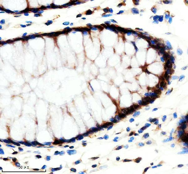
One human-reactive anti-REST antibody has real IHC data from paraffin-embedded colon, colon cancer and thyroid cancer sections (catalog: A03852-3 reactivity; A03852-3 IHC captions).
A03852-3 is listed for human IHC (catalog: A03852-3 applications/reactivity). Its captions document staining in paraffin-embedded human colon, colon cancer and thyroid cancer sections; no IF figure is supplied (A03852-3 IHC/IF image captions).
Which to pick: For tissue IHC, choose A03852-3, a rabbit antibody listed for human IHC at 2–5 μg/ml (catalog: A03852-3 host/applications/reactivity/dilution); its captions document paraffin sections with EDTA pH 8 retrieval (A03852-3 IHC captions). The fixative is unreported (A03852-3 IHC captions). For IF/ICC or cross-species work, the payload provides no validated option: A03852-3 has no listed IF/ICC application and is human-reactive only (catalog: A03852-3 applications/reactivity).