This website uses cookies to ensure you get the best experience on our website.


- Table of Contents
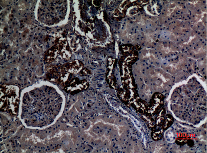
Use kidney distal tubules as a high-staining reference and adipose adipocytes as a no-detection comparator (HPA tissue IHC). This guide covers expected renal and squamous staining patterns (HPA tissue IHC) and the catalog antibody’s 1:100 IHC dilution (datasheet).

Expected localisation, validated protocols, controls and antibodies — the at-a-glance facts below, then the full design guide.
| Expected localisation | Renal basolateral membrane; squamous membrane and cytoplasm (HPA tissue IHC) | |
| Staining pattern | Distal tubules: basolateral rim; squamous cells: membrane and cytoplasm (HPA tissue IHC) | |
| Antigen retrieval | Citrate pH 6.0 HIER, 95–98 °C, 20 min (rule: cytoplasmic / membrane antigen) | |
| Positive control | Cervix+4 more · see all | |
| Negative control | Adipose tissue+4 more · see all |
| Fixation | Keep paraffin fixation consistent (standard IHC practice; not target-specific); Matched tissue-IHC evidence does not establish the fixation claim. Validate the specimen-specific method before use. (selected-SKU IHC image A03655) | |
| Caveat | Renal staining is basolateral despite apical annotation (HPA tissue IHC; UniProt) | |
| Regulation | Regulation unreported (UniProt) | |
| Isoform / epitope | No annotated isoforms; epitope side varies across 12 TM spans (UniProt) |
The catalog antibody protocol is followed by three published RHCG immunoperoxidase protocols for mouse brain, rat kidney, and human and rat kidney sections (PMC6106833; PMC4321814; PMC2692438).
| Sample | Paraffin-embedded human kidney tissue; fixative not specified (datasheet A03655) |
| Fixation | Image fixative and duration unreported (datasheet A03655); verify before use. |
| Sectioning | 4–5 µm sections on charged slides (standard) |
| Deparaffinisation | Xylene, graded ethanol series to water (standard) |
| Antigen retrieval | Heat-induced epitope retrieval in citrate buffer, pH 6.0, 20 min at 95–98 °C (standard rule: cytoplasmic / membrane antigen) |
| Peroxidase block | 3% H2O2, 10 min, room temperature (standard) |
| Blocking | 10% normal serum of the secondary host, 30 min, room temperature (standard) |
| Primary antibody | Rabbit anti-RHCG, 1:100 (datasheet A03655) |
| Primary incubation | Overnight at 4 °C (standard) |
| Detection | HRP-polymer secondary, DAB chromogen 5–10 min (standard) |
| Counterstain | Hematoxylin, blue, dehydrate and mount (standard) |
| Expected result | RHCG-positive staining in squamous epithelial cells of cervix (HPA tissue IHC: High). HPA tissue profile: Selective basolateral membrane expression in renal tubules with membranous and cytoplasmic expression in squamous epithelia. No signal in the no-primary control. |
RHCG is a 12-transmembrane protein found at the cell membrane, including apical and basolateral sites (UniProt Q9UBD6 topology and subcellular location). In tissue IHC, expect selective basolateral staining in renal tubules and membranous plus cytoplasmic staining in squamous epithelia (HPA: tissue IHC profile; Enhanced reliability). Interpret location within the identified cell type; a membrane signal alone does not establish a correct result.
| Distinct basolateral staining in kidney distal tubules (HPA: kidney distal tubules, High; tissue IHC profile). | This matches the observed renal IHC pattern (HPA: selective basolateral membrane expression in renal tubules). UniProt also lists apical membrane, basolateral membrane, and subapical vesicles; use the HPA tissue pattern when judging this paraffin-section result (UniProt Q9UBD6 subcellular location). |
| Membranous staining with some cytoplasmic signal in squamous epithelial cells (HPA: tissue IHC profile). | This can be the expected tissue pattern in cervix, esophagus, oral mucosa, tonsil, or vagina, where HPA reports High staining in squamous epithelial cells (HPA: tissue IHC). Do not reject the cytoplasmic component solely because RHCG is a membrane protein (HPA: tissue IHC profile; UniProt Q9UBD6 subcellular location). |
| Predominantly nuclear staining in an otherwise positive tissue (HPA: tissue IHC profile; UniProt Q9UBD6 subcellular location). | A nuclear pattern is unsupported by the supplied RHCG localisation evidence and raises concern for nonspecific staining (HPA: tissue IHC profile; UniProt Q9UBD6 subcellular location). Review the cell outlines and a no-primary control before scoring it as RHCG (standard IHC practice). |
| Strong signal in adipocytes or adrenal glandular cells (HPA: Not detected in these cells). | These cell types are poor matches for the HPA tissue pattern; cross-reactivity or endogenous chromogenic activity is possible (HPA: tissue IHC; standard IHC practice). Check whether the signal persists without primary antibody, then compare its compartment and morphology with a positive control (standard IHC practice). |
| No signal in kidney distal tubules or a listed squamous epithelium (HPA: High in these cell types). | A negative result in a known-positive control makes the run difficult to interpret (HPA: kidney and squamous epithelia, High; standard IHC practice). Check tissue identity, the IHC-validated antibody, retrieval, and detection controls before calling the study tissue negative (standard IHC practice). |
| Tissue and cell selection (HPA: tissue IHC). | HPA reports High staining in kidney distal tubules and squamous epithelial cells of cervix, esophagus, oral mucosa, tonsil, and vagina (HPA: tissue IHC). Salivary glandular cells are Low, so they provide a weaker visual benchmark (HPA: tissue IHC). |
| Compartment differs by tissue (HPA: tissue IHC profile). | Kidney staining is described as selective basolateral membrane expression, whereas squamous epithelia show membranous and cytoplasmic expression (HPA: tissue IHC profile). Apply the appropriate compartment expectation to each cell type rather than requiring an identical pattern across tissues. |
| Membrane topology (UniProt Q9UBD6 topology). | RHCG has 12 annotated transmembrane segments, with cytoplasmic and extracellular regions (UniProt Q9UBD6 topology). An antibody's epitope location is not supplied here; topology alone cannot establish which retrieval condition will work in paraffin sections. |
| Glycosylation and processing (UniProt Q9UBD6). | UniProt lists one glycosylation site at residue 48 and one chain spanning residues 1–479, with no signal peptide or propeptide annotated (UniProt Q9UBD6). These annotations do not establish a distinct shed-fragment staining pattern or a fixation effect. |
| IHC validation (HPA: antibody validation). | Two rabbit polyclonal antibodies, HPA041874 and HPA043317, carry Enhanced IHC validation (HPA: antibodies). HPA describes the tissue profile as consistent with RNA expression; this supports the reported IHC pattern but does not validate every staining result in a new run (HPA: Enhanced reliability). |
| IF/ICC evidence (HPA: subcellular and antibody records). | For the IF/ICC question, HPA's subcellular summary says “Membrane,” but lists no ICC-IF image cell lines or main location, and neither listed antibody has an ICC validation entry (HPA: subcellular; HPA: antibodies). Treat an IF/ICC localisation result as provisional; the supplied evidence supports an IHC tissue comparison, not an IF/ICC protocol. |
| Situation | Likely cause | Next action |
|---|---|---|
| Kidney control shows no distal-tubule signal (HPA: kidney distal tubules, High). | The run may have a tissue-identification, primary-antibody, retrieval, or detection problem; the supplied sources do not identify a RHCG-specific fixation sensitivity (standard IHC practice; HPA: tissue IHC). | Confirm distal tubules on the section, verify that the antibody is IHC-validated, and inspect the run's positive and detection controls (standard IHC practice). Reassess the study tissue only after the expected kidney control pattern appears (HPA: tissue IHC profile). |
| Squamous epithelium looks cytoplasmic as well as membranous (HPA: tissue IHC profile). | That combination is reported for squamous epithelia and need not represent background by itself (HPA: tissue IHC profile). | Check whether signal tracks the squamous epithelial cells and whether a no-primary section stays clear (HPA: squamous epithelial cells, High; standard IHC practice). Judge the result against the squamous pattern, not the kidney basolateral pattern (HPA: tissue IHC profile). |
| Most cells have an even brown haze (standard chromogenic IHC practice). | Diffuse background can obscure the selective cell and compartment pattern; it does not match HPA's described RHCG tissue distribution (HPA: tissue IHC profile). | Inspect a no-primary control, blocking and wash steps, and chromogen development (standard IHC practice). Score RHCG only where identifiable cells show the expected distribution above background (HPA: tissue IHC profile; standard IHC practice). |
| Adipocytes or adrenal glandular cells stain strongly (HPA: Not detected). | A mismatch with HPA-negative cell types raises concern for cross-reactivity or endogenous detection activity; staining alone cannot distinguish them (HPA: tissue IHC; standard IHC practice). | Compare a no-primary section and a known-positive tissue; investigate endogenous enzyme activity if signal persists without primary antibody (standard chromogenic IHC practice). Recheck cell identity and localisation before assigning RHCG positivity (HPA: tissue IHC profile). |
| Staining is mainly nuclear (UniProt Q9UBD6 subcellular location). | Nuclear localisation is absent from the supplied RHCG locations and from the observed HPA tissue profile (UniProt Q9UBD6 subcellular location; HPA: tissue IHC profile). | Do not score nuclear-only staining as the expected RHCG pattern. Inspect the no-primary control and compare membrane and cytoplasmic signal in a known-positive section (standard IHC practice; HPA: tissue IHC profile). |
| An IF/ICC image shows a membrane signal (HPA: subcellular summary, Membrane). | A membrane signal is compatible with the HPA summary, but the supplied HPA record has no ICC-IF image cell lines and no ICC validation entry for either listed antibody (HPA: subcellular; HPA: antibodies). | Use the separate IF/ICC guide for assay design. In this IHC guide, base the interpretation on the validated tissue IHC patterns and do not infer IF/ICC performance from them (HPA: tissue IHC; HPA: antibodies). |

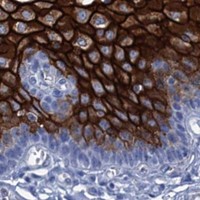


Comprehensive Human Protein Atlas IHC scoring per tissue (reliability: Enhanced — High consistency between antibody staining and RNA expression data.). Rows are taken directly from the HPA tissue chart — click any row's HPA link to view the source.
| Tissue | Cell type | Level | Evidence | Source |
|---|---|---|---|---|
| Cervix | Squamous epithelial cells | High | Protein (IHC) | HPA → |
| Esophagus | Squamous epithelial cells | High | Protein (IHC) | HPA → |
| Kidney | Distal tubules | High | Protein (IHC) | HPA → |
| Oral mucosa | Squamous epithelial cells | High | Protein (IHC) | HPA → |
| Skin | Arrector pili muscle cells | Low | Protein (IHC) | HPA → |
| Tissue | Cell type | Level | Evidence | Source |
|---|---|---|---|---|
| Adipose tissue | Adipocytes | Not detected | Protein (IHC) | HPA → |
| Adrenal gland | Glandular cells | Not detected | Protein (IHC) | HPA → |
| Appendix | Glandular cells | Not detected | Protein (IHC) | HPA → |
| Bone marrow | Hematopoietic cells | Not detected | Protein (IHC) | HPA → |
| Breast | Adipocytes | Not detected | Protein (IHC) | HPA → |
Troubleshoot RHCG staining in paraffin sections by checking tubular membrane localisation, tissue controls, and the limits of the antibody’s documented IHC evidence.
A03655 has an IHC image from paraffin-embedded human kidney (IHC image caption). IF is listed, but no IF image is supplied (catalog: applications; IF image alts).
A03655 is listed for IHC and IF, with Human, Mouse, and Rat reactivity (catalog: applications; reactivity). Its IHC image shows paraffin-embedded human kidney at 1:100; no IF image is supplied (IHC image caption; IF image alts).
Which to pick: For tissue IHC, choose A03655 based on its paraffin-embedded human kidney image at 1:100; the fixative is unreported (IHC image caption). For IF/ICC, A03655 lists IF at 1:50, but supplies no IF image or ICC application claim; its host is Rabbit and clonality is unreported (catalog: applications, IF dilution, IF image alts, host, clone). For cross-species work, A03655 lists Human, Mouse, and Rat reactivity, while its pictured IHC sample is human kidney only (catalog: reactivity; IHC image caption).