RMI2 / RecQ-mediated genome instability protein 2 · IHC design guide

Design Immunohistochemistry for RMI2

Plan chromogenic RMI2 IHC in paraffin sections with the catalog antibody at 0.5–1 μg/mL (datasheet A08685). Use tonsil germinal center cells as a high-staining control and interpret nuclear and cytoplasmic staining with the reported uncertainty in mind (HPA tissue IHC).

Evidence assembled Oct 2026 · For research use; verify linked source records and product datasheet before use
Immunohistochemistry protocol sheet for RMI2 (IHC for RMI2): expected localisation Nuclear (UniProt); nuclear and cytoplasmic tissue staining (HPA tissue IHC), antibody A08685, validated IHC image, and IHC protocol steps
Printable RMI2 IHC protocol sheet — expected localisation Nuclear (UniProt); nuclear and cytoplasmic tissue staining (HPA tissue IHC), antibody A08685, controls and protocol steps. Open the full RMI2 IHC guide →

RMI2 Immunohistochemistry Experimental Design Guide

Expected localisation, validated protocols, controls and antibodies — the at-a-glance facts below, then the full design guide.

Must know before staining
Expected localisation Nuclear (UniProt); nuclear and cytoplasmic tissue staining (HPA tissue IHC)
Staining pattern Nuclear and cytoplasmic staining in several tissues (HPA tissue IHC)
Antigen retrieval Citrate pH 6 HIER, heat-mediated (datasheet A08685)
Positive control ⓘ Placenta+4 more · see all
Negative control ⓘ None in HPA (detected in all 45 tissues); use no-primary + isotype controls
Important caveats
Reasons your staining may differ from the expected pattern.
Fixation Keep fixation consistent across tissue sections (standard IHC practice; not target-specific)
Caveat Tissue staining reliability is uncertain (HPA tissue IHC)
Regulation Lymphoid tissue-enhanced RNA (HPA tissue RNA)
Isoform / epitope 2 isoforms; epitope differences unspecified (UniProt)
Section 1

Recommended RMI2 IHC & IF Protocols

The catalog antibody’s IHC-P protocol (datasheet A08685) is accompanied by three published RMI2 IHC protocols (PMC8516379; PMC10594864; PMC10134310).

Recommended immunohistochemistry (IHC-P) protocol parameters
SampleParaffin-embedded human intestinal cancer tissue; fixative not specified (datasheet A08685)
FixationImage fixative and duration unreported (datasheet A08685); verify before use.
Sectioning4–5 µm sections on charged slides (standard)
DeparaffinisationXylene, graded ethanol series to water (standard)
Antigen retrievalHeat retrieval: Citrate pH 6, 20 min (datasheet A08685)
Peroxidase block3% H2O2, 10 min, room temperature (standard)
Blocking10% goat serum (datasheet A08685)
Primary antibodyRabbit anti-RMI2, 0.5-1μg/ml (datasheet A08685)
Primary incubationOvernight at 4 °C (datasheet A08685)
DetectionStreptavidin-biotin complex (SABC), DAB chromogen (datasheet A08685)
CounterstainHematoxylin, blue, dehydrate and mount (standard)
Expected resultRMI2-positive staining in trophoblastic cells of placenta (HPA tissue IHC: High). HPA tissue profile: Cytoplasmic and nuclear expression in several tissues. No signal in the no-primary control.
💡Decision noteStart with heat-mediated citrate retrieval at pH 6 for the catalog antibody (datasheet A08685); the breast cancer protocol reports sodium citrate retrieval without specifying pH (PMC10134310).
Section 2

What Is the Expected RMI2 Staining Pattern?

RMI2 is a nuclear protein that colocalizes with BLM at DNA repair foci and has no transmembrane segment (UniProt Q96E14). In paraffin section IHC, expect nuclear staining, particularly in placental trophoblastic cells and tonsillar germinal center cells, where HPA reports high staining (HPA tissue IHC). HPA also reports cytoplasmic staining in several tissues, but rates its tissue IHC profile Uncertain, pending external verification (HPA tissue IHC).

What am I looking at on my slide?
Nuclear stain in placental trophoblastic cells or tonsillar germinal center cells.This fits the expected compartment and HPA's high staining in those cells (UniProt Q96E14; HPA tissue IHC). Score the specified cells and compartment separately; a positive area alone does not establish that every stained structure represents RMI2 (standard IHC practice).
Predominantly cytoplasmic stain, with little or no nuclear stain.Treat this as equivocal: HPA reports cytoplasmic and nuclear tissue staining, while UniProt places RMI2 in the nucleus (HPA tissue IHC; UniProt Q96E14). Review cell morphology and controls before assigning a cytoplasmic result; HPA rates its tissue IHC profile Uncertain (HPA tissue IHC).
Strong stain in cells outside the expected cell population, without corresponding stain in the expected cells.Possible explanations include cross-reactivity or endogenous chromogenic detection activity (standard IHC practice). Compare cell identity and distribution with the reported high trophoblastic and germinal center staining; the HPA tissue profile remains Uncertain (HPA tissue IHC).
Diffuse stain across nuclei, cytoplasm, and tissue spaces.A widespread haze does not resolve the nuclear distribution reported for RMI2 (UniProt Q96E14). Review blocking, antibody concentration, detection background, and wash steps using slide controls (standard IHC practice); diffuse staining alone cannot establish target specificity.
No stain in placental trophoblastic cells or tonsillar germinal center cells.These are HPA's high staining cell populations, so their absence calls for a run and specimen review (HPA tissue IHC). Check tissue preservation, retrieval conditions, antibody and detection controls, and counterstain before interpreting a negative result (standard IHC practice).
💡Expected RMI2 appearanceCall a result consistent with RMI2 when nuclear stain is clear in trophoblastic cells or tonsillar germinal center cells, which HPA scores High; isolated diffuse or unexpected cell staining is equivocal (UniProt Q96E14; HPA tissue IHC).
How each factor affects the staining
Tissue and cell selectionPlacental trophoblastic cells and tonsillar germinal center cells are the strongest supplied IHC examples; adrenal glandular and bone marrow hematopoietic cells are Medium (HPA tissue IHC). Select and score the named cell population, since tissue labels alone do not identify the stained cells (standard IHC practice).
Low staining comparatorsHPA lists oral squamous epithelium, salivary glandular cells, cardiomyocytes, fibroblasts, and adipocytes as Low; it lists no negative population (HPA tissue IHC). Low staining can provide context, but it is not a validated absence control (standard IHC practice).
Protein location and formsRMI2 is nuclear, has no transmembrane segment, and has two listed isoforms (UniProt Q96E14). UniProt lists no signal peptide or propeptide and a chain spanning residues 2–147 (UniProt Q96E14). These annotations do not identify an antibody epitope or predict isoform-specific staining.
Antibody evidenceThe listed antibody, HPA040995, is rated Uncertain for IHC and Supported for ICC (HPA antibodies). HPA rates the overall tissue IHC profile Uncertain because staining and RNA expression show medium consistency; external verification is pending (HPA tissue IHC). Interpret compartment and cell specificity with that limit in mind.
IF/ICC: What pattern should be expected?HPA places the main ICC-IF signal in nuclear speckles (Supported) and an additional cytosolic signal at Uncertain confidence; its image cell lines are A-431, U-251MG, and U2OS (HPA subcellular). This is an IF interpretation cue, not a paraffin IHC protocol or proof that cytosolic IHC staining is specific.
Why is my staining missing, weak or wrong?
SituationLikely causeNext action
The high staining reference cells are blank.A run or specimen problem may obscure staining; HPA's high examples are trophoblastic and germinal center cells (HPA tissue IHC).Check tissue identity and morphology, a run control, retrieval record, primary antibody step, and chromogenic detection reagents before scoring the slide negative (standard IHC practice).
The stain is cytoplasmic only.HPA describes some cytoplasmic tissue staining, but UniProt assigns RMI2 to the nucleus; HPA tissue IHC is Uncertain (HPA tissue IHC; UniProt Q96E14).Score nuclear and cytoplasmic signal separately, inspect the high staining cell populations, and compare a no-primary control before attributing cytoplasmic color to RMI2 (standard IHC practice).
Color appears in unexpected cells or persists without primary antibody.Cross-reactivity is possible in unexpected cells; color without primary antibody suggests endogenous detection activity or reagent background (standard IHC practice).Use the no-primary control to assess detection background; review blocking and the chromogenic detection system, then reassess the named HPA cell populations (standard IHC practice; HPA tissue IHC).
The slide has uniform haze or obscured nuclei.Excess background can prevent a compartment call (standard IHC practice).Review antibody concentration, blocking, wash steps, and counterstain; require readable cell boundaries and nuclei before scoring localization (standard IHC practice).
Low staining tissue is completely blank.HPA calls the listed comparator cell populations Low, and supplies no negative population; a blank result there alone cannot validate the run (HPA tissue IHC).Evaluate a high staining example in its specified cells alongside the low staining tissue, then check the run control if the expected high cells are also blank (HPA tissue IHC; standard IHC practice).
IHC and IF images show different secondary compartments.HPA reports cytoplasmic tissue IHC and an Uncertain additional cytosolic ICC-IF location, while nuclear speckles are Supported in ICC-IF (HPA tissue IHC; HPA subcellular).Report each assay and compartment separately with its HPA confidence level; avoid treating the IF cytosolic observation as validation of cytoplasmic paraffin IHC staining (HPA tissue IHC; HPA subcellular).

Sample controls for RMI2 IHC & IF

🧪Run tonsil first and look for staining in germinal center cells (HPA: High in tonsil germinal center cells). HPA detects RMI2 in all 45 scored tissues, so there is no supported negative tissue; use no-primary and isotype controls, and treat cells without nuclear staining on the positive slide as a background reference, not confirmed RMI2-negative cells (HPA: no negative tissue rows; UniProt Q96E14: nucleus).
Positive control tissue: Placenta (Trophoblastic cells, HPA High)
Negative control tissue: None in HPA: RMI2 is detected in all 45 scored tissues. Use a no-primary (secondary-only) and an isotype control instead.
ICC-IF cell lines (HPA subcellular resource): HPA ICC-IF images show RMI2 in A-431, U-251MG, U2OS, with annotated localisation: Nuclear speckles (supported) (HPA subcellular).
Technical controls: Include a no-primary, secondary-only control and an isotype control matched to the primary antibody’s host species and clonality; a knockout sample or validated peptide block can provide a biological specificity control (standard IHC practice). Check endogenous peroxidase in tonsil before chromogenic detection (standard IHC practice).
⚠️Feasibility: No target-specific fixation window or fixation effect is reported, and the selected A08685 tissue-IHC caption does not state the fixative (selected-SKU caption: fixative not stated). The paraffin-section example uses heat retrieval in citrate buffer at pH 6 for 20 minutes, but the evidence does not establish whether RMI2 staining depends on that retrieval step (selected-SKU caption). Whether frozen sections or IF would be easier is unreported; IF/ICC images support a mainly nuclear-speckle location, while any cytosolic signal needs cautious interpretation (HPA subcellular: nuclear speckles supported; cytosol uncertain).

HPA tissue IHC evidence for RMI2

Comprehensive Human Protein Atlas IHC scoring per tissue (reliability: Uncertain — Medium consistency between antibody staining and RNA expression data. Pending external verification.). Rows are taken directly from the HPA tissue chart — click any row's HPA link to view the source.

Positive expression · recommended positive controls

TissueCell typeLevelEvidenceSource
Placenta Trophoblastic cells High Protein (IHC) HPA →
Tonsil Germinal center cells High Protein (IHC) HPA →
Adrenal gland Glandular cells Medium Protein (IHC) HPA →
Appendix Glandular cells Medium Protein (IHC) HPA →
Bone marrow Hematopoietic cells Medium Protein (IHC) HPA →

Undetected expression · recommended negative controls

TissueCell typeLevelEvidenceSource
None in HPA: RMI2 is detected in all 45 scored tissues. Use a no-primary (secondary-only) and an isotype control instead.
Section 3

Advanced RMI2 IHC Tips

Troubleshoot RMI2 staining in paraffin sections by checking retrieval, controls, cellular localisation and how staining is scored.

What retrieval condition should I start with for RMI2 IHC?
Use heat mediated citrate buffer at pH 6 for 20 minutes as the starting retrieval condition for RMI2 paraffin section IHC (datasheet A08685). The selected tissue image used that condition before overnight incubation with 1 µg/mL antibody at 4°C, so keep the remaining steps comparable when assessing retrieval (datasheet A08685). If staining is weak, compare a modestly longer retrieval time on matched sections while checking whether tissue structure and nuclear detail remain intact (standard IHC practice). Include a section processed without primary antibody to distinguish retrieval related background from specific staining (standard IHC practice).
Could fixation explain weak or uneven RMI2 staining?
Target specific fixation sensitivity is unknown: the selected RMI2 caption describes a paraffin section but does not state its fixative (datasheet A08685). Record the fixative and fixation duration for each specimen, then compare matched sections using the same citrate pH 6, 20 minute retrieval condition (datasheet A08685; standard IHC practice). When fixation histories differ, examine nuclear morphology and staining distribution before attributing intensity differences to RMI2 expression (standard IHC practice). A fixation effect should be assessed by comparing similarly sampled material processed under documented conditions; the reported tissue staining pattern cannot establish one (standard IHC practice).
How should I assess nuclear and cytoplasmic RMI2 staining?
Score nuclear staining separately because RMI2 is annotated in the nucleus and colocalises with BLM at nuclear DNA repair foci (UniProt Q96E14). Cytoplasmic staining needs careful interpretation: tissue IHC reports both nuclear and cytoplasmic expression, while the additional cytosolic location in cell imaging is uncertain (HPA tissue IHC; HPA subcellular). Check whether apparent nuclear signal lies within intact nuclei using the counterstain, and compare cells away from section folds and damaged edges (standard IHC practice). Record nuclear and cytoplasmic patterns independently across matched sections; a diffuse chromogen deposit alone cannot establish a repair focus (standard IHC practice; UniProt Q96E14).
Can this antibody distinguish RMI2 isoforms or epitope loss?
RMI2 has 2 annotated isoforms, but the supplied antibody caption does not identify the recognised epitope or demonstrate isoform selectivity (UniProt Q96E14; datasheet A08685). Do not interpret a negative paraffin section as loss of a particular isoform without an epitope map and suitable isoform controls (standard IHC practice). RMI2 has no annotated transmembrane segment, and its recorded modifications include N terminal acetylation and phosphorylation at residue 7; neither fact establishes how this antibody behaves after retrieval (UniProt Q96E14). If staining differs between samples, first compare processing and cellular localisation before proposing epitope specific biology (standard IHC practice).
How can IF help assess the RMI2 pattern seen by IHC?
Use IF as a separate localisation check: RMI2 is annotated in the nucleus, and cell imaging supports a mainly nuclear speckle pattern (UniProt Q96E14; HPA subcellular). For multiplexing, pair RMI2 with a validated marker for the expected cell population, such as trophoblastic cells in placenta or germinal center cells in tonsil, and confirm that signals occupy the same cells (HPA tissue IHC; standard IF practice). Choose spectrally separated fluorophores and assess an unstained sample for tissue autofluorescence before judging overlap (standard IF practice). Because RMI2 has no transmembrane segment and the expected signal is intracellular, permeabilisation must allow antibody access to nuclear epitopes; optimise it with appropriate controls (UniProt Q96E14; standard IF practice).
What should I check when RMI2 DAB staining is widespread?
The selected IHC image used 10% goat serum blocking, a biotinylated secondary antibody, a streptavidin biotin detection complex and DAB (datasheet A08685). Run a no primary control and inspect whether deposits persist in tissue regions, vessel contents or damaged areas; persistent signal calls for a review of blocking, detection and wash steps (standard IHC practice). Include a peroxidase blocking step when using enzyme based DAB detection, and assess endogenous biotin as a possible contributor with this detection system (standard IHC practice; datasheet A08685). Compare background under the same exposure and development conditions before reducing antibody concentration or DAB development time (standard IHC practice).
How should I quantify RMI2 staining across tissue samples? ⚠ ANSWER MARKED FOR VERIFICATION
Define the cell population and compartment before scoring: nuclear localisation is annotated, while tissue IHC also reports cytoplasmic staining (UniProt Q96E14; HPA tissue IHC). For nuclear chromogenic staining, report the percentage of positive nuclei and an H score based on intensity categories; count cells using consistent thresholds across sections (standard IHC practice). If spatial abundance matters, report positive cells per mm² of viable tissue and state how the analysed area was selected (standard IHC practice). Normalise comparisons to the same eligible cell population and tissue area, excluding folds, necrosis and poorly preserved regions, and keep nuclear and cytoplasmic scores separate (standard IHC practice).
Which findings would make an RMI2 positive result convincing?
A convincing result shows reproducible staining in intact cells, with a nuclear component consistent with RMI2 localisation and appropriate control behaviour (UniProt Q96E14; standard IHC practice). High staining in placental trophoblastic cells or tonsillar germinal center cells can inform sample selection, but the tissue IHC evidence is rated uncertain and awaits external verification (HPA tissue IHC). Treat isolated edge staining, necrotic deposits or signal in a no primary control as possible artefacts; check endogenous enzyme activity when interpreting DAB (standard IHC practice). Cytoplasmic staining alone warrants caution because tissue IHC reports it, whereas the additional cytosolic localisation in cell imaging is uncertain (HPA tissue IHC; HPA subcellular).
Boster reagents

Best RMI2 / RecQ-mediated genome instability protein 2 IHC Antibodies

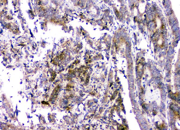
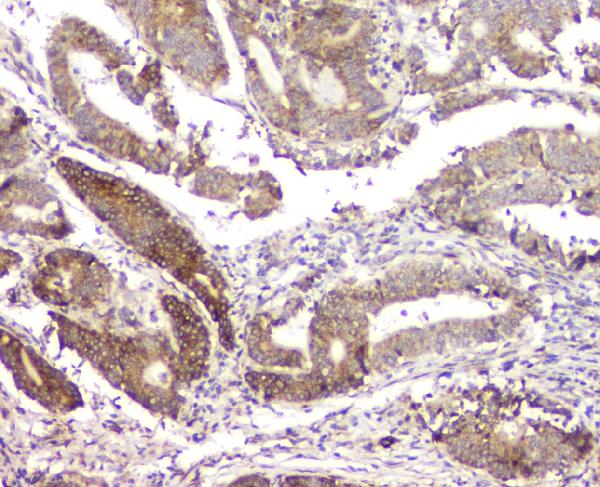
A08685 has IHC images of paraffin sections from human intestinal cancer, human mammary cancer and rat liver (A08685 image captions). Its listed reactivity is human, mouse and rat (catalog: reactivity).

Real IHC data IHC analysis of RMI2 using anti-RMI2 antibody (A08685). RMI2 was detected in paraffin-embedded section of human intestinal cancer tissue. Heat mediated antigen retrieval was performed in citrate buffer (pH6, epitope retrieval solution) for 20 mins. The tissue section was blocked with 10% goat serum. The tissue section was then incubated with 1μg/ml rabbit anti-RMI2 Antibody (A08685) overnight at 4°C. Biotinylated goat anti-rabbit IgG was used as secondary antibody and incubated for 30 minutes at 37°C. The tissue section was developed using Strepavidin-Biotin-Complex (SABC)(Catalog # SA1022) with DAB as the chromogen.
Anti-RMI2 Antibody ®
Cat # A08685

A08685 will render with its own human intestinal cancer paraffin-section IHC image (A08685 card image caption). Additional captions show human mammary cancer and rat liver paraffin sections (A08685 image captions).

Which to pick: Choose A08685 for paraffin-section IHC based on its human and rat tissue images; the fixative is unreported (A08685 image captions). For IF/ICC, A08685 lists ICC and IHC-F, but supplies no IF image (catalog: applications; IF image alts). For cross-species work, its listed reactivity covers human, mouse and rat, with IHC images shown for human and rat only (catalog: reactivity; A08685 image captions).

Each figure is that product's own IHC / IF validation image from its datasheet.

References

  1. UniProt Consortium. UniProt entry Q96E14 (RMI2_HUMAN, RecQ-mediated genome instability protein 2).
  2. Human Protein Atlas. RMI2 tissue IHC expression (reliability: Uncertain).
  3. Human Protein Atlas. RMI2 subcellular location (ICC-IF): Mainly localized to the nuclear speckles. In addition localized to the cytosol..
  4. Human Protein Atlas. RMI2 antibody validation summary (1 antibodies).
  5. The Clinical Significance of RMI2 in Hepatocellular Carcinoma. Technology in cancer research & treatment 2021 — PMC8516379.
  6. A combined bioinformatics and experimental approach identifies RMI2 as a Wnt/β-catenin signaling target gene related to hepatocellular carcinoma. BMC cancer 2023 — PMC10594864.
  7. RMI2 is a novel prognostic and predictive biomarker for breast cancer. Cancer medicine 2023 — PMC10134310.
  8. Androgen receptor inhibitor-induced "BRCAness" and PARP inhibition are synthetically lethal for castration-resistant prostate cancer. Science signaling 2017 — PMC5855082.
  9. PubMed PMID:14702039 — UniProt-cited evidence.
  10. PubMed PMID:15616553 — UniProt-cited evidence.
  11. PubMed PMID:15489334 — UniProt-cited evidence.