This website uses cookies to ensure you get the best experience on our website.


- Table of Contents
Plan RPL10 paraffin-section IHC around granular cytoplasmic staining (HPA tissue IHC). This guide covers fixation consistency (standard IHC practice), the 2–5 μg/mL primary antibody range (datasheet A04190-3), and cautious scoring because staining may reflect more than one gene (HPA tissue IHC).

Expected localisation, validated protocols, controls and antibodies — the at-a-glance facts below, then the full design guide.
| Expected localisation | Granular cytoplasmic tissue staining (HPA tissue IHC) | |
| Staining pattern | Ubiquitous granular cytoplasmic staining (HPA tissue IHC) | |
| Antigen retrieval | EDTA pH 8.0 HIER, heat-mediated (datasheet A04190-3) | |
| Positive control | Adrenal gland+4 more · see all | |
| Negative control | None in HPA (detected in all 45 tissues); use no-primary + isotype controls |
| Fixation | Keep fixation consistent across sections. (standard IHC practice; not target-specific) | |
| Caveat | Staining may reflect products of more than one gene (HPA tissue IHC) | |
| Regulation | No expression regulation annotated (UniProt) | |
| Isoform / epitope | No isoforms annotated; mature chain spans residues 2–214 (UniProt) |
The catalog antibody’s IHC-P protocol (datasheet A04190-3) is accompanied by three published RPL10 IHC protocols covering ovarian, colorectal, and testicular sections (PMC5858496; PMC8927999; PMC11872710).
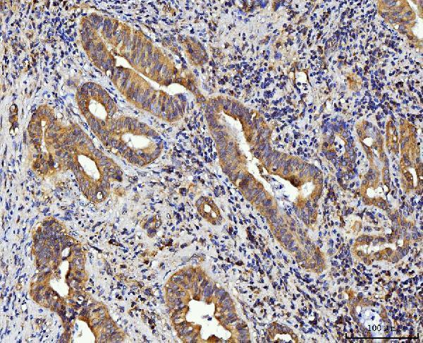
| Sample | Paraffin-embedded human gall bladder adenosquamous carcinoma tissue; fixative not specified (datasheet A04190-3) |
| Fixation | Image fixative and duration unreported (datasheet A04190-3); verify before use. |
| Sectioning | 4–5 µm sections on charged slides (standard) |
| Deparaffinisation | Xylene, graded ethanol series to water (standard) |
| Antigen retrieval | Heat retrieval: EDTA pH 8.0 (datasheet A04190-3); 20 min, 95–100 °C (standard) |
| Peroxidase block | 3% H2O2, 10 min, room temperature (standard) |
| Blocking | 10% goat serum (datasheet A04190-3) |
| Primary antibody | Rabbit anti-RPL10, 2-5 μg/ml (datasheet A04190-3) |
| Primary incubation | Overnight at 4 °C (datasheet A04190-3) |
| Detection | HRP-conjugated secondary, DAB chromogen (datasheet A04190-3) |
| Counterstain | Hematoxylin, blue, dehydrate and mount (standard) |
| Expected result | RPL10-positive staining in glandular cells of adrenal gland (HPA tissue IHC: High). HPA tissue profile: Ubiquitous granular cytoplasmic expression. No signal in the no-primary control. |
RPL10 is a cytoplasmic component of the large ribosomal subunit and has no transmembrane segment (UniProt P27635). In paraffin-section IHC, expect granular cytoplasmic staining across many cell types, including the high-staining examples listed by HPA (HPA tissue IHC: ubiquitous granular cytoplasmic expression). HPA rates tissue staining reliability Enhanced, while reporting medium agreement with RNA expression and caution that the antibodies may detect protein from more than one gene (HPA tissue IHC: reliability description).
| Granular cytoplasmic staining in adrenal glandular cells, marrow hematopoietic cells, or bronchial epithelium (HPA tissue IHC: High in each). | This fits the observed IHC pattern and UniProt cytoplasmic location (HPA tissue IHC: ubiquitous granular cytoplasmic expression; UniProt P27635). Compare intensity with nearby cells and controls; High is an HPA observation, not a required score for every section (HPA tissue IHC: High in listed cells; general IHC practice). |
| Predominantly nuclear, sharply membranous, or extracellular signal, with little cytoplasmic staining (UniProt P27635: cytoplasm; HPA tissue IHC: granular cytoplasm). | These compartments do not fit the supplied IHC pattern or the lack of a transmembrane segment (HPA tissue IHC: granular cytoplasm; UniProt P27635: topology). Treat them as possible staining artefacts and assess control sections and detection background before interpreting them as RPL10 (general IHC practice). |
| A cell population outside the reported pattern stains much more strongly than adjacent known-positive cells (HPA tissue IHC: listed High cell populations). | Consider cross-reactivity or endogenous detection activity, especially if the signal has the wrong compartment (general IHC practice; UniProt P27635: cytoplasm). HPA warns that its tissue staining may include protein from more than one gene, so morphology alone cannot establish target specificity (HPA tissue IHC: reliability description). |
| Broad, even chromogen covers cells and tissue spaces without distinct cytoplasmic granules (HPA tissue IHC: granular cytoplasm). | This obscures the reported pattern and is compatible with nonspecific background rather than an interpretable positive result (HPA tissue IHC: granular cytoplasm; general IHC practice). Review the negative control, blocking, antibody dilution, washes, and detection chemistry as general IHC checks (general IHC practice). |
| No signal appears in an otherwise intact section of HPA-listed high-staining tissue (HPA tissue IHC: High in listed cells). | Target-specific fixation effects are not established by the supplied assay evidence. Verify with a matched IHC source before attributing a result to fixation. |
| Cell population and tissue | HPA reports High staining in adrenal glandular, marrow hematopoietic, bronchial epithelial, caudate neuronal, and cortical glial cells, among others; soft-tissue fibroblasts are Low (HPA tissue IHC: positive and low lists). Use those distinctions when choosing comparison areas, without treating Low as absent (general IHC practice). |
| Compartment | UniProt places RPL10 in the cytoplasm as a 60S ribosomal-subunit component; HPA describes granular cytoplasmic tissue staining (UniProt P27635: subcellular location and subunit; HPA tissue IHC: profile). For chromogenic IHC, score the cellular pattern separately from overall stain intensity (general IHC practice). |
| Antibody evidence and specificity | HPA lists IHC as Enhanced for CAB010339 and Supported for HPA011311, while tissue reliability is Enhanced with medium RNA agreement and a multi-gene detection caution (HPA antibodies: IHC status; HPA tissue IHC: reliability description). Keep that specificity limit in mind when assigning weak or unexpected color to RPL10. |
| Protein processing and topology | UniProt lists no signal peptide, propeptide, or transmembrane segment and annotates the mature chain as residues 2–214 (UniProt P27635: processing and topology). The supplied record therefore gives no basis to predict a secreted or membrane-restricted IHC pattern. |
| ICC-IF comparison | HPA reports enhanced endoplasmic-reticulum and cytosol localization by ICC-IF, whereas its tissue IHC profile is granular cytoplasmic (HPA ICC-IF: main location; HPA tissue IHC: profile). The ICC-IF observation can inform compartment interpretation, but it does not define a paraffin-section protocol. |
| Situation | Likely cause | Next action |
|---|---|---|
| No chromogenic signal in a known-positive comparison section (HPA tissue IHC: listed High cell populations). | A run-level detection or antibody-use problem is possible; the supplied sources do not identify an RPL10-specific fixation effect (general IHC practice; HPA tissue IHC: source scope). | Confirm positive-control staining, reagent activity, and the IHC-validated antibody's paraffin-section instructions; review retrieval only as a general IHC variable (general IHC practice). |
| Nuclear or sharply membranous color dominates (UniProt P27635: cytoplasm and no transmembrane segment). | The location conflicts with the supplied tissue pattern and may reflect nonspecific staining or detection artefact (HPA tissue IHC: granular cytoplasm; general IHC practice). | Compare negative and known-positive controls, then assess blocking and detection conditions before scoring the color as target staining (general IHC practice). |
| Unexpected cells stain strongly while HPA-listed high-staining cells are weak (HPA tissue IHC: positive list). | Cross-reactivity or endogenous detection activity is possible; HPA also cautions about detection of protein from more than one gene (general IHC practice; HPA tissue IHC: reliability description). | Check cell identity and compartment, compare an appropriate negative control, and report the specificity uncertainty rather than assigning the signal solely to RPL10 (general IHC practice; HPA tissue IHC: reliability description). |
| Diffuse color masks cytoplasmic granules (HPA tissue IHC: granular cytoplasmic profile). | Nonspecific background can obscure a cell-level pattern; the payload does not identify a target-specific cause (general IHC practice). | Inspect negative controls, blocking, washes, primary-antibody dilution, and chromogen development using the catalog antibody's IHC-P instructions (general IHC practice). |
| Staining varies across the section despite similar cell populations (HPA tissue IHC: ubiquitous granular cytoplasmic profile). | Section or run variability is possible, but HPA's tissue levels do not establish an RPL10-specific retrieval or fixation mechanism (general IHC practice; HPA tissue IHC: source scope). | Review section quality and processing records, then compare matched control sections before interpreting regional loss as biology (general IHC practice). |
| Q: Should IF/ICC show the same fine pattern as paraffin IHC? | HPA reports endoplasmic-reticulum and cytosol localization in ICC-IF and granular cytoplasmic staining in tissue IHC; these are different assay observations (HPA ICC-IF: main location; HPA tissue IHC: profile). | A: Expect broadly cytoplasmic localization in either assay, but evaluate IF/ICC detail using its own guide and controls; do not transfer an IF/ICC protocol to this IHC design (UniProt P27635: cytoplasm; general IHC/IF practice). |


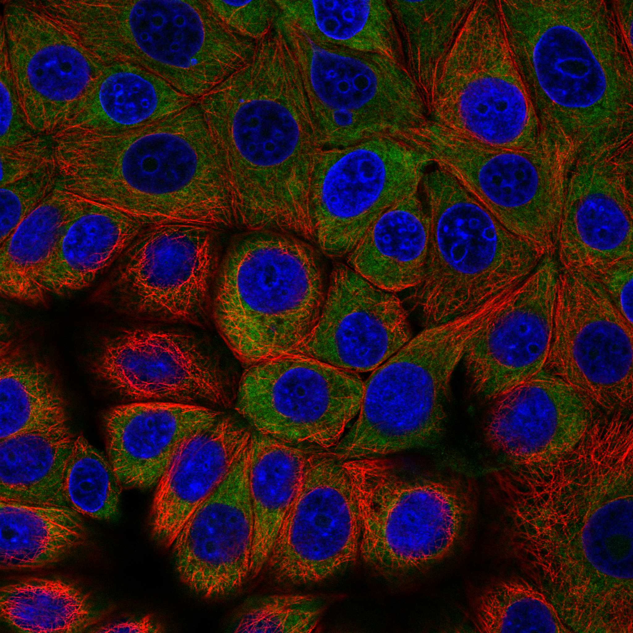
Comprehensive Human Protein Atlas IHC scoring per tissue (reliability: Enhanced — Medium consistency between antibody staining and RNA expression data. Caution, targets protein from more than one gene.). Rows are taken directly from the HPA tissue chart — click any row's HPA link to view the source.
| Tissue | Cell type | Level | Evidence | Source |
|---|---|---|---|---|
| Adrenal gland | Glandular cells | High | Protein (IHC) | HPA → |
| Appendix | Endocrine cells | High | Protein (IHC) | HPA → |
| Bone marrow | Hematopoietic cells | High | Protein (IHC) | HPA → |
| Breast | Glandular cells | High | Protein (IHC) | HPA → |
| Bronchus | Respiratory epithelial cells | High | Protein (IHC) | HPA → |
| Tissue | Cell type | Level | Evidence | Source |
|---|---|---|---|---|
| None in HPA: RPL10 is detected in all 45 scored tissues. Use a no-primary (secondary-only) and an isotype control instead. | ||||
Troubleshoot RPL10 staining in paraffin sections by checking retrieval, processing, compartment, controls and scoring before interpreting differences between samples.
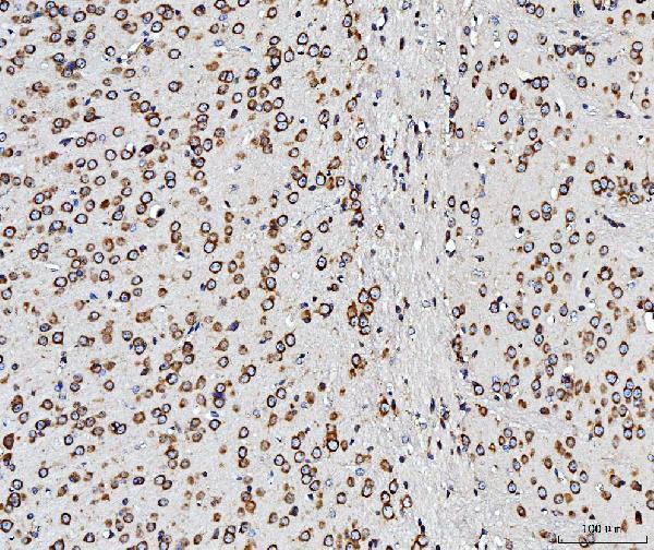
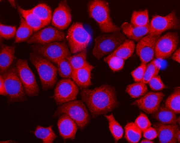
The IHC-validated antibody A04190-3 has IHC images from paraffin-embedded human gall bladder adenosquamous carcinoma and mouse and rat brain, plus an IF image from MCF-7 cells (A04190-3 image captions).
A04190-3 is shown in paraffin-embedded human gall bladder adenosquamous carcinoma and mouse and rat brain sections (A04190-3 IHC image captions). A04190-3 is also shown by IF in MCF-7 cells (A04190-3 IF image caption).
Which to pick: Choose A04190-3 for paraffin-section IHC; its IHC captions show staining in human, mouse and rat tissue, while the fixative is unreported (A04190-3 IHC image captions). Choose the same SKU for IF/ICC based on its listed applications and MCF-7 IF image (catalog applications; A04190-3 IF image caption). Its listed reactivity covers human, mouse and rat; clonality is unreported (catalog reactivity; catalog clone field).